

 PDF
PDF ePub
ePub Citation
 Print
Print


INTRODUCTION
Obstructive left main coronary artery (LMCA) disease is associated with a high rate of morbidity and mortality as a result of compromised myocardial blood supply; therefore, revascularization by coronary artery bypass grafting (CABG) surgery has been regarded as standard treatment. Over the past 20 years, there have been considerable therapeutic developments in the technique of percutaneous coronary intervention (PCI) for the treatment of obstructive coronary artery disease (CAD), involving improvements in stent technology, procedural techniques and refinement, periprocedural anticoagulation, concomitant antiplatelet agents, and cardiovascular medication.1)2)
Several randomized clinical trials (RCTs) have been conducted to evaluate the potential therapeutic role of PCI as an alternative to standard CABG. With the introduction of first-generation drug-eluting stents (DESs), RCTs demonstrated that stenting achieved similar rates of mortality and hard clinical endpoints and a lower rate of stroke, although the rate of repeat revascularization was seen to be higher.3)4)5)6) The development of second-generation DES was associated with improved efficacy and safety profiles compared with first-generation DES.7)8) Subsequent RCTs were conducted and PCI has achieved greater clinical recognition as a reasonable therapeutic modality.9)10) These data may impact on future clinical guidelines for myocardial revascularization and will ultimately will lead to greater use of PCI worldwide. Importantly, when undertaking PCI of the LMCA, there is increasing awareness of the need to achieve optimal procedural outcomes through the use of available technologies, including safer and more effective stents, intravascular imaging, and physiological assessment.
This review provides an update on the current management of LMCA disease with an emphasis on clinical data and procedural knowledge to support the use of PCI in a growing proportion of patients.
EVALUATION OF LMCA DISEASE
Contemporary evaluation of LMCA disease
Most patients with significant LMCA disease are symptomatic as a result of compromised blood supply to a large area of the myocardium. However, significant LMCA disease can be identified incidentally in stable patients undergoing coronary angiography. In the absence of significant LMCA stenosis or relevant clinical symptoms, the hemodynamic significance of incidental or intermediate LMCA lesions warrants further evaluation. Current clinical practice guidelines rely on angiographic lesion severity as the sole determinant of risk and for the de facto threshold for revascularization decision-making. However, this approach may be outdated in the current era of clinically proven noninvasive and invasive modalities and revascularization based solely on the angiographic appearance of LMCA stenosis of intermediate severity (50–70%) is not appropriate.11) In addition to the unwarranted surgical risk, premature CABG for potentially noncritical lesions may ultimately prove harmful to patients due to low graft patency rates and an accelerated rate of obstruction of bypassed native coronary vessels that may be up to 6-fold higher and which makes subsequent PCI technically challenging if required for symptom relief.12) In the contemporary clinical setting, more detailed evaluation of the anatomic severity and hemodynamic significance of clinically ambiguous LMCA lesions can be obtained by intracoronary imaging with intravascular ultrasound (IVUS), or physiologically using pressure wire assessment of the fractional flow reserve (FFR).
IVUS characterizes the vessel size and the distribution of the plaques within the LMCA and its daughter branches, enabling accurate minimal lumen area (MLA) measurements at a cross-sectional level. Evaluation of IVUS in a prospective study showed that a MLA ≥6 mm2 assessed by this technique is a safe value for deferring revascularization of the LMCA.13) Another study proposed a smaller MLA of ≤4.5 mm2 as a useful surrogate of functional significance in patients with isolated ostial and shaft intermediate LMCA stenosis.14) Ideally, a dual pullback from the left anterior descending artery (LAD) and left circumflex artery (LCX) should be used to fully characterize the LMCA bifurcation and avoid overestimation of, for example, the MLA at the LCX ostium from a LAD pullback.
Physiological guidance by means of FFR or instantaneous wave-free ratio (iFR) may be helpful for the evaluation of intermediate or ambiguous LMCA lesions.15) In this setting, the visual-functional mismatch between coronary angiography and FFR can be as high as 30–40%,11)16) and deferring LMCA PCI based on FFR values >0.75 or 0.80 has been shown to be safe.16)17) Recently, deferring revascularization of LMCA disease with an iFR >0.89 has also shown favorable outcomes.18)19) However, specific outcome studies evaluating iFR in LMCA disease are required to determine whether iFR could be widely adopted as the sole determinant of revascularization in patients with intermediate LMCA lesions.
MEDICAL TREATMENT FOR LMCA DISEASE
In the COURAGE trial,20) an initial strategy of optimal medical therapy vs. initial revascularization showed similar long-term outcomes in patients with stable CAD, excluding LMCA disease. The use of guideline-directed medical treatment (GDMT) and lifestyle interventions should be encouraged in patients with LMCA disease, as they are for patients with non-LMCA disease. However, the safety of deferred revascularization in patients with stable LMCA disease is not fully understood. Current clinical practice guidelines strongly recommend revascularization in all patients with ≥50% stenosis of the LMCA.21) This class IA recommendation is based on post hoc analysis of a few historical RCTs involving patients with chronic stable angina, demonstrating the superiority of surgical revascularization over medical treatment in terms of 5–10-year survival.22)23) However, in the context of updated GDMT, the medical management of patients in these early RCTs was not adequate as only 66% of patients were treated with beta blockers and only 19% received aspirin. In addition, the current widespread use of disease-modifying pharmacological interventions (i.e., statins, inhibitors of the renin-angiotensin-aldosterone system, and more effective antiplatelet agents, such as P2Y12 inhibitors) might alleviate adverse cardiovascular events as secondary prevention.
Patients with LMCA disease may have a diverse risk spectrum. Low-risk patients, such as those with 50–70% stenosis or preserved left ventricular (LV) function, have shown more favorable survival while receiving medical therapy alone than have patients with high-risk features (such as >70% stenosis, poor LV function, elevated LV end-diastolic pressure, or prior myocardial infarction [MI]), with 3-year survival rates of 66% vs. 41%, respectively.24)25) The study design of most contemporary large RCTs comparing clinical outcomes after medical vs. revascularization therapy has excluded patients with LMCA disease. Therefore, it has not yet been established whether optimal medical therapy represents a safe and appropriate alternative to revascularization in some selected LMCA groups of low-risk patients with stable LMCA disease.
PCI FOR THE TREATMENT OF LMCA DISEASE
In the 40 years history of PCI, the use of coronary stenting overcame the inherent limitations of balloon angioplasty and expanded the therapeutic role of PCI for LMCA disease. However, although PCI with bare-metal stents has demonstrated technical feasibility and acceptable clinical outcomes in highly selected, elective, low-risk patients,26)27)28) a substantial risk of angiographic and clinical restenosis has hampered the wider use of PCI for patients with LMCA disease. In the mid-2000s, the introduction of DES was associated with superior efficacy with respect to restenosis and repeat revascularization and therefore the use of PCI for the treatment of LMCA has increased significantly.29)30)31) In addition to significant improvements in stent technology, improved interventional techniques and adjunctive pharmacotherapy have progressively enhanced PCI outcomes in patients with LMCA disease.2) Contemporary second- and third-generation DES employ improved technology and engineering, including thinner strut platforms, easier delivery profiles, biocompatible or bioresorbable polymers, and more effective antiproliferative drugs.32) In the contemporary PCI setting of LMCA disease, one issue of clinical interest is that there are substantial differences between contemporary DES with regard to efficacy and safety outcomes. A recent merged analysis involving 4,470 patients with LMCA disease who underwent PCI with second-generation DES showed no significant differences in the 3-year rate of target-vessel failure among different DES.33) In addition, the incidence of definite stent thrombosis was extremely low (<1.0%) for all types of DES.
PCI strategy and technique
LMCA PCI conducted by experienced operators (i.e., those who have performed at least 15 LMCA PCIs per year for at least 3 consecutive years) is associated with better short- and long-term outcomes than LMCA PCI performed by less experienced operators.34) PCI of the LMCA ostium or shaft is a straightforward procedure that is associated with a lower requirement for repeat revascularization than PCI of the distal LMCA bifurcation.35) The ostium of the LMCA lacks the tunica adventitia and is richer in smooth muscle cells and elastic tissue than any other portion of the LMCA and its branches, which requires attention to ensure that stent expansion is adequate.36)
For LMCA bifurcation disease, many challenging technical issues remain. Generally, provisional stenting has been advocated as the preferred approach in bifurcation lesions, as it is technically simpler with at least similar outcomes to a systematic 2-stent strategy.37)38)39) In practice, the single-stent crossover technique has been used more frequently, in as many as 60% of all LMCA bifurcation treatments.40) However, this was recently challenged by two RCTs that compared the double-kissing (DK) crush technique with the culotte and provisional stenting strategies for the treatment of true distal LMCA bifurcations.41)42)43) In both studies, the DK crush technique significantly reduced the primary composite ischemic endpoint.
Selection of a single- or 2-stent technique should be based on disease involvement of the LCX ostium, as side-branch compromise after crossover stenting is frequent. Therefore, to determine the choice of a single- or 2-stent strategy, IVUS provides accurate information for both main- and side-branch disease status in LMCA bifurcation lesions. After main-stent crossover from the proximal LAD to the LM, geometric changes in the LCX ostium were related mainly to carina shift, reduction of MLA, and increased eccentricity of the external elastic membrane and carina angle between the LAD and the LCX.44) In cases in which the LCX ostium is significantly compromised (>50%) after provisional stenting, FFR measurement should be considered first before further treatment of the LCX.
IVUS is also useful for procedural optimization of PCI of the distal LMCA bifurcation. After stent implantation, IVUS guidance ensures adequate expansion at the level of the ostial LAD, the ostial LCX, the polygon of confluence (e.g., the convergence zone of the LMCA, LAD, and LCX), and the distal LMCA.45) The best IVUS-MSA (minimal stent area) criteria that predicted angiographic in-stent restenosis were 5.0 mm2 for the LCX ostium, 6.3 mm2 for the LAD ostium, 7.2 mm2 for the POC, and 8.2 mm2 for the distal LMCA. The 2-year major adverse coronary event-free survival rate was significantly lower in patients with underexpansion of at least one segment vs. lesions with no underexpansion (90±3% vs. 98±1%, p<0.001); post-stenting underexpansion was an independent predictor for major adverse cardiac events.
CLINICAL STUDIES COMPARING PCI AND CABG FOR LMCA DISEASE
Randomized clinical trials
Key clinical trials comparing PCI and CABG from the first-generation DES era to the second-generation DES era are summarized in Table 1. The SYNTAX study was a key pivotal trial; in the LMCA subgroup, no significant differences were seen in the 5-year rate of major adverse cardiac and cerebrovascular events (MACCEs), mortality, or MI between PCI and CABG.4) However, the 5-year rate of repeat revascularization was higher after PCI and the rate of stroke was higher after CABG. The first LMCA-specific RCT (RECOMBAT) showed that the 5-year rate of MACCEs, death, MI, or stroke was similar between PCI and CABG, but the rate of target-vessel revascularization was significantly higher after PCI.6)46) These trials prompted the initiation of two additional large-scale RCTs, EXCEL and NOBLE, which involved the use of contemporary DES.9)10) In the EXCEL study, the primary composite endpoint of death, stroke, or MI at 3 years was similar between PCI and CABG (p value for non-inferiority=0.02; p value for superiority=0.98).9) PCI was associated with a lower incidence of major periprocedural adverse events (i.e., major arrhythmias, infections, reoperations, bleeding, or transfusions). PCI was also associated with a more rapid recovery and greater improvement in quality of life at 30 days than was CABG, although both procedures resulted in similar quality of life and angina relief at 3 years.47) In the NOBLE trial, the primary composite endpoint of all-cause mortality, nonprocedural MI, stroke, or repeat revascularization at 5 years was significantly higher after PCI than after CABG (29% vs. 19% exceeding the limit for non-inferiority, respectively). The difference in favor of CABG was statistically significant (p value for superiority=0.007) and was driven by significantly higher rates of nonprocedural MI, repeat revascularization, and stroke in the PCI arm.10)
Table 1
Randomized clinical trials of percutaneous coronary intervention vs. coronary artery bypass grafting for left main coronary artery disease
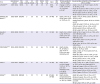
| Trial | Recruitment period | PCI/CABG | F/U (years) | SS (mean) | ACS (%) | Distal (%) | MVD (%) | Stent | IMA (%) | Primary endpoint (PCI vs. CABG) | Key secondary endpoints at the longest F/U (PCI vs. CABG) |
|---|---|---|---|---|---|---|---|---|---|---|---|
| LEMANS3)64) | 2001–2004 | 52/53 | 10 | NR | NR | 58 | 91 | BMS, DES | 81 | Change in LVEF at 1 year: 3.3±6.7% vs. 0.5±0.8%, p=0.047 | • Death, CVA, MI, or RR at 10 years: 52.2% vs. 62.5%, p=0.42 |
| • Death at 10 years: 21.6% vs. 30.2%, p=0.41 | |||||||||||
| • CVA at 10 years: 4.3% vs. 6.3%, p=0.58 | |||||||||||
| • MI at 10 years: 8.7% vs. 10.4%, p=0.68 | |||||||||||
| • RR at 10 years: 26.1% vs. 31.3%, p=0.39 | |||||||||||
| SYNTAX-Left MAIN4)65) | 2005–2007 | 357/348 | 5 | 30 | 30 | 61 | 68 | DP-PES | 97 | Death, CVA, MI, or RR at 1 year: 15.8% vs. 13.6%, p=0.4 | • Death, CVA, MI, RR at 5 years: 36.9% vs. 31.0%, p=0.12 |
| • Death/CVA/MI at 5 years: 19.0% vs. 20.8%, p=0.57 | |||||||||||
| • Death at 5 years: 12.8% vs. 14.6%, p=0.53 | |||||||||||
| • CVA at 5 years: 1.5% vs. 4.3%, p=0.03 | |||||||||||
| • MI at 5 years: 8.2% vs. 4.8%, p=0.10 | |||||||||||
| • RR at 5 years: 26.7% vs. 15.5%, p<0.001 | |||||||||||
| Boudriot et al.5) | 2003–2009 | 100/101 | 1 | 23 | NR | 72 | 41 | DP-SES | 99 | Death, MI, or RR at 1 year: 19.0% vs. 13.9%, p for non-inferiority=0.19 | • Death or MI at 1 year: 5.0% vs. 7.9%, p for non-inferiority<0.001 |
| • Death at 1 year: 2.0% vs. 5.0%, p for non-inferiority<0.001 | |||||||||||
| • MI at 1 year: 3.0% vs. 3.0%, p for non-inferiority=0.002 | |||||||||||
| • RR at 1 year: 14.0% vs. 5.9%, p for non-inferiority=0 | |||||||||||
| PRECOMBAT6)46) | 2004–2009 | 300/300 | 5 | 25 | 45 | 64 | 73 | DP-SES | 94 | Death, stroke, MI, ID-TLR at 1 year: 8.7% vs. 6.7%, p for non-inferiority=0.01 | • Death, stroke, MI, or ID-TLR at 5 years: 17.5% vs. 14.3%, p=0.26 |
| • Death, stroke, or MI at 5 years: 8.4% vs. 9.6%, p=0.66 | |||||||||||
| • Death at 5 years: 5.7% vs. 7.9%, p=0.32 | |||||||||||
| • Stroke at 5 years: 0.7% vs. 0.7%, p=0.99 | |||||||||||
| • MI at 5 years: 2.0% vs. 1.7%, p=0.76 | |||||||||||
| • RR at 5 years: 13% vs. 7.3%, p=0.02 | |||||||||||
| EXCEL9) | 2010–2014 | 948/957 | 3 | 21 | 24 | 81 | 51 | DP-EES | 99 | Death, stroke, or MI at 3 years: 15.4% vs. 14.7%, p for non-inferiority=0.02, p=0.98 for superiority | • Death, stroke, MI, or IDR at 3 years: 23.1% vs. 19.1%, p for non-inferiority=0.01 |
| • Death at 3 years: 8.2% vs. 5.9%, p=0.11 | |||||||||||
| • Stroke at 3 years: 2.3% vs. 2.9%, p=0.37 | |||||||||||
| • MI at 3 years: 8.0% vs. 8.3%, p=0.64 | |||||||||||
| • IDR at 3 years: 12.6% vs. 7.5%, p<0.001 | |||||||||||
| NOBLE10) | 2008–2015 | 592/592 | 5 | 22 | 17 | 81 | NR | BP-BES, DP-SES | 93 | Death, stroke, nonprocedural MI, RR at 5 years: 29% vs. 19%, p=0.0066 | • Death at 5 years: 12% vs. 9%, p=0.77 |
| • Stroke at 5 years: 5% vs. 2%, p=0.073 | |||||||||||
| • Nonprocedural MI at 5 years: 7% vs. 2%, p=0.004 | |||||||||||
| • RR at 5 years: 16% vs. 10%, p=0.032 |
ACS = acute coronary syndrome; BMS = bare-metal stent; BP-BES = biodegradable-polymer biolimus-eluting stent; CABG = coronary artery bypass grafting; CVA = cerebrovascular accident; DES = drug-eluting stent; DP-EES = durable-polymer everolimus-eluting stent; DP-SES = durable-polymer sirolimus-eluting stent; DP-PES = durable-polymer paclitaxel-eluting stent; F/U = follow-up; IDR = ischemia-driven revascularization; ID-TLR = ischemia-driven target lesion revascularization; IMA = internal mammary artery; LVEF = left ventricular ejection fraction; MI = myocardial infarction; MVD = multivessel disease; NR = not reported; PCI = percutaneous coronary intervention; RR = repeat revascularization; SS = SYNTAX score.
There may be several explanations for the inconsistent results seen in the EXCEL and NOBLE studies.48) First, different types of DES were used. In EXCEL a thin-strut, fluoropolymer-based CoCr-EES was employed, which was associated with the lowest risk of stent thrombosis of all available DES.49) The NOBLE study used first-generation, thicker-strut, stainless-steel, sirolimus-eluting Cypher stents (11%) or the biolimus-eluting Biomatrix Flex stent (89%). A substantial difference in the rate of definite stent thrombosis (0.7% in EXCEL vs. 3% in NOBLE) suggests the differential performance of stenting for LMCA disease. Secondly, the soft clinical endpoint of repeat revascularization was adopted as the key component of the primary endpoint in the NOBLE study. The majority of previous studies have consistently shown that the rate of repeat revascularization is significantly higher after PCI than after CABG. Therefore, the selection of this primary composite outcome may unfairly penalize the PCI stratum. The SYNTAX trial showed that the increase in repeat revascularization in the PCI group did not directly translate into an increase in the incidence of death or MI.50) Thirdly, the definitions used for components of the primary composite outcomes differed between the studies, particularly the definition of MI. The Society for Cardiovascular Angiography and Interventions -defined clinically relevant MI definition was used in EXCEL,51) while periprocedural MI was disregarded in NOBLE. Finally, in the NOBLE study, the rate of stroke was more than two times higher after PCI than after CABG, which is not in agreement with the findings of previous clinical trials comparing PCI and CABG. This observation lacks a clear explanation and biologic plausibility and is, therefore, likely to be due to a chance effect.52)
Meta-analyses
In a meta-analysis of the four largest studies of LMCA revascularization with follow-up available at 3–5 years, incorporating data from the EXCEL and NOBLE trials, the hazard ratio (HR) for death, stroke, or MI with PCI compared with CABG was neutral (1.06) in a random-effects model (p=0.60).53) Based on individual patient data reconstruction, the Kaplan-Meier estimates of death, stroke, or MI at 5 years were 18.3% for PCI and 16.8% for CABG (p=0.52). No statistically significant subgroup interaction for this combined outcome was noted across studies based on the generation of DES used for PCI (p value for interaction=0.25). There were no significant differences in the pooled effects for death (HR, 1.04; p=0.77) and cardiac death (HR, 1.00; p=0.99). The endpoints of MI and stroke also did not differ between the PCI and CABG groups (HR, 1.48; p=0.17 and 0.87; p=0.72, respectively), but these outcomes were confounded by high heterogeneity across the trials. Repeat revascularization was consistently higher following PCI in all trials, leading to a pooled HR of 1.70 (p<0.001). In another meta-analysis, including all the six trials available to date, missing data were collected by the principal investigators, enabling further subgroup analyses.54) PCI was found to significantly reduce death, MI, or stroke by 36% within 30 days. PCI reduced periprocedural MI by 33%, but this effect was offset by 93% more spontaneous MIs beyond 30 days after the procedure. Cardiac death differed in relation to angiographic complexity in that it tended to be lower with PCI among patients with low SYNTAX scores and higher in patients with high SYNTAX scores.
A recent large-scale, pooled analysis of individual patient data reported a comparable treatment effect for PCI and CABG with regard to all-cause mortality up to 5 years in selected patients participating in RCTs.55) This analysis included 11 RCTs involving 11,518 patients who were assigned to undergo PCI (n=5,753) or CABG (n=5,765). The 5-year rate of all-cause mortality was 11.2% after PCI and 9.2% after CABG (HR, 1.20; 95% CI, 1.06–1.37; p=0.004). Interestingly, the 5-year all-cause mortality differed significantly between the two interventions in patients with multivessel disease (11.5% after PCI vs. 8.9% after CABG; HR, 1.28; 95% CI, 1.09–1.49; p=0.002), including in those with diabetes (15.5% vs. 10.0%, respectively; HR, 1.48; 95% CI, 1.19–1.84; p=0.0004), but not in those without diabetes (8.7% vs. 8.0%, respectively; HR, 1.08; 95% CI, 0.86–1.36; p=0.49). By contrast, the 5-year rate of all-cause mortality was similar between the two groups in patients with LMCA disease (10.7% after PCI vs. 10.5% after CABG; HR, 1.07; 95% CI, 0.87–1.33; p=0.52), regardless of diabetes status and SYNTAX score.
Registries
Large registries of patients undergoing revascularization for LMCA disease in different geographical regions are useful resources to generalize the findings from RCTs into the daily clinical scenario. The MAIN-COMPARE, IRIS-MAIN, PRECOMBAT-2, and DELTA-2 registries cover a treatment period of 2000–2015,2)56)57)58)59) and are summarized in Table 2. Compared with PCI patients enrolled in the EXCEL trial, the mean SYNTAX score in these registries tended to be higher. In the DELTA-2 study of LMCA PCI with second-generation DES (n=3,986), Kaplan-Meier estimates of events at 2 years were 9.5% and 16.7% for death or target-vessel revascularization, respectively.56) HRs for PCI vs. CABG with respect to the composite of death, stroke, or MI were similar between the PCI and CABG groups.
Table 2
Contemporary large observation registries of percutaneous coronary intervention vs. coronary artery bypass grafting for left main coronary artery disease

| Study | Enrolment period | Number | SS (mean) | ACS (%) | Distal (%) | MVD (%) | Stent | IMA (%) | Key outcome (PCI vs. CABG) | Adjusted outcome (PCI vs. CABG) |
|---|---|---|---|---|---|---|---|---|---|---|
| MAIN-COMPARE (Wave 2)57)58) | 2000–2006 | PCI, 784 | PCI, NR | PCI, 63 | PCI, 57 | PCI, 58 | G2-DES | NR | Death, Q-wave MI, or stroke at 5 years: 12.7% vs. 16.3%, p=0.02 | HR, 0.99; 95% CI, 0.73–1.36; p=0.99 |
| CABG, 690 | CABG, NR | CABG, 76 | CABG, 53 | CABG, 88 | ||||||
| PRECOMBAT-259) | 2009–2010 | PCI, 334 | PCI, 21 | PCI, 45 | PCI, 72 | PCI, 57 | G2-DES | NR | Death, MI, stroke, or ischemia-driven TVR at 540 days: 8.9% vs. 6.7%, p=0.23 | HR, 0.84; 95% CI, 0.51–1.40; p=0.51 |
| CABG, 272 | CABG, 27 | CABG, 54 | CABG, 60 | CABG, 75 | ||||||
| IRIS-MAIN (Wave 3)2) | 2007–2013 | PCI, 1,658 | PCI, NR | PCI, 55 | PCI, 65 | PCI, 64 | G2-DES | 95 | Death, stroke, or MI at 3 years: NR | HR, 0.91; 95% CI, 0.68–1.21; p=0.50 |
| CABG, 704 | CABG, NR | CABG, 57 | CABG, 72 | CABG, 91 | ||||||
| DELTA-256) | 2006–2015 | PCI, 3,986 | PCI, 27 | PCI, 36 | PCI, 85 | PCI, 74 | G2-DES | NR | Death, CVA, or MI at 501 days: 10.3% vs. 11.6%, p=NR | HR, 0.73; 95% CI, 0.55–0.98; p=0.03 |
| CABG, 901 | CABG, 38 | CABG, 65 | CABG, 58 | CABG, 94 |
ACS = acute coronary syndrome; CABG = coronary artery bypass grafting; CI = confidence interval; CVA = cerebrovascular accident; DES = drug-eluting stent; G2 = second-generation; HR = hazard ratio; IMA = internal mammary artery; MI = myocardial infarction; MVD = multivessel disease; NR = not reported; PCI = percutaneous coronary intervention; SS = SYNTAX score; TVR = target vessel revascularization.
Recently, an observation study of the MAIN-COMPARE registry reported 10-year comparative outcomes of PCI and CABG for LMCA disease.60) Overall, there was no significant difference in the adjusted risk of death and the composite outcome between the PCI group and the CABG group up to 10 years. The risk of target-vessel revascularization was significantly higher in the PCI group. However, in the cohort comparing DES and concurrent CABG, DES was associated with a higher risk of death (HR, 1.35; 95% CI, 1.00–1.81) and the composite outcome of death, Q-wave MI, or stroke (HR, 1.46; 95% CI, 1.10–1.94) than was CABG after 5 years. In the DES era, the application of PCI for LMCA disease was substantially expanded and widely performed in patients with a broader range of clinical and anatomical complexities.61) Therefore, the results of this study can be interpreted as follows: CABG is associated with superior long-term outcomes compared with multivessel PCI in patients with high clinical and anatomical complexity. It is interesting to note that the suggestion of superiority of CABG over PCI during long-term follow-up was also seen in the EXCEL and NOBEL trials, which reported a trend towards late catch-up or crossover in the rate of death or the composite endpoint of death, stroke, or MI favoring CABG over PCI during the late period of follow-up. Therefore, longer-term follow-up is necessary to examine additional differences between PCI and CABG over time.
Revascularization guidelines
Existing clinical practice guidelines continue to advocate CABG surgery as the singular class I indication for myocardial revascularization of LMCA disease. However, more recent RCTs and registry studies support PCI as a reasonable alternative in selected patients with less complex LMCA anatomy.
As new evidence has become available, guideline recommendations for LMCA revascularization have slowly evolved over time in both Europe and the US (Table 3). Recently, the 2018 European Society of Cardiology guidelines incorporated compelling data from the EXCEL and NOBLE trials, as well as the results of the pooled analysis.62) The 2018 European guideline indicates the same class of recommendation, but all evidence levels have been upgraded to level A. For PCI in LMCA with intermediate anatomical complexity, the previous class IIa recommendation was maintained in view of the incomplete 5-year follow-up in the two largest RCTs in this setting. In the future, the guideline will propose less restrictive indications for PCI, thereby expanding the patient pool that might be eligible for PCI. In addition, given that SYNTAX score was not an important factor for decision-making regarding optimal revascularization and to differentiate the comparative outcomes between CABG and PCI in the EXCEL and NOBLE studies, it may be debated whether the SYNTAX score can play a pivotal role in decision-making regarding LMCA revascularization.
Table 3
Secular change of myocardial revascularization guidelines for left main coronary artery disease

| Guideline | Class of recommendation | Level of evidence | |
|---|---|---|---|
| 2005 ACC/AHA/SCAI66) | III—PCI is not recommended in patients with unprotected LMCA disease and eligibility for CABG | C | |
| 2005 ESC/EACTS67) | IIb—Stenting for unprotected LMCA disease should only be considered in the absence of other revascularization options | C | |
| 2009 ACC/AHA/SCAI68) | IIb—PCI of the LMCA with stents as an alternative to CABG may be considered in patients with anatomic conditions that are associated with a low risk of PCI procedural complications and clinical conditions that predict an increased risk of adverse surgical outcomes | B | |
| 2010 ESC/EACTS69) | IIa—LMCA isolated or þ 1VD, ostium/shaft | B | |
| IIb—LMCA isolated or þ 1VD, distal bifurcation | |||
| IIb—LMCA þ 2VD or 3VD, SYNTAX score ≤32 | |||
| III—LMCA þ 2VD or 3VD, SYNTAX score ≥33 | |||
| 2011 ACCF/AHA/SCAI21) | IIa—For SIHD patients when both of the following are present: | B | |
| • Anatomic conditions associated with a low risk of PCI procedural complications and a high likelihood of good long-term outcomes (e.g., a low SYNTAX score [#22], ostial or trunk left main stenosis) | |||
| • Clinical characteristics that predict a significantly increased risk of adverse surgical outcomes (e.g., STS-predicted risk of operative mortality >5%) | |||
| IIb—For SIHD patients when both of the following are present: | B | ||
| • Anatomic conditions associated with a low-to-intermediate risk of PCI procedural complications and an intermediate-to-high likelihood of good long-term outcomes (e.g., low-intermediate SYNTAX score of <33, bifurcation left main stenosis) | |||
| • Clinical characteristics that predict an increased risk of adverse surgical outcomes (e.g., moderate-severe chronic obstructive pulmonary disease, disability from previous stroke, or previous cardiac surgery; STS-predicted risk of operative mortality >2%) | |||
| III: HARM—For SIHD patients (vs. performing CABG) with unfavorable anatomy for PCI who are good candidates for CABG | B | ||
| 2014 ESC/EACTS70) | I—LMCA with a SYNTAX score ≤22 | B | |
| IIa—LMCA with a SYNTAX score 23–32 | |||
| III—LMCA with a SYNTAX score ≥33 | |||
| 2014 ACC/AHA/AATS/PCNA/SCAI/STS71) | IIa—For SIHD patients when both of the following are present: | B | |
| • Anatomic conditions associated with a low risk of PCI procedural complications and a high likelihood of good long-term outcomes (e.g., a low SYNTAX score [≤22], ostial or trunk left main stenosis) | |||
| • Clinical characteristics that predict a significantly increased risk of adverse surgical outcomes (e.g., STS-predicted risk of operative mortality >5%) | |||
| IIb—For SIHD patients when both of the following are present: | B | ||
| • Anatomic conditions associated with a low-to-intermediate risk of PCI procedural complications and an intermediate-to-high likelihood of good long-term outcome (e.g., low-intermediate SYNTAX score of <33, bifurcation left main stenosis) | |||
| • Clinical characteristics that predict an increased risk of adverse surgical outcomes (e.g., moderate-severe chronic obstructive pulmonary disease, disability from previous stroke, or previous cardiac surgery; STS-predicted risk of operative mortality >2%) | |||
| III: HARM—For SIHD patients (vs. performing CABG) with unfavorable anatomy for PCI and who are good candidates for CABG | B | ||
| 2018 ESC/EACTS62) | I—LMCA with a SYNTAX score ≤22 | A | |
| IIa—LMCA with a SYNTAX score 23–32 | |||
| III—LMCA with a SYNTAX score ≥33 | |||
AATS = American Association for Thoracic Surgery; ACC = American College of Cardiology; ACCF = American College of Cardiology Foundation; AHA = American Heart Association; CABG = coronary artery bypass grafting; EACTS = European Association for Cardio-Thoracic Surgery; ESC = European Society of Cardiology; LMCA = left main coronary artery; PCI = percutaneous coronary intervention; PCNA = Preventive Cardiovascular Nurses Association; SCAI = Society for Cardiovascular Angiography and Interventions; SIHD = stable ischemic heart disease; STS = Society of Thoracic Surgeons; VD = vessel disease.
The heart team approach
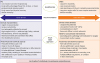
Regardless of which method of revascularization is used, current guidelines highlight the importance of a ‘heart team’ approach to the management of LMCA disease. The heart team evaluates the risks and benefits of PCI, surgery, or medical treatment alone, taking into account the patient's informed preference (Figure 1). In general, PCI offers more rapid recovery and a lower early adverse event rate, whereas CABG offers more durable revascularization. However, the relative outcomes of PCI vs. CABG can be attributed to a complex interplay of patient comorbidities, coronary anatomic complexity, and ventricular function, in addition to other less tangible factors such as operator expertise and compliance with medication. The complexity and extent of coexisting CAD with the intention of achieving complete revascularization should also be considered by the heart team. Previous evaluation has shown that major adverse cardiovascular events are higher in patients with incomplete revascularization than in those with complete revascularization regardless of the revascularization strategy.63) The heart team approach is critical when evaluating the risks and benefits of surgery in high- and extreme-risk populations. Additional clinical factors that are not included in most risk models also need to be considered by the heart team when making management recommendations, including frailty, cognitive status, surgical recovery and social support, quality of life, life expectancy, patient preference, and any potential concerns regarding tolerance or adherence with long-term dual antiplatelet therapy.
Figure 1
Heart team approach for LMCA revascularization.
Figure adapted with permission from Park et al.48)
CABG = coronary artery bypass grafting; CTO = chronic total occlusion; EF = ejection fraction; DAPT = dual antiplatelet therapy; LM = left main; MI = myocardial infarction; MVD = multivessel disease; PCI = percutaneous coronary intervention.
CONCLUSIONS
Over the past 20 years, significant advancements in stent technology, technical refinement, image and physiological guidance, and adjunctive drug therapy have led to progressive improvements in outcomes following PCI in patients with LMCA disease. In the contemporary clinical setting, LMCA PCI has become a viable option in daily practice not only for patients with less complex clinical and anatomic characteristics (i.e., isolated left main disease, ostial or shaft left main disease, or additional less complex CAD), but also for patients with complex clinical and anatomic characteristics (i.e., distal LMCA bifurcation or those with acute MI or unsuitability for CABG).
Which approach will be of most benefit to individual patients with LMCA disease should be decided by the local heart team, which comprises a general cardiologist, interventional cardiologist, and cardiac surgeon. The heart team will consider the clinical circumstances, any technical issues, and the likelihood of safely achieving complete revascularization with each procedure. It will also be important to consider the patient's own preference once the procedures have been explained in full.
 XML Download