

 PDF
PDF ePub
ePub Citation
 Print
Print


INTRODUCTION
Atherosclerosis is a chronic inflammatory disease process, which accounts for majority of the cases of coronary artery disease (CAD). It is a dynamic process which leads to plaque formation over time and in some cases progress to plaque disruption leading to acute coronary syndromes (ACSs).1) Traditionally, invasive coronary angiography (ICA) has been the mainstay for diagnosing and evaluating the severity of atherosclerosis. However, since atherosclerosis is typically asymptomatic until it becomes severe enough to cause ischemia, coronary computed tomography angiography (CCTA) has evolved as a robust tool for identification and risk stratification of asymptomatic atherosclerosis as well as diagnosing CAD in symptomatic patients. Given that CCTA is a non-invasive imaging modality, it has been widely adopted as a safe and reliable technology and its application continues to expand.
In this review, we focus on the role of CCTA in diagnosis and prognostication of symptomatic and asymptomatic coronary atherosclerosis, as well as the recent advancements in the realm of identifying plaque morphology.
CORONARY ARTERY CALCIUM (CAC) SCORE
Coronary artery calcification is strongly associated with overall atherosclerotic burden and is a powerful prognostic tool above and beyond clinical risk scores.2)3) The following section will briefly review the clinical interpretation, prognostic value and appropriate clinical use of CAC scoring.
The process of intimal calcification has long been associated with coronary atherosclerosis. Based on this principle, detection of coronary artery calcification using CCTA has evolved as a widespread screening tool for atherosclerotic heart disease in asymptomatic individuals and has proven pivotal in guiding treatment.4)5) CAC score is reported using the Agatston scoring system, calculated as the product of the calcium density factor, stratified by Hounsfield unit (HU), multiplied by the area of the calcification. The sum of the calcium score of each calcification within all of the tomographic slices is then summed up to give the total CAC score.6) The CAC score is widely applicable since it can be performed rapidly (using semi-automated software), at low a radiation dose (1–2 mSev) and does not require the administration of contrast.
CAC score has great utility as a screening tool as it can detect coronary atherosclerosis with high sensitivity and has a high negative predictive value (NPV) for ruling out CAD.7) A CAC of 0 has a very high NPV for mortality of 0.6%.8) A CAC of > 100 signifies presence of mild CAD and > 400 signifies severe disease. A higher calcium score directly correlates with higher risk of all-cause mortality and cardiovascular (CV) events independent of traditional cardiac risk factors in the Framingham risk score.9)10) On the other hand, recent data from the Multi-Ethnic Study of Atherosclerosis (MESA) study suggests that calcium volume may be superior to the Agatston CAC, as calcium HU density had an inverse association with CV outcomes.11)12)
In addition to prognostication of CAD, CAC has also been shown to impact treatment strategies and guide lifestyle modifications. Studies have demonstrated that high baseline CAC is associated with a 2 to 3-fold increase in the odds for initiation of CV preventative medications including aspirin and statins.13) Although the data on the impact of high CAC on lifestyle modifications is more controversial, some studies have noted a favorable effect on exercise and dietary changes.14) Based on this data, the 2010 American College of Cardiology Foundation/American Heart Association guidelines recommend that measurement of CAC is reasonable for CV risk assessment in asymptomatic adults at intermediate risk (10% to 20% 10-year risk) (class IIA; level of evidence B). Measurement of CAC in low to intermediate risk (6% to 10% 10-year risk) is a lower strength of recommendation (class IIB; level of evidence B) and CAC is not recommended (class III) in low risk (<6% 10-year risk) individuals.
Major limitations of CAC score include poor evaluation of the degree of angiographic stenosis and prediction of ischemia.15)16)17) Coronary calcium score is also limited in the ability to identify non-calcified plaque, as well as to monitor plaque progression in response to treatment.18) Additionally, the applicability of CCTA in symptomatic patients is limited, since a calcium score of zero does not necessarily portend a similar reassuring prognosis, as it would in asymptomatic patients.19)
IDENTIFICATION OF CAD AND DEGREE OF STENOSIS
Diagnostic accuracy of CCTA compared to invasive coronary angiography (ICA)
Coronary computed tomographic angiography has been widely accepted as a reliable non-invasive modality for identification of CAD and evaluation of severity of coronary plaque. Compared with non-contrast CAC score, CCTA offers the advantage of detecting both calcified and non-calcified plaque as well as monitoring plaque progression and evaluating plaque morphology. With advancement in technology, the radiation dose associated with CCTA has significantly decreased and when compared to functional testing with myocardial perfusion imaging (MPI), the cumulative dose of radiation is comparable or even lower depending on the patient's body habitus.20) The following section will review the accuracy and the role of CCTA in diagnosis of CAD.
Several studies have evaluated the diagnostic accuracy of CCTA for evaluating the severity of coronary atherosclerosis versus the gold standard of ICA. Comparison of these two modalities in CORE 64, a prospective multicenter trial of 405 symptomatic patients with suspected CAD and CAC of ≤ 600, found the sensitivity and specificity of CCTA to be 85% and 90% respectively for detecting coronary stenosis.21) In the ACCURACY trial, a trial of 230 patients with chest pain, sensitivity and specificity for ≥70% stenosis was 94% and 83%, respectively.22) A more notable finding was a NPV of 99% and a positive predictive value (PPV) of only 48%. The high NPV of 97% was reproduced in another prospective multicenter trial, and on a vessel based analysis, the authors found that lesions in the right coronary artery (RCA) and left circumflex artery (LCx) were more often undetected compared to left main coronary artery (LMCA) or left anterior descending artery (LAD). All patients with 3-vessel disease or left main CAD were appropriately identified in this study. Notably, CCTA did overestimate lesion severity in 245 vessels lending to a generally lower specificity of 64% and a PPV of 86%. In essence, given the high sensitivity and NPV of CCTA compared to coronary angiography, it can be concluded that a negative scan is very helpful in excluding obstructive CAD.23)
Utility of CCTA in prognosticating patients with CAD
In addition to diagnosis and localization of CAD, CCTA also provides important prognostic information. Based on the high sensitivity and NPV of CCTA, a negative scan is very helpful in excluding obstructive CAD and has been associated with an excellent prognosis and a very low event rate of <1%; comparable to healthy low-risk individuals.23) In the first large single-center study evaluating the association of CAD detected on CCTA with all-cause mortality in patients with suspected CAD and chest pain, Min et al.23) concluded that the extent and severity of CAD on CCTA correlated directly with all cause mortality. In this study, at the mean follow-up period of 15, CCTA predictors of death included proximal LAD stenosis and number of vessels with ≥50% and ≥70% stenosis (p<0.0001 for all). On the other hand, patients with <50% stenosis had the highest survival at 99.7%. Min et al.24) reproduced these findings in a larger 2 center study of 5,330 patients, and found that over 2.3±0.6 years of follow-up, individuals with increasing numbers of vessels with obstructive stenoses (≥70%) experienced increased risk of death compared to those without obstructive CAD, with 1-vessel (hazard ratio [HR], 2.23; 95% confidence interval [CI], 1.34–3.72), 2-vessel (HR, 3.29; 95% CI, 1.62–6.71), or 3-vessel (HR, 7.35; 95% CI, 3.79–14.29) (p<0.001 for all). Within the large international multicenter COronary CT Angiography EvaluatioN For Clinical Outcomes: an InteRnational Multicenter (CONFIRM) registry, among 24,775 patients, Min et al.29) found these results to be true in both older (≥65 years) and younger (<65 years) subgroups, demonstrating the generalizability of this finding across age, gender and ethnicity. In a meta-analysis by Bamberg et al.,25) significant CAD (defined as presence of ≥1 significant coronary stenosis) was associated with a 10-fold higher risk of CV events and a 6 times higher risk of death independent of CAC score. The authors concluded that each diseased coronary segment lends a 23% higher risk for adverse CV outcomes.
Although obstructive CAD confers a poor prognosis, the presence and extent of non-obstructive plaque also predicts incident mortality beyond patient demographic data and traditional CAD risk factors.26) Among patients with non-obstructive CAD, Bittencourt et al.27) found that patients with extensive plaque (>4 segments) experienced a higher rate of CV death or myocardial infarction, comparable with those who have non-extensive disease. Even among patients with obstructive CAD, greater extent of coexisting non-obstructive plaque was associated with higher event rate. The role of CCTA in identifying non-obstructive CAD is important, because most patients with non-obstructive CAD would have normal functional stress testing and would likely not be referred for ICA or initiated on CV medications. To summarize, in CCTA, both plaque burden and stenosis severity, particularly in proximal segments, carry incremental prognostic value.28)
In addition to prognosticating clinical outcomes, CCTA is also useful in prognosticating the impact of invasive treatment strategy. Min et al.29) evaluated the benefit of revascularization in patients without prior CAD within the CONFIRM registry, and found that only patients with high risk CAD (defined as 2-vessel obstructive CAD with proximal LAD involvement, 3-vessel CAD, and left main CAD) exhibited survival benefit from revascularization when compared to standard medical therapy, while those with lesser disease severity did not.
ASSESSMENT OF CORONARY ATHEROSCLEROTIC PLAQUE MORPHOLOGY WITH CCTA
As our knowledge and understanding of atherosclerosis has evolved, it is now well established that in addition to the degree of coronary stenosis, plaque morphology plays an important role in prognostication and predicting plaques that will be associated with future adverse events.30) The following section discusses the evolving role of CCTA in characterization of plaque morphology.
Virmani et al.1) and investigators of Providing Regional Observations to Study Predictors of Events in the Coronary Tree (PROSPECT) trial have identified several important features of the high-risk or “vulnerable” plaque.” Larger plaque volume (especially in non-calcified plaque) has been associated with worse prognosis.31) In addition to the plaque volume, large lipid rich necrotic core with a thin fibrous cap (<65 µm) and coronary microcalcifications have been identified as features of high risk plaque using invasive imaging technology like intravenous ultrasound (IVUS) and optical coherence tomography (OCT).32) Given that these vulnerable plaques often times do not cause critical stenosis prior to rupture, recognition of plaque morphology in non-obstructive CAD in vivo can be a very helpful tool for risk stratification.30) Although in vivo assessment of plaque morphology is well established with invasive techniques like IVUS and OCT33) they carry inherent risks of an invasive procedures.34) Therefore, there has been significant interest in using CCTA as a non-invasive imaging modality, to evaluate plaque characteristics and morphology.
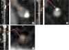
Although CCTA lacks the spatial resolution to visualize cap thickness, several high-risk plaque features that predict plaque vulnerability have been identified on CCTA. In a study of 38 patients with ACS, Motoyama et al.35) found that coronary plaques that were more likely to rupture if they had the following features on CCTA: 1) positive remodeling, 2) presence of non-calcified or low attenuation plaque measured to be <30 HU, and 3) spotty calcification (Figure 1). They also found that although each of these characteristics individually predicted ACS, the presence of all three of these features in a plaque had incremental effect on the PPV (exact number 95%) for the presence of culprit lesion, and the absence of all three of these features had a high NPV (100%) for the absence of culprit lesion. Of these three individual plaque characteristics, they identified positive remodeling as the most important feature in the culprit lesions.35) Additionally, the presence of a napkin ring sign, as characterized by a low attenuation core (<30 HU) surrounded by a rim like area of higher (>130 HU) attenuation has been proposed as an alternative marker of high-risk plaque.36) In a prospective study of 895 patients by Otsuka et al.,36) the napkin ring sign was an independent predictor of ACS. In another study evaluating thin-cap fibroatheroma (TCFA) vs. non-TCFA plaque on OCT, the TCFA plaques were 11-fold more likely to have the ring-like attenuation of a napkin ring sign on CCTA. Although the histopathologic correlate of the napkin ring sign is not well defined, a small study postulated that the central low attenuation area represents the lipid-rich necrotic core and the rim of higher attenuation may correspond to the fibrous plaque of a fibroatheroma.37) When compared to patients with stable angina, patients with ACS had significantly greater number of non-calcified plaques and spotty calcifications; a finding which lends to the higher risk associated with these plaque features.38) Furthermore, these high-risk plaque features have been shown to predict a higher risk of CV events independent of the degree of stenosis.39) Even in patients without the evidence of ischemia on an exercise MPI stress test, the presence of these high-risk plaque features in plaques with >50% stenosis was associated with higher risk of ACS.40) Chang et al.,41) in the recently completed Incident COronary EveNts Identified by Coronary Tomography (ICONIC) trial, will elucidate the prognostic significance of high risk plaque features for precursors of ACS versus non-events in a nested case control study within the CONFIRM registry.
Figure 1
CCTA based plaque characteristics associated with high-risk plaque on multiplanar reconstruction and corresponding and short-axis en face axial view. (A) Positive remodeling (red arrows) characterized by remodeling index (reference vessel size/stenosis vessel size) ≥1.1. (B) Spotty calcification (red arrows) characterized by calcification <3 mm. (C) Low attenuation plaque (red arrows) characterized by <30 HUs.
CCTA = coronary computed tomography angiography; HU = Hounsfield unit.
Akin to IVUS and OCT, there has been significant interest in using CCTA for quantitative assessment of plaque subcomponents (e.g. calcified and non-calcified plaque volumes). Several studies have compared IVUS to CCTA and have found an overall good correlation between the two modalities.42)43) Comparing 76 plaques measured on CCTA with IVUS, the plaque volume determined by CCTA was highly correlated (r=0.98; p<0.001). CCTA tended to overestimate calcified and mixed plaque volume compared to IVUS (4±19 mm3; p=not significant) and underestimate noncalcified plaque volume (9±11 mm3; p<0.001).44) Voros et al.45) found that the minimal lumen diameter may be underestimated up to 21%, and that there was 39% overestimation of percentage diameter stenosis due to calcification. It should be noted that distinction of plaque components is affected by calcium blooming artifact and partial volume artifact, and the overlap of HU densities between fibrous and lipid plaque46) Given the limited ability of CCTA in evaluation of soft tissue, the measurement of non-calcified plaque may be impacted, due to less accurate assessment of the outer vessel border and distinction of coronary lumen.47)48) Despite improvements in CCTA technology, it is unlikely that there will be perfect correlation between these two modalities due to underlying differences in spatial resolution and techniques. However, CCTA holds great promise in developing as a reliable modality for plaque quantification and characterization.
Several studies have also demonstrated the relationship of the above mentioned atherosclerotic plaque characteristics (APCs) with invasive fractional flow reserve (FFR) measurements. In a study of 58 patients with intermediate lesions (30% to 69%) the area under the receiver operator characteristics curve for ischemic FFR was highest (p=0.001) for total atherosclerotic plaque volume (0.85), followed by minimum luminal area (0.78), minimum luminal diameter (0.75), diameter stenosis (0.68), and area stenosis (0.66).49) In a larger study of 252 patients, assessing the relationship of APCs to ischemia based on FFR, a 5% increase in plaque volume was associated with a 50% increased risk of ischemia (FFR≤0.80). In multivariable analyses, a stepwise increased risk of ischemia was observed for 1 (odds ratio [OR], 4.0; p<0.001) and ≥2 (OR, 12.1; p<0.001) APCs. These findings were APC dependent, with positive remodeling (OR, 5.3; p<0.001) and low attenuation plaque (OR, 2.1; p=0.038) associated with ischemia, but not spotty calcification.50)
THE EMERGING ROLE OF CCTA IN EVALUATION OF PLAQUE PROGRESSION
Based on the ability of CCTA to quantify and characterize plaque, there has also been significant interest in using this non-invasive modality to evaluate plaque progression. A retrospective study evaluating the impact of statins on plaque morphology found a significant reduction in non-calcified, low attenuation plaque in patients prescribed statins compared to controls at one year follow up.51) In another prospective randomized controlled trial evaluating the impact of atorvastatin in patients with human immunodeficiency virus, a significant reduction in the non-calcified coronary plaque and high-risk plaque was noted in the statin group versus placebo.52) Within the Rule Out Myocardial Infarction/Ischemia Using Computer Assisted Tomography (ROMICAT) trial, a substudy of 69 patients presenting to the emergency room with acute chest pain and no ACS underwent a repeat CCTA after 2 years. On repeat CCTA, these patients exhibited an increase in the plaque burden of 13%, driven by an increase in the non-calcified plaque with no significant increase in the calcified plaque.53) Similarly, a substudy of 32 patients in the PROSPECT trial, underwent serial CCTA after 3 years and were noted to have a 5.8% increase in plaque volume over 3 years, as well as positive remodeling.54) Evaluation of plaque progression will continue to develop as the detection of plaque morphology evolves. Clinically, monitoring treatment with visualization of plaque progression holds potential to individualize therapies for atherosclerosis rather than relying on surrogates like lipid profiles. Chang et al.41) will soon complete the Progression of AtheRosclerotic PlAque DetermIned by Computed TomoGraphic Angiography Imaging (PARADIGM) trial, a cohort study of serial CCTA, large-scale clinical trial evaluating the correlation between the presence of high-risk plaque features and plaque progression in more than 2,000 patients was recently completed and will help to better characterize the measures of plaque progression on CCTA. In the pilot study for the PARADIGM trial 467 patients underwent serial CCTA with a median interval period of 3.2 years and had corresponding lipid profiles.55) The authors found that intensive lipid control (low-density lipoprotein below 70 mg/dL) corresponded with significant attenuation of plaque volume progression on CCTA.
CONCLUSION
CCTA is a safe, accurate and reliable non-invasive imaging modality for diagnosing and screening patients at low to intermediate risk for CAD. CCTA is also a cost-effective and an efficient test for guiding management in patients with symptoms concerning for CAD. Given the high NPV of CCTA for ruling out CAD, it has become an integral part of routine clinical practice. In the future, CCTA holds potential for characterizing and identifying high-risk plaque as well as monitoring plaque progression for individualizing treatment.
 XML Download