

 PDF
PDF ePub
ePub Citation
 Print
Print


Introduction
Severe tricuspid regurgitation (TR) causes cyanosis and heart failure in newborns, and can be fatal. This condition may be associated with congenital tricuspid valve deformity with or without Ebstein's malformation. The management strategy for severe TR in neonates remains controversial. A few cases of a ruptured tricuspid valve papillary muscle have been reported in the setting of severe TR in neonates. We report a case of a neonate who had profound TR caused by a ruptured tricuspid valve papillary muscle and required urgent surgical treatment.
Case
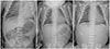
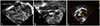
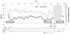
A male neonate with normal prenatal screening was born by vaginal delivery following 39+4 weeks of gestation with 3240 g body weight. The Apgar scores were 8 and 9 at 1 and 5 minutes, respectively. At 30 minutes of life, the newborn became cyanotic with oxygen saturation of less than 50% despite oxygen supplementation. Emergency endotracheal intubation was performed and epinephrine was administered via the endotracheal tube due to bradycardia. After cardiopulmonary resuscitation, the patient was transferred to the Seoul National University Hospital. On admission, the patient's initial capillary blood gas analysis showed pH 6.694 and pCO2>130.0 mm Hg. The chest radiograph showed a tension-pneumothorax of the right lung. A chest tube was inserted immediately (Fig. 1). The initial echocardiography demonstrated normal segmental heart anatomy with moderate TR with a velocity of 3 m/sec. The direction of shunt flow through the foramen ovale was mainly from the right to left atrium. The flow through the ductus arteriosus was invisible. He presented with features of severe pulmonary hypertension, including profound hypoxemia, with oxygen saturation less than 50%, despite the administration of 100% oxygen and nitric oxide. He required resuscitation with intravenous inotropic drugs, high frequency ventilation, and intermittent cardiac massage. Other management, including the infusion of sodium bicarbonate, diuretics, and complete sedation, failed to improve the hypoxemia and low cardiac output. Moderate systemic hypothermia and topical cooling of the head were applied for myocardial and brain protection. At 30 hours of life, repeated echocardiography showed progressive and profound TR secondary to a flail anterior leaflet of the tricuspid valve (Fig. 2), which was caused by a rupture of the papillary muscle of the tricuspid valve. The forward flow from the right ventricle toward the pulmonary artery was nearly absent because of the severe TR, mimicking pulmonary valve atresia. The mild degree of right ventricular dilatation suggested that the TR might be an acute process. The neonate continued to deteriorate, despite maximum efforts. Blood pressure was 24/12 mm Hg, pulse rate was 34 beats/min, body temperature was 35.0℃, and SpO2 was 34%. Capillary blood gas analysis revealed a pH 7.192, pCO2 39.9 mm Hg, and base excess -13 (Fig. 3). An emergency cardiac operation was planned. To stabilize the neonate's condition, venoarterial extracorporeal membrane oxygenation (ECMO) was applied. With ECMO support, the patient's vital signs stabilized. At surgery, the flail anterior leaflet of the tricuspid valve was found. There was no supporting subvalvular structure and the head of the papillary muscle was torn. The papillary muscle appeared to have ischemic changes. The right ventricle and right atrium showed normal morphology. The head of the ruptured papillary muscle was reimplanted into the septal endocardium. Two artificial chordae were interposed between the unsupported leaflet of the tricuspid valve and the papillary muscle. Tricuspid valve annuloplasty was not necessary because the valve annulus was not dilated. Subsequently, the neonate was weaned from cardiopulmonary bypass without difficulty. After the repair, his cyanosis disappeared, and his cardiac output improved dramatically. Postoperative echocardiography showed a trivial TR with good ventricular contractility. Epinephrine, milrinone, dopamine, dobutamine, and ventilation with nitric oxide were tapered slowly, and extubation was performed on the 11th postoperative day. He was discharged 20 days postoperatively and was prescribed sildenafil and enalapril. The echocardiography performed at 7 months of age showed normal function of the tricuspid valve. During the follow-up period of 15 months, the patient showed normal development without any neurological sequela.
Discussion
Isolated TR in neonates is uncommon, but this is an important cause of heart failure and intractable hypoxia in newborns. Congenital TR occurs in neonates with structural abnormalities of the tricuspid or pulmonary valves and in those with a structurally normal heart.1) The flail tricuspid leaflet caused by a ruptured tricuspid valve papillary muscle in a structurally normal heart is an extremely rare condition of unguarded TR in neonates, which is associated with a high perinatal mortality, despite medical treatment.2) However, prompt surgical repair in patients with TR caused by a ruptured papillary muscle may result in a dramatically successful recovery.3) Therefore, it cannot be overemphasized that the accurate etiological diagnosis of neonatal symptomatic TR is essential.
To our knowledge, nine cases have been previously reported. One case of fatal TR due to a ruptured tricuspid valve papillary muscle was attributed to prenatal hypoxic insult and subsequent myocardial ischemic damage. It was described in 1988 and the newborn died of cardiopulmonary failure after 12 hours of life. The autopsy showed ischemic myocardial infarction and necrosis in the anterior papillary muscle supporting the tricuspid valve.1) The anterior papillary muscle supporting the tricuspid valve is perfused by the distal coronary circulation, but is vulnerable to ischemic injury during the diastolic phase setting the right ventricular pressure.4)5)
Papillary muscle rupture can be attributed to ischemic etiologies, including hypoxemia, infection, rhesus isoimmunization, coagulopathy, premature closure of the ductus arteriosus in utero,6) and maternal connective tissue disease,7) as well as non-ischemic etiologies, such as trauma and infective endocarditis.8) In our case, the etiology was unclear. There was no perinatal history to explain the fetal hypoxia. The fetal echocardiograms of our patient taken at the gestational ages of 21, 27, and 32 weeks were reviewed; however, there was no discernible abnormality of the tricuspid valve apparatus. Most cases of neonatal papillary muscle rupture result from an unrecognized period of intrauterine fetal hypoxia before birth.4) In the tricuspid valve structural anomalies, such as Ebstein's anomaly, the cardiac chamber is remodeled with eventual right heart enlargement to augment the stroke volume. Conversely, papillary muscle rupture may result in a relatively acute process of severe TR and sudden cardiopulmonary collapse presenting as a rapidly decompensating cardiac failure refractory to medical therapy.4)
Our case showed intractable hypoxia and circulatory collapse with a right to left shunt through a patent foramen ovale and severe TR. Emergency ECMO was applied for hemodynamic stabilization. Subsequently, the tricuspid valve repair surgery was performed by reimplantation of the ruptured papillary muscle. In most neonatal cases of severe TR, such as Ebstein's anomaly, urgent surgery is not recommended because of the high mortality and morbidity rates, as well as the expected course of gradual regression of pulmonary hypertension and subsequent increase in TR. However, in the present case, early surgical intervention was deemed necessary, and it was found to improve survival.9) ECMO has been proven as a reliable method for cardiopulmonary support in neonates, and it was an effective treatment before surgery in this case.4) With a timely diagnosis and proper surgical treatment, ECMO, as a bridge treatment, can save the neonate's life without further complications.
 XML Download