

 PDF
PDF ePub
ePub Citation
 Print
Print


Introduction
Adult-onset Still's disease (AOSD) is a systemic inflammatory disorder that presents daily spiking fevers, leukocytosis, evanescent rash and arthritis. It is a rare disease of unknown etiology, and few studies have reported on the association of myocarditis with AOSD. Myocarditis should be diagnosed by endomyocardial biopsy (EMB). However, few studies report on using EMB during the acute phase of myocarditis with AOSD.
Case
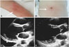
The patient was a 36-year-old female with a high-grade fever and epigastric abdominal pain. A laboratory test showed elevated C-reactive protein, and a general practitioner prescribed levofloxacin and ceftriaxone. Symptoms did not improve after five days and the patient was admitted to a hospital, where minocycline and doripenem were prescribed to treat a suspected case of peritonitis. Symptoms worsened, and the patient developed dyspnea and hypoxia. A week after the appearance of first symptoms, the patient was transferred to our hospital with a fever of 39.2℃, heart rate of 120/min and blood pressure of 88/63 mm Hg. Physical examination revealed a systolic heart murmur in the apex area, abdominal tenderness, hepatomegaly and poorly-marginated urticaria-like erythema on the anterior chest, abdomen and both arms (Fig. 1A and B). The electrocardiogram showed normal sinus rhythm and T-wave inversion at precordial leads, and a chest X-ray showed cardiomegaly and pulmonary edema. A blood test revealed leukocytosis (white blood cell count 11800/µL, neutrophil 90%), elevated C-reactive protein (36.0 mg/dL), creatinine level (1.05 mg/dL), total bilirubin (2.3 mg/dL), troponin T (0.077 ng/mL), brain natriuretic peptide (4220.0) and elevated ferritin (3500 ng/mL). Echocardiography revealed diffuse hypokinesis of the left ventricle {ejection fraction (EF) 20%}, moderate mitral regurgitation, severe tricuspid regurgitation and massive pericardial effusion (Fig. 1C).
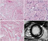
Broad spectrum antibiotic therapy with meropenem, ciprofloxacin, minocycline and vancomycin was initiated, in addition to catecholamine support with norepinefline and vasopressin for shock and non-invasive positive pressure ventilation for respiratory failure. All microbial cultures and specific antibody for infectious agents were negative. An autoantibody panel with anti-nucleocid-antibody, rheumatoid factor and anti-double stranded deoxyribonucleic acid antibody were all negative. When symptoms failed to improve, coronary angiography, right heart catheterization and EMB were performed. There was no indication of coronary artery stenosis, mean pulmonary artery pressure was 23 mm Hg, pulmonary capillary wedge pressure 19 mm Hg, left ventricular end-diastolic pressure 21 mm Hg and cardiac index 1.72 mL/min/m2. EMB revealed fibrosis and infiltration of inflammation cells, mainly composed of neutrophils (Fig. 2A, B, and C). The patient received a diagnosis of active acute myopericarditis associated with cardiogenic shock and heart failure. Inotropes (dobutamine 2 µg/kg/min) were initiated, resulting in improved cardiac index. Clinical manifestation of a spiking fever unresponsive to antibiotics, leukocytosis, elevated ferritin levels and skin rash led to a diagnosis of AOSD with acute myocarditis using Yamaguchi criteria. Four days after admission to our hospital, pulse therapy of intravenous methylprednisolone 500 mg was performed twice daily for three days, followed by a daily dose of prednisone 60 mg (1 mg/kg/day). Steroid therapy was effective for fever and pericardial effusion, and left ventricular EF improved (EF 60%) (Fig. 1D). Patient did not need catecholamine and ventilator support five days after steroid treatment. Carvedilol 2.5 mg, enalapril 1.25 mg and spironolactone 25 mg daily were prescribed for heart failure and colchicine 0.5 mg daily for pericarditis. Fourteen days after admission, cardiac-magnetic resonance imaging (MRI) was performed and revealed high signal intensity at the basal to middle portion of the left ventricular wall with short-TI inversion recovery and at the medial layer with gadolinium-delayed enhancement (Fig. 2D). Steroid dosage was gradually decreased, and the asymptomatic patient was discharged 32 days after admission with instructions to continue prednisone 25 mg daily.
Discussion
Yamaguchi criteria are the most popular of several proposed diagnostic criteria for AOSD, with fever, typical rash, arthralgia and leucocytosis as major criteria, and sore throat, lymphadenopathy, liver dysfunction, negative rheumatoid factor and antinuclear antibody as minor criteria.1) The patient in this study met Yamaguchi criteria for AOSD, in addition to pericarditis and myocarditis. Cardiac involvement in AOSD is typically pericarditis, due to serosal disturbance. Myocardial involvement in AOSD is rare. Therefore, echocardiography is an important tool in diagnosing clinical signs of heart failure and left ventricular and pericardial effusion. Myocarditis has been diagnosed by patient clinical course, but a definitive diagnosis can only be confirmed by EMB.2) Data from an AOSD patient with myocarditis showed that EMB had been performed six weeks after onset, and results showed mononuclear cell infiltration and fibrosis.3) In this study, EMB was performed ten days after the suspected onset of heart failure and found cardiac muscle degeneration, fibrosis and infiltration of neutrophil cells. The etiology of myocarditis with AOSD is uncertain. However, neutrophil leukocytosis is an important Yamaguchi criterion for AOSD and assisted in the diagnosis of myocarditis associated with AOSD in our patient.
Cardiac MRI as an alternative non-invasive tool to evaluate myocarditis has been reported.4)5)6) In this study, cardiac-MRI showed high signal intensity at the basal to middle portion of the left ventricle with short-TI inversion recovery and at the medial layer with gadolinium-delayed enhancement. Cardiac-MRI may be a useful diagnostic tool in myocarditis associated with AOSD. However, EMB is the gold standard. In this study, the relationship between AOSD and myocarditis could only be inferred by EMB with neutrophil infiltration.
Studies suggested high-dose intravenous corticosteroids for treatment of myocarditis with AOSD, and intravenous immunoglobulin, tumor necrosis factor-α antagonist and anti-interleukin-1 inhibitor anakinra for relapsing or resistant cases.7)8)9) However, there were no clinical studies on the treatment appropriate for management of myocarditis in AOSD. Our patient received corticosteroid therapy to produce a dramatic positive response.
This study is possibly the first to report myocarditis in AOSD diagnosed by neutrophil infiltration in the myocardium. With AOSD and cardiac dysfunction, diagnostic evaluation with EMB should be performed if myocarditis is suspected, followed by appropriate treatment.
 XML Download