

 PDF
PDF ePub
ePub Citation
 Print
Print


Introduction
Takotsubo cardiomyopathy, also known as transient left ventricular (LV) apical ballooning syndrome, consists of transient LV dysfunction. It is characterized by regional wall motion abnormalities that extend beyond a single epicardial vascular distribution, chest pain or dyspnea, new electrocardiographic (ECG) abnormalities (ST-segment elevation and/or T-wave inversion), and minor elevations in serum levels of cardiac enzymes in the absence of significant coronary artery dise-ase. In the majority of patients, the apical akinesis completely resolves within a month.1)2) Patients affected by the disease are predominantly postmenopausal women who experience emotional or physical stress and subsequently develop chest pain and dyspnea.3-5) The pathophysiological mechanism is not com-pletely understood, but it is likely to be associated with catecholamine overload.6)
Herein we report a case of a 28-year-old woman with takotsubo-like severe LV dysfunction associated with Cesarean section. We also discuss plausible mechanisms and the clinical importance of our case.
Case
On May 19th 2009, a 28-year-old woman was admitted to the emergency department after a Cesarean delivery at a local obstetric clinic. It was reported that her oxygen saturation was 93% immediately after delivery but could not be maintained at this level. General anesthesia has been induced using vecuronium bromide, diprivan, and succinylcholine and maintained with sevoflurane. There were no other complications such as major bleeding or shock during surgery and she was in- and extubated uneventfully.
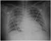
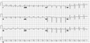
The patient had unremarkable past medical and obstetrical histories. She had no allergies and no family history of heart disease. It was her first-ever episode of dyspnea. She was complaining of dizziness. At the time of admission, she was fully conscious, but was in respiratory distress. Her blood pressure was 142/82 mmHg, heart rate was 138 bpm, body temperature was 36.8℃, respiratory rate was 17/minutes. Mild rale on both lower lung field was detected by auscultation. A chest radiograph showed pulmonary edema (Fig. 1). The twelve-lead ECG performed on admission revealed ST segment elevation at lead III and T-wave inversion at leads I and aVL (Fig. 2). Laboratory studies upon admission were: white blood cells 14,290 cells/µL, hemoglobin 14.0 g/dL, hematocrit 40.6%, and platelets 275,000 cells/&L. Arterial blood gas analysis values were: pH 7.42, pCO2 29 mmHg, pO2 69 mmHg, bicarbonate 19 mmol/L and O2 saturation 94% with 7 L/minute of nasal oxygen supply. A baseline cardiac enzyme study showed troponin-T and creatine kinase-MB isoform (CK-MB) to be 0.50 ng/mL and 18.2 ng/mL, respectively, but repeated measurements 9 hours later showed decreased levels: 0.23 ng/mL and 10.7 ng/mL, respectively. There was a further decrease of CK-MB on day 3 (6.6 ng/mL). Echocardiographic evaluation on hospital day 1 revealed markedly reduced global LV systolic function with an ejection fraction of 23% and impairment of the midventricular and apical contractility sparing the base (Fig. 3A and B).
After admission, she was treated with furosemide, spironolactone, and captopril. Her symptoms gradually improved during the hospital stay. An echocardiography taken 5 days later showed improved hypokinesia of the midventricular region. The patient was discharged on hospital day 8 in stable condition. A follow-up echocardiography at the outpatient clinic 2 weeks after her baseline examination revealed nearly normalized cardiac function (Fig. 3C and D).
Discussion
We report a case of a 28-year old woman with transient severe LV dysfunction after Cesarean delivery. Considering the characteristics of her clinical presentation and the period in which it developed, peripartum cardiomyopathy was at first part of the differential diagnosis. Peripartum cardiomyopathy is a disease occurring during pregnancy or during the postpartum period. The criteria for the diagnosis are: development of heart failure in the last month of pregnancy or within the first 5 postpartum months, absence of a determinable etiology for the cardiac failure, and absence of demonstrable heart disease prior to the last month of pregnancy. In most cases, the onset is in the first 3 months of the postpartum period. 7)
Our case is very similar to peripartum cardiomyopathy. However, the most probable diagnosis of this patient was takotsubo cardiomyopathy, which was mainly based on the characteristic echocardiographic findings. Indeed, the second and third diagnostic criteria for peripartum cardiomyopathy also apply to takotsubo cardiomyopathy. However, because coronary angiography was not performed, this case cannot meet the complete diagnostic criteria of takotsubo cardiomyopathy.
There are an increasing number of reports on takotsubo cardiomyopathy in the setting of a medical procedure such as spinal anesthesia, general anesthesia, and surgery. Although takotsubo cardiomyopathy is common in elderly women, young patients can also be affected in these iatrogenic conditions.8-10) In this regard, pregnant women who are undergoing Cesarean delivery can be a vulnerable population. Therefore, physicians involved in Cesarean deliveries need to be prepared to suspect, diagnose, and manage this unexpected event.
Transient LV dysfunction has been reported to arise at the time of induction and intubation during general anethesia. However, the relationship between a specific anesthetic and takotsubo cardiomyopathy has been unclear. In the described case, the patient's symptoms appeared after successful in- and extubation. In our patient, the circumstances under which the symptoms started during the surgery made the Cesarean section an iatrogenic stress and a possible responsible factor. Alternatively, the emotional and physical stress of the delivery could have induced the disease. If we have conducted tests for myocardial inflammation associated with infection or autoimmunity, it could have been helpful to completely rule out other causes and finally diagnose takotsubo cardiomyopathy.
In conclusion, our patient had takotsubo-like severe LV dysfunction that occurred in the postpartum period. It is difficult at times to distinguish takotsubo-like severe LV dysfunction from peripartum cardiomyopathy. A common pathophysiological mechanism may underlie these two diseases.

 XML Download