

 PDF
PDF ePub
ePub Citation
 Print
Print


Introduction
Although atherosclerosis manifests clinically in middle and late adulthood, it is well-known that it has a long asymptomatic phase of development, which begins early in life, often during childhood. In most children, atherosclerotic vascular changes are minor and can be minimized or prevented with a healthy lifestyle. However, in some children the process is accelerated because of risk factors or specific diseases. Identification of children who are at risk for atherosclerosis may allow early intervention to decrease the atherosclerotic process, preventing or delaying cardiovascular diseases (CVD), such as myocardial infarction, stroke, and peripheral vascular disease.
Atherosclerotic Changes in Child
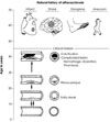
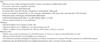
Atherosclerosis begins in childhood as an accumulation of fatty streaks-lipid-engorged macrophages (foam cells) and T lymphocytes in the intima of the arteries. Fatty streaks may or may not progress, and may regress. In some people, lipid accumulation is more pronounced with time, and the accumulated lipid becomes covered by a fibromuscular cap to form what is termed a fibrous plaque. Temporally, between the fatty streak and the fibrous plaque, transitional stages of atherosclerosis exist that are not identifiable by gross examination alone. With time, fibrous plaques enlarge and undergo calcification, hemorrhage, ulceration or rupture, and thrombosis. Thrombotic occlusion precipitates clinical disease such as myocardial infarction, stroke, or gangrene depending on which artery is affected (Fig. 1).1)
As detailed below, autopsy studies in children and young adults who had died of non-cardiovascular causes have conclusively demonstrated early development of atherosclerosis.
Attention was first drawn to the early origin of atherosclerosis by an autopsy study conducted on young soldiers killed in the Korean War.2) Their average age was 22 years, and over 70% of them had evidence of atherosclerosis in their coronary arteries. Postmortem coronary angiography and dissection of hearts from 105 United States soldiers killed in Vietnam demonstrated that 45% had some evidence of atherosclerosis and 5% had gross evidence of severe coronary atherosclerosis.3) Another study demonstrated a very high incidence of lipid-laden macrophages in the intima of the aorta and coronary arteries of young American children killed in motor accidents, with over 50% of children aged 10-14 years having some evidence of early atherosclerosis.4) A nation-wide autopsy-based study of atherosclerosis in young Japanese (1 month-39 years) disclosed the presence of fatty streaks in 29% of aortas in those aged <1 year and in 3.1% of coronary arteries of children aged 1-9 years.5) Another examination 13 years later revealed an increased prevalence and extent of coronary artery lesions in autopsied subjects who died in their third and fourth decade of life.6) In the Bogalusa Heart Study,7) the extent of fatty streaks and fibrous plaques in the aorta and coronary arteries were examined in 204 young patients 2-39-years-of-age. The prevalence of fatty streaks in the coronary arteries increased with age from approximately 50% at 2-15-years-of-age to 85% at 21-39-years-of-age, and the prevalence of raised fibrous-plaque lesions increased with age from 8% at 2-15-years-of-age to 69% at 26-39-years-of-age. The prevalence and the extent of atherosclerosis was greater with increasing age, body mass index (BMI), blood pressure, and levels of serum total cholesterol (TC) and low-density lipoprotein cholesterol (LDL-C). The degree of involvement increased with worsening severity and greater numbers of risk factors. In the Pathobiological Determinants of Atherosclerosis in Youth (PDAY) study,8) the right coronary arteries and aortas were examined during autopsy in 2,876 individuals aged 15-34 years. Raised fatty streaks were present in the abdominal aortas of approximately 20% of those aged 15-19 years and approximately 40% of subjects 30-34-years-of-age, and in the right coronary arteries of approximately 10% of 15-19-year-old subjects and approximately 30% of those aged 30-34 years at the time of death. The percent of intimal surface involved with raised fatty streaks increased with age and was associated with a high level of non-high-density lipoprotein cholesterol (HDL-C) and low HDL-C, hypertension, obesity, and impaired glucose tolerance. An intravascular ultrasound study determined that 17% of otherwise healthy heart donors <20 years of age, 37% of those aged 20-29 years, 60% of those aged 30-39 years, 71% of those aged 40-49 years, and 85% of those ≥50-years-of-age had evidence of coronary atherosclerosis.9)
Noninvasive Assessment of Preclinical Atherosclerosis
Until recently, our understanding of the childhood antecedents of adult CVD was limited mainly to autopsy studies and pathologic findings in teenagers and young adults who died from accidental causes. Recent advances in noninvasive techniques have allowed detection of early changes in physiology and visualization of anatomical and mechanical abnormalities that reflect the preclinical biology of atherogenesis.
In adults, noninvasive measures of atherosclerosis have become established as valid and reliable tools for refining the cardiovascular risk to target individuals who need early intervention. Noninvasive ultrasound studies of the neonatal aorta and fetal and early childhood postmortem studies indicate that impaired fetal growth, in utero exposure to maternal hypercholesterolemia, and diabetic macrosomia may all be important risk factors for vascular changes consistent with the earliest physical signs of atherosclerosis.10) With limited pediatric data, the use of these techniques in children and adolescents largely has been reserved for research purposes.11)
Functional measures
Endocardial function can be evaluated by measuring changes in blood vessel diameter in response to specific stimuli using the technique of flow-mediated dilation (FMD). FMD measures the nitric oxide-mediated vasodilation produced by increased flow after a period of ischemia (e.g., ischemia induced by an inflated blood pressure cuff) by brachial artery ultrasonography. Abnormalities in endothelial function have been noted in children affected with a variety of conditions. Decreased FMD has been observed in children with type 1 diabetes mellitus, a family history of premature coronary disease,12) and Kawasaki disease (KD).13)14)
Structural measures
Carotid intima-media thickness (cIMT) has been used extensively in children and young adults with risk factors for CVD. Several large studies have shown that increased IMT correlates well with traditional cardiovascular risk factors that include excess weight, dyslipidemia, and hypertension.15-17) In addition, increased IMT is found in adolescents with familial hyperlipidemia and in children with a parent with a history of premature myocardial infarction.12)18) Young adults with increased IMT have an increased likelihood of a cardiovascular events that include myocardial infarction and stroke.
In addition to cIMT, some pediatric studies have measured IMT in the aorta (aIMT). This technique has proved useful even in neonates and young children. Increased aortic wall thickness has been associated with low birth weight,19) intrauterine growth restriction,20) maternal smoking,21) and familial hypercholestrolemia.22)
The combined thickness of carotid artery intima and media is associated with coronary heart disease risk factors. The intima is more directly involved in atherosclerosis than the media, and the thickness of the intima alone measured by high-resolution ultrasonography may aid in detecting early vascular changes of atherosclerosis.23)
Serum LDL-C level may be more closely associated with the carotid intimal thickness than IMT for patients with coronary atherosclerosis.24)
Arterial stiffness can be assessed noninvasively by measuring a parameter known as the pulse wave velocity (PWV) between two major arteries located in the upper body (i.e., carotid or brachial artery) and the lower body (i.e., femoral or ankle). PWV reflects the time needed for the pulse wave to travel a given distance along the blood vessel. Stiffer arteries produce a higher PWV.
Arterial stiffness normally varies among adolescents and young adults based upon gender, age, and ethnicity. After adjusting for age and gender, arterial stiffness in adolescents and young adults is associated with CVD risk factors that include BMI, hypertension, and elevations in serum triglyceride (TG), homocysteine, and fasting insulin concentrations.25-27)
PWV may have an advantage compared to other noninvasive techniques in the primary care setting due to its simplicity of use and shorter time required for a measurement.
Computed tomography (CT) allows localization and quantification of calcium deposits in coronary arteries. However, the paucity of data regarding calcification in early atherogenesis, and the potentially harmful effects of early radiation exposure currently limits the clinical use of CT in children.
Magnetic resonance imaging (MRI) is increasing being used to assess early atherosclerosis, since it allows characterization of cardiovascular structure, function, and blood flow without exposure to ionizing radiation. It has been used to measure atheromatous plaques and characterize structure in the peripheral circulation.
Both CT and MRI are currently being combined with positron emission tomography and fluorescent imaging to evaluate plaque biology in more detail.28)
Risk Factors for Atherosclerosis in Children
In adults, large prospective population-based studies have shown that a higher risk of CVD is associated with multiple risk factors that include obesity, hypertension, dyslipidemia, diabetes, smoking, and family history of CVD. While data linking these risk factors to cardiovascular events are limited in children, these risk factors are associated with acceleration of atherosclerosis in children. These risk factors do not generally occur in isolation but are usually found concurrently.
Overweight/Obesity
The prevalence of children who are clinical overweight or obsess is increasing rapidly globally. Children and adolescents who are overweight/obese are more likely to be overweight/obese as adults. In autopsy studies, BMI was positively correlated with more extensive atherosclerotic changes in the aorta and coronary arteries during childhood.7)8)
Childhood obesity raises the risk of other risk factors that are associated with heart disease in early adulthood, such as high blood pressure or diabetes mellitus. In one study, children between the ages of 6 and 19 years with metabolic syndrome showed an increased risk of CVD {odds ration: 14.6, 95% confidence interval: 2.8-45.3} at 25 year follow-up compared to the general school population.29) Generally, overweight children are inactive and may have obstructive sleep apnea, both of which are associated with CVD in adults.
Hypertension
Children and adolescents with hypertension are more likely to have hypertension as adults. In adults, hypertension is a well-established risk factor for CVD that include myocardial infarction and stroke. Although similar direct evidence linking hypertension with CVD is lacking, hypertension has been linked to increased IMT and arterial stiffness, suggestive of accelerated atherosclerosis.
Dyslipidemia
Dyslipidemia is a disorder of lipoprotein metabolism that results in increased TC, high LDL-C, low HDL-C, and high TG. The National Cholesterol Education Program (NCEP) defines dyslipidemia in children as values that are greater than the 95th percentile and defined cutoff points.30) Subsequently, the National Health and Nutrition Examination Surveys (NHANES) presented age- and gender-specific lipoprotein threshold concentrations for adolescents.31) The predictive capacity of both NCEP and NHANES cut points were similar predicting high common carotid artery IMT in adulthood.32) Adolescent lipid levels were more strongly associated with high IMT in adulthood than change in lipid levels. Overweight or obese adolescents with dyslipidemia had increased cIMT in adulthood compared with those who did not have both risk factors, leading the authors to suggest that dyslipidemia screening could be limited to overweight or obese adolescents.
Although dyslipidemia is an established risk factor for CVD in adults, no long-term studies directly link dyslipidemia in childhood with subsequent CVD, with the possible exception of children with monogenetic causes of dyslipidemia such as familial hypercholesterolemia. Autopsy studies demonstrated an increase atherosclerotic lesions in the coronary artery and aorta with increasing serum LDL-C and decreasing HDL-C.7)8) Individuals who displayed elevations in non-HDL-C, L-DLC, and TC: HDL-C in children are more likely to have increased IMT in adulthood.33)
Family history
A family history of CVD is an independent risk factor for coronary vascular events, especially in young adults. Children whose parents or grandparents had a heart attack or stroke at an early age have twice the risk of developing CVD in middle-aged adults.34) Healthy offsprings of parental history of premature acute myocardial infarction can present with lipid disturbances and increased carotid IMT compared to healthy children with no parental history of coronary disease.35)
Smoking
Smoking and passive exposure to tobacco smoke increases the risk of CVD. Teenagers who smoke are likely to continue smoking into adulthood, thus increasing their risk of early CVD. Smoking is associated with a much greater extent of fatty streaks and raised lesions in the abdominal aorta.36) Although the mechanisms in cigarette smoking-related cardiovascular dysfunction are largely unknown, smoking increases inflammation, thrombosis, and oxidation of LDL-C. Recent experimental and clinical data support the hypothesis that cigarette smoking exposure increases oxidative stress as a potential mechanism for initiating cardiovascular dysfunction.37)
High-Risk Diseases
Pediatric diseases or events associated with high-risk for CVD include familial hypercholesterolemia, types 1 and 2 diabetes mellitus, chronic kidney disease, cardiac transplantation, KD, chronic inflammatory disease, survivors from childhood cancer, and congenital heart disease. The American Heart Association has issued recommendations for cardiovascular risk management in these high-risk diseases.40)41)
Risk Assessment
In adults without known CVD, a multivariable assessment is used to estimate their risk for cardiovascular events and to guide therapeutic choices aimed at risk reduction (Framingham Risk Score). No such system is used in clinical pediatric practice. As described below, a grading system has been proposed based on data from the PDAY study.42)43)
Pathobiological Determinants of Atherosclerosis in Youth Risk Scores
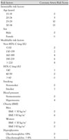
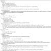
The PDAY study of autopsy findings in young people developed a risk score that provides a simple way to calculate weighted effects of the major established risk factors on atherosclerosis (Table 1). Risk scores computed from the modifiable risk factors were associated with prevalence of microscopically demonstrable lesions of atherosclerosis and with the extent of the earliest detectable gross lesion (fatty streaks) in the coronary artery and aorta. When tested in young persons in the Coronary Artery Risk Development in Youth Adults (CARDIA) study, this risk score predicted calcium in the coronary arteries as long as 15 years after the risk factors were measured.44)
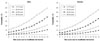
The severity of childhood atherosclerosis increases as the number of CVD risk factors. Children with one or more known atherosclerotic risk factors should be screened for additional risk factors. Fig. 2 shows the estimated probability of advanced atherosclerotic lesions in the coronary arteries by the PDAY modifiable risk score (5-year age group for men and women).43)
Screening Tests
Screening every child with tests to look for atherosclerosis risk factors is not currently recommended. The most current recommendation is to screen children and adolescents who have one or more of the following risks:
The first screening should take place after 2 years of age but no later than 10 years of age. The following screening items should be included:
Primary Prevention of Atherosclerotic Cardiovascular Disease in Childhood
The existing evidence indicates that primary prevention of atherosclerotic CVD should begin in childhood. It would be most effective to control risk factors early in life. The following guidelines are based on the recommendations by the American Heart Association (AHA).40)41)45-47)
Management of Children and Adolescents With High-Risk Diseases
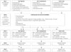
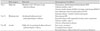
Certain pediatric disease states are associated with dramatically accelerated atherosclerosis, with clinical coronary events occurring in childhood or very early adult life. Intensive cardiovascular risk reduction is of critical importance in such children. The AHA developed recommendations for cardiovascular risk management in high-risk pediatric settings. The recommendations were peer reviewed and then endorsed by the American Academy of Pediatrics.40)41) As summarized in Table 4, the eight identified diseases or states have been categorized in three risk tiers ("high", "moderate", "at risk") based upon risk for CVD.
For children at the highest risk (tier I), the intervention strategy regards the diagnosis as a "coronary heart disease equivalent" with recommendations for risk reduction similar to secondary prevention guidelines for adults with established coronary disease. For tier II, complete risk factor assessment is recommended with specific defined therapeutic goals. For children with diagnoses in tier III, the focus is on complete risk factor assessment with therapeutic goals as defined for children in general.
 XML Download