

 PDF
PDF ePub
ePub Citation
 Print
Print


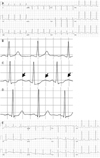
A 31-year-old woman presented with chest discomfort in the 22nd week of pregnancy. She had been treated with an intravenous infusion of ritodrine hydrochloride (0.25 mg/min) for preterm labor the prior day. On physical examination, the body temperature was 36.3℃, the blood pressure was 117/68 mmHg, the pulse was 96 beats/min, and the respiratory rate was 20 breaths/min. Electrocardiography (ECG) showed ST-T changes resembling ischemia in the inferolateral leads (Fig. A) with a heart rate of 98 beats/min. An ECG taken the preceding day revealed no ST-T changes (Fig. B). The ECG in limb lead II showed flattening of the T wave, slight ST depression, and prominent U waves, as in hypokalemia (Fig. C). QT prolongation and a U wave were prominent in the precordial leads (Fig. A). Urgent echocardiography to detect cardiac function and ischemic changes showed normal cardiac function without wall motion abnormalities. After discontinuing the ritodrine infusion, the ECG returned to normal the following day (Fig. D and E) and her chest discomfort resolved. Her serum potassium level, which had been 4.1 mEq/L the previous day, was 3.4 mEq/L, and returned to 4.1 mEq/L after discontinuation of the ritodrine infusion. The serum fasting glucose level was 81 mg/dL, and 80 mg/dL the preceding day. The plasma calcium was 8.8 mg/dL and the magnesium level was 1.9 mg/dL.
Ritodrine, a beta 2-selective adrenergic drug, is used as a tocolytic drug for management of preterm labor.1) One of the most common side effects of ritodrine is hypokalemia because of a marked increase in plasma insulin, which results in stimulation of the cellular uptake of potassium.2)3) Ritodrine-induced hypokalemia shows typical ECG changes resembling hypokalemia typical of other causes. As in this case, typical ECG changes associated with a ritodrine infusion may not be compatible with the absolute serum level of potassium.
 XML Download