

 PDF
PDF ePub
ePub Citation
 Print
Print


Introduction
Acute myocardial infarction is a potentially fatal event and a common causes of death in adults. Sudden coronary artery occlusion results in ischemic related death of cardiomyocytes.1) The human heart has low regenerative ability. The inflammatory response and cytokine release from the myocardium are essential components of the host response to acute myocardial infarction, and play a crucial role in cardiac repair. Tumor necrosis factor-alpha (TNF-α) and interleukin-6 (IL-6) are increased after an acute myocardial infarction and can regulate myocyte survival and induce additional cellular inflammatory responses.2)3) Chemokines stimulate the recruitment of inflammatory leukocytes to the infarct related myocardium.4) Monocyte chemoattractant protein (MCP-I)/CCL2 has a potent effect on macrophage recruitment and myofibroblast accumulation in healing myocardium and also plays an important role in postinfarct left ventricular healing.5) Transforming growth factor (TGF)-β also plays a crucial role in cardiac repair by the suppression of inflammation.6) Timely resolution of the inflammation and inhibition of the inflammatory response and recovery from tissue injury of the infarcted area are essential for optimal healing of the myocardium. Here we review recent researches and evolving concepts of the inflammatory responses and cardiac repair after acute myocardial infarction.
Inflammatory Response
The complement cascade, reactive oxygen species (ROS) and cytokine cascade mediated pathway play an important role in the post infarction inflammatory response.
Complement cascade activation
Hill and Ward were the first to report that ischemic myocardial injury can induce activation of the complement cascade in a rat infarct model.7) Pinckard et al.8) also showed that ischemic myocardial necrosis was associated with the release of subcellular membrane constituents that are triggered by the early acting components of the complement cascade (C1, C4, C2 and C3) in patients with an acute myocardial infarction. Yasojima et al.9) demonstrated that complement gene expression was upregulated by ischemia and reperfusion in the rabbit heart. Complement activation was reported to induce neutrophil and monocyte recruitment in the ischemic myocardium.10) Weisman et al.11) reported that infusion of soluble human complement receptor type 1 (sCR1) significantly suppressed post-infarct inflammation and necrosis in a rat model of myocardial ischemia and reperfusion. In spite of promising experimental results, recent clinical trials testing the effects of complement inhibition, in patients with acute myocardial infarction, showed disappointing results. Administration of the human anti-C5 monoclonal antibody pexelizumab, in patients with an acute myocardial infarction, had no effect on infarct size and clinical outcome.12-14)
Reactive oxygen species
Meldrum et al.15) demonstrated that H2O2 alone induced myocardial TNF-α mediated cardiac injury by a p38 mitogen-activated protein kinase (MAPK)-dependent mechanism. Reactive oxygen intermediates may generate a leukotatic stimulus that includes, complement activation, induction of hemorrhagic shock-induced P-selectin expression, chemokine upregulation and an increase in the endothelial intercellular adhesion molecule (ICAM)-1 ability to bind neutrophils.16-19) The use of the antioxidant enzymes superoxide dismutase and catalase reduced the infarct size in dogs with myocardial ischemia and reperfusion.20) However, there have been some failed studies of antioxidant treatment used to prevent myocardial ischemic injury.21)22) Two clinical studies using recombinant human superoxide dismutase in patients with an acute myocardial infarction undergoing percutaneous coronary intervention or thrombolysis showed no significant improvement of left ventricular function.23)24)
Cytokine amplication
Cytokines can self-amplify through a positive feedback loop targeting the nuclear factor (NF)-κB. Upregulation of TNF-α in the infarct myocardium can upregulate the levels of TNF-α in the neighboring normal myocardium, leading to amplified cytokine effects.3) TNF-α stimulates expression of proinflammatory cytokines, chemokines and adhesion molecules by leukocytes and endothelial cells and regulates extracellular matrix metabolism by reducing collagen synthesis and by enhancing matrix metalloprotease (MMP) activity in cardiac fibroblasts;25) other adhesive cytokines such as monocyte chemoattractant protein (MCP)-1 is also induced in the ischemic and reperfused canine myocardium. Kumar et al.26) suggested a significant role for MCP-1 in monocyte trafficking in reperfused myocardium.
Cytokine and chemokine upregulation
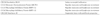
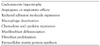
Chemokine upregulation is a noted feature of the postinfarction inflammatory response (Table 1).27) Recent investigators have demonstrated strong induction of several chemokines in the ischemic myocardium supporting their role in leukocyte recruitment.4) MCP-1 upregulation has been demonstrated in a mouse model.28) Frangogiannis reported that a MCP-1 -/- infarct mouse model had decreased mesenger ribonucleic acid (mRNA) expression of the cytokine TNF-α, IL-1β, TGF-β and IL-10 and showed defective macrophage differentiation.27) Induction and release of cytokines such as TNF-α and IL-6 are rapidly released in the central zone during a myocardial infarction; however, they are usually maximal in the border zone.3)29) This robust upregulation may return to baseline levels if the infarction is small, and if the infarction is large and the inflammatory response is excessive, there can be sustained cytokine upregulation, corresponding to a chronic remodeling phase.
Cellular inflammatory response to myocardial infarction
The neutrophils
Neutrophils are recruited during the initial stage of cardiac ischemic injury. Neutrophil transmigration in the infarcted myocardium requires adhesive interactions with activated vascular endothelial cells. Neutrophils may secrete oxidants and proteases and possibly express mediators capable of amplifying cell recruitment.30) Neutrophil depletion in animals undergoing reperfused myocardial infarction has been reported to significantly decrease the infarct size suggesting that a significant amount of myocardial injury may be induced by neutrophil dependent mechanisms.31)32)
However, the mechanisms associated with neutrophil-induced myocardial ischemic injury have not been identified. Jaeschke et al.33) suggested that neutrophils may directly injure parenchymal cells through release of specific toxic products. The selectin family consists of L-selectin, P-selectin and E-selectin. P-selectin expression occurs rapidly in endothelial cells during cardiac ischemic injury. Experimental study has suggested that monoclonal antibodies against P-selectin reduced myocardial necrosis, preserving coronary endothelial function and attenuating neutrophil infiltration in ischemic and reperfused myocardium.34) However, there have been inconsistent results of selectin-related interventions in experimental models of myocardial ischemia.35)36)
The mononuclear cells
As previously mentioned, MCP-1/CCL2 plays an important role in monocyte recruitment to the infarcted myocardium.5) Cytokines, such as TGF-β, free radical oxygen, complement, and the CC chemokine may also play a role in monocyte infiltration. Infiltration of monocytes into the infarcted myocardium is followed by maturation and differentiation of these blood-derived cells into macrophages.
Cardiac Repair After Myocardial Infaction
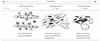
Healing of an infarction can be divided into three overlapping phases: the inflammatory phase, the proliferative phase and the maturation phase (Fig. 1).37) The acute repair process is mediated by cytokines and inflammatory cells in the infarcted myocardium. The induction of pro-inflammatory mediators and leukocyte infiltration play a crucial role in phagocytosis and removal of necrotic cells and matrix debris from infarcted myocardium. Moreover, they promote tissue repair and scar formation.38) The strategy for optimal infarct healing requires inhibition of cytokines and chemokine synthesis after myocardial infarction.
Transforming growth factor-β as a key regulator in cardiac repair
TGF-β is a multifunctional cytokine that controls proliferation and cellular differentiation in most cells. TGF-β has been shown to be significantly upregulated in an experimental rat model of myocardial infarction; in addition, TGF-β mRNA and protein was significantly increased at the infarct border zone.39)40) During infarct healing, TGF-β may play a role in the suppression of chemokine and cytokine synthesis and is thought to be a key mediator of the transition from inflammation to fibrosis.41) Lefer et al.42) reported that TGF-β injections reduced myocardial ischemic injury mediated by proinflammatory cytokines such as TNF-α during the inflammatory phase of myocardial healing. Anti-TGF-β treatment before or after coronary artery ligation increased mortality and worsened the left ventricular remodeling in mice with non-reperfused myocardial infarction.43) The inhibition of TGF-β signaling by injection of a TGF-β II receptor resulted in reduction of left ventricular remodeling by modulation of cardiac fibrosis; early TGF-β inhibition increased mortality and left ventricular dilatation.44)45) Youn et al.46) reported that the angiotensin converting enzyme inhibitor and angiotensin receptor blockade resulted in decreased TGF-β mRNA expression after nontransmural infarction in the rat.
The role of other cytokines in cardiac repair
There are three IL-1 molecules consisting of IL-1α, IL-β and IL-1 Ra that are specific receptor antagonists.48) Bujak et al.49) demonstrated that IL-1 signaling is essential for activation of inflammatory and fibrogenic pathways in the healing infarct and plays an important role in the pathogenesis of remodeling after infarction. IL-10 exerts potent anti-inflammatory effects and modulates MMP expression.50-52) However, Zymek et al.53) and our group reported that IL-10 signaling plays a noncritical role in the suppression of inflammatory mediators, resolution of the inflammatory response and fibrous tissue deposition following myocardial infarction in the mouse. This result may be due to the involvement of multiple overlapping regulatory mechanisms controlling various proinflammatory pathways activated in the infarcted myocardium.
The role of proteins in cardiac repair
CD44 is a cell surface glycoprotein involved in cell-cell interaction and cell adhesion and migration. CD44-hyaluronan interactions play a role in leukocyte extravasation at the inflammatory site and serves as a key factor in the resolution of inflammation through removal of matrix breakdown products and clearance of apoptotic neutrophils.54-56) Huebener et al.57) tested the role of CD44 in infarct healing and demonstrated that CD44 mRNA levels were significantly induced in the infarcted heart; CD44 null mice showed enhanced and prolonged inflammation in the infarcted heart followed by decreased myofibroblast infiltration, reduced collagen deposition and diminished proliferative activity. Huebener et al.57) concluded that CD44 is critically involved in infarct healing by regulating the inflammatory and fibrotic response. Thrombospondin (TSP)-1 is an adhesive glycoprotein involved in cell to cell and cell to matrix interaction with potent angiostatic properties; it is a TGF-β activator.58) TSP-1 showed strikingly selective localization in the infarct border zone, and TSP-1 knockout animals had markedly increased macrophage and myofibroblast density in the infarct and in remodeling of noninfarcted myocardial areas, and was more extensive in the postinfarction remodeling than in the wild-type mice. Frangogiannis et al.59) concluded that the selective endogenous expression of TSP-1 at the infarct border zone may serve as a "barrier," limiting expansion of granulation tissue and protecting the noninfarcted myocardium from fibrotic remodeling. Smad is an essential protein component of the TGF-β pathway.60) Hao et al.61) showed that TGF-β mRNA was significantly increased in the infarct scar compared to viable myocardium, and that Cardiac Smad 2, 3 and 4 proteins were significantly increased in the border and scar tissues when compared to viable myocardium. These results indicate that TGF-beta/Smad signaling may be involved in the remodeling of the infarct scar. Optimal cardiac repair requires containment of the inflammation in the infarcted area. Extension of the inflammation into the non-infarcted area could result in expansion of the neutrophil infiltration and worsening of the remodeling. Currently, investigators are testing potential treatment strategies for prevention of the expansion of the inflammatory response into viable myocardium.
Conclusions
The inflammatory response and cytokines such as TNF-α and IL-6 are integral components of the host response to tissue injury and play an important role after acute myocardial infarction. Furthermore, TGF-β plays an important role in cardiac repair after myocardial infarction. Many experimental studies have shown a dramatic reduction in infarct size with the use of anti-inflammatory treatment and inhibition of cytokine signaling. However, treatment with specific cytokine inhibitors has been unsuccessful in clinical practice; explanations include: the animal models have fundamental differences compared to the human disease process and the inflammatory cascade is a complex network of multiple overlapping regulatory mechanisms that controls various pro-inflammatory pathways. The properties of cytokines in the network include redundancy, pleiotropy, synergistic activity, and the antagonistic effects on each other. We are now only beginning to elucidate the roles and significance of various cytokines and growth factors in healing of a myocardial infarction.
If we get the knowledge of inflammatory response to ischemic myocardial injury and role of cytokines after myocardial infarction, the effective inflammation-related interventions may allow us to promote improved healing and cardiac remodeling after myocardial infarction.
 XML Download