

 PDF
PDF ePub
ePub Citation
 Print
Print


Introduction
Patent ductus arteriosus (PDA) is a congenital heart disease that results in left-to-right shunting; it is associated with increased left ventricular (LV) preload.1)2) Over the last 30 years, treatment of most PDAs has been accomplished by transcatheter interventional closure using various devices, such as detachable coils, Nit-Occluders, and Amplatzer duct occluders,3-8) although video-assisted thoracoscopic surgery has been introduced for large PDAs.9) Previous studies have reported an immediate change in LV performance followed by recovery within a few months after successful transcatheter closure of PDAs in children.2)10) However, some cases with larger ductus size and higher pulmonary pressure had immediate LV dysfunction {fractional shortening (FS) below 29%} after interventional PDA closure.
Echocardiography is the most commonly used method for evaluating LV function; such studies include 2-dimensional (2-D), M-mode, conventional Doppler, and tissue Doppler technology. The purpose of this study was to investigate changes in LV function before and after percutaneous transcatheter PDA closure using conventional and tissue Doppler echocardiography (TDE). In addition, we evaluated pre-closure factors associated with immediate LV dysfunction after the procedure.
Subjects and Methods
Study population
Forty-three pediatric patients were enrolled in this study. They were all diagnosed with PDAs, and they all underwent successful percutaneous transcatheter PDA closure between January 2005 and May 2007. Patients of unsuitable size for interventional closure or with additional congenital heart defects were excluded. Echocardiography was performed to detect combined cardiac defects and to analyze ventricular function. Study approval was obtained from the institutional ethics committee at the Kyungpook National University School of Medicine, and written informed consent was obtained from the patients' parents in all cases.
Two-dimensional and tissue Doppler echocardiography
Echocardiography was performed with the patient in the supine position using an Acuson Sequoia (Siemens Medical Solution, Mountain View, CA, USA) with a 4 MHz transducer at pre-closure, post-closure (the next or second day after the procedure), and follow-up (3 months after the procedure). Some patients required sedation for echocardiography. The LV end-diastolic dimension (LVEDD) and LV end-systolic dimension (LVESD) were measured in the parasternal long-axis view. Fractional shortening (FS) was calculated as {(LVEDD-LVESD)/LVEDD}×100. We defined FS below 29% as "Abnormal" 11) and considered FS changes 10% greater than the pre-closure level as "Change".2) For diastolic functional analysis, the mitral inflow signal was acquired from three cardiac cycles in the apical four-chamber view. The pulsed Doppler sample volume was 2 mm, placed at the mitral valve tip. The early peak flow velocity (E) and atrial filling velocity (A) were measured three times and averaged, and the E/A ratio was calculated. In addition, the peak systolic S', peak early diastolic E', and peak late diastolic A' velocities were obtained from the TDE using the pulsed wave Doppler method at the septal corner, and the lateral portion of the mitral annulus and the E'/A' and E/E' ratios were calculated. The TDE was recorded from the apical four-chamber view, and the sample volume was 2 mm.
Cardiac catheterization
All patients received a first generation cephalosporin injection (50 mg/kg) 30 minutes before the procedure, were sedated with ketamine, midazolam, or propofol during the procedure, and also received a dose of 50-100 unit/kg of heparin after vein and artery puncture. Hemodynamic information was obtained from a standard cardiac catheterization, and the pulmonary blood flow/systemic blood flow (Qp/Qs), pulmonary artery pressure/systemic artery pressure (Pp/Ps), and pulmonary artery resistance/systemic artery resistance (Rp/Rs) ratios were calculated before PDA closure using the Fick principle. A lateral aortogram was performed at the distal aortic arch before PDA closure, followed by PDA sizing in an appropriate image view. Percutaneous transcatheter closure of the PDA was performed using antegrade or retrograde technique, according to the device type. Nearly complete or complete closure was confirmed by repeat aortogram after the procedure.
Statistical analysis
Statistical analysis was carried out using the statistical package for social science (SPSS) software program for Windows (version 12; SPSS, Chicago, Illinois, USA). Continuous variables were compared using the Student t-test and are expressed as mean values±standard deviation. Changes in echocardiographic parameters were compared using the paired t-test. Correlation between two continuous variables was assessed using linear regression analysis. Multiple stepwise linear regression analysis was used to identify pre-closure determinants of post-closure FS below 29%. Univariate analysis was performed first, and a p<0.05 was considered statistically significant. Multivariate analysis was then performed using variables that were significant on univariate analysis; a p<0.1 was considered significant. The best cutoff value for a FS below 29% post-closure was identified based on the receiver operating characteristic curve analysis.
Results
Clinical characteristics
The clinical characteristics of the patients are summarized in Table 1. There were 14 boys and 29 girls, with a median age of 24 months at the time of the procedure (range 6-97 months). The mean narrowest PDA diameter measured by the aortogram was 2.0±1.3 mm (range 1.9-5.5 mm). All patients underwent percutaneous transcatheter closure using devices such as Amplatzer duct occluders, Nit-Occluders, and detachable coils. PDA occlusions were performed through an antegrade approach using the Amplatzer duct occluder (AGA Medical Corp., Golden Valley, MN, USA) or Nit-Occluder in 26 patients and a retrograde approach using a detachable coil in 13 patients. Both the Nit-Occluder and the detachable coil were used in four patients.
Two-dimensional and tissue Doppler echocardiography
Compared to pre-closure values, the FS was significantly decreased post-closure (p<0.01), but recovered at follow-up when it returned to pre-closure values (Table 2). The LVEDD was significantly decreased post-closure (p<0.01) and showed a persistent decrease at follow-up. However, there was no significant difference between the post-closure and follow-up values. The LVESD was no different in a comparison of pre- and post-closure values, despite the significant decrease at follow-up. There was no significant interval change in the E, A, or E/A ratio based on the mitral inflow signal (p> 0.1). The TDE of the septum and mitral lateral annulus showed that the S' was significantly decreased post-closure and the A' was significantly decreased at follow-up (Table 2). The S' values at post-closure and follow-up, the E' values at all examination times, and the A' values at pre- and post-closure were all similar (p>0.05).
Parameters associated with left ventricular dysfunction
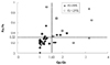
In order to identify the factors associated with a post-closure decrease in the FS below 29%, stepwise multiple linear regression analysis was performed (Table 3). Univariate stepwise linear regression analysis showed that the LVEDD and the pulmonic end size obtained from the lateral aortogram, Qp/Qs, Pp/Ps, and Rp/Rs ratios were associated with a FS below 29% at post-closure examination (p<0.05). Among these parameters, the Qp/Qs and Pp/Ps ratios were statistically significant factors on multivariate stepwise linear regression analysis for post-closure FS below 29% (p<0.1). The receiver operating characteristic curve analysis showed that a Qp/Qs ratio over 1.60 and a Pp/Ps ratio over 0.32 were the best cutoff values for post-closure FS below 29%, with a sensitivity of 86% and a specificity of 84%.
Table 4 lists the post-closure FS values, FS changes between pre- and post-closure, and Qp/Qs and Pp/Ps ratios. Changes in FS between pre- and post-closure were below 10%, 10-20%, and over 20% in 21 patients, 8 patients, and 14 patients, respectively. Changes in FS of over 10% were considered to be a "Change", and 22 patients had such changes. A significant "Change" in FS was noted in 40% (14/35) of patients with post-closure FS equal to or greater than 29% and in 100% (8/8) of patients with post-closure FS below 29%. The Qp/Qs ratio was over 1.60 in 12 of 43 patients and below 1.60 in 31 patients. Fifty-eight percent (7/12) of patients with a Qp/Qs ratio over 1.60 had a post-closure FS below 29% compared with 3% (1/31) of patients with a Qp/Qs ratio below 1.60. In addition, the Pp/Ps ratio was over 0.32 in 12 of 43 patients and below 0.32 in 31 patients. Among patients with a Pp/Ps ratio over 0.32, the post-closure FS was below 29% in 50% (6/12) of them, compared with 6% (2/31) of the patients with a Qp/Qs ratio below 1.60.
Eight patients had a Qp/Qs ratio over 1.60 and a Pp/Ps ratio over 0.32 (Fig. 1). Five of eight patients had LV "dysfunction" with post-closure FS below 29% and FS change of over 20%. Post-closure FS below 29% was observed in one patient with a Qp/Qs ratio below 1.60 and a Pp/Ps ratio over 0.32, and in two patients with Qp/Qs ratios over 1.60 and Pp/Ps ratios below 0.32. No patients with Qp/Qs ratios below 1.60 and Pp/Ps ratios below 0.32 had post-closure FS below 29%.
Discussion
Transcatheter interventional closure of PDAs has been practiced with various devices since 1967.12) Recently, with the introduction of the Amplatzer ductal occluder into clinical practice, transcatheter closure has become possible for even very large PDAs.13)
Because a PDA is a congenital heart defect with a left-to-right shunt, it is associated with an increased LV preload. PDA closure causes an immediate reduction in ventricular preload.1)2)14) Prior reports15) have shown that patients with PDAs differ from normal control subjects in LV volume and function as measured by two- and three-dimensional echocardiography at baseline. These changes caused by the PDA generally resolve by six months after percutaneous closure. Galal et al.2) demonstrated that patients with large PDAs (larger than 3.1 mm) had significant deterioration in FS immediately after PDA closure, which was followed by FS recovery within six months.
Compared to the values before PDA closure, in this study, a significant decrease in the LVEDD was noted; there was no difference in the LVESD immediately after PDA closure. However, at follow-up, an additional decrease in the LVEDD and a significant decrease in the LVESD were detected. An immediate decrease in the LVEDD and a late decrease in the LVESD resulted in a change in the FS when pre-closure, post-closure, and follow-up values were compared. Although FS is load-dependent and an acute change in LV volume loading after PDA closure is the primary underlying cause of this change in FS; FS below 29% after PDA closure was unusual and was considered to reflect LV dysfunction.
This study showed that LV dysfunction was present in eight patients immediately after PDA closure. Among these eight patients with LV dysfunction, seven patients had FS changes over 20%, five had Qp/Qs ratios over 1.60 and Pp/Ps ratios over 0.32, and two patients each had only Qp/Qs ratios over 1.60 or Pp/Ps ratios over 0.32. All of the patients were medicated with digitalis or angiotensin converting enzyme (ACE) inhibitors, but none showed clinical symptoms of ventricular dysfunction. In one case with a post-closure FS below 29% and a FS change below 20%, the FS was 28% pre-closure, 23% post-closure, and 25% at follow-up, and the Qp/Qs and Pp/Ps ratios were 1.60 and 0.33, respectively. This patient may have had a cardiomyopathy concurrent with the PDA. However, further diagnostic evaluations such as a cardiac biopsy were not performed, and the patient has been undergoing regular follow-up.
The relationship of the Qp/Qs and Pp/Ps ratios with FS was thought to be explained by larger amounts of shunting, higher pulmonary artery pressure, greater changes in the FS, and significant LV dysfunction. Therefore, based on our findings, although the patients had normal ventricular function before the procedure, patients with Qp/Qs ratios over 1.60 and Pp/Ps ratios over 0.32 at catheterization are predicted to be at risk for deterioration of ventricular function after the procedure. These patients require early evaluation of ventricular function using echocardiography.
Jeong et al.16) reported persistent LV systolic dysfunction (EF<50%) after PDA closure in adult PDA patients with low pre-closure LV EF. In contrast to Jeong's study, our study showed recovery of the FS at follow-up, despite the immediate post-closure LV systolic dysfunction (FS<29%). This discrepancy might be due to differences in the populations studied (adults vs. children). A longer duration of LV volume overload may cause changes in LV contractility and may lead to incomplete recovery after reduction in the loading condition. The ventricular remodeling status is different according to patient age. In this study, the tissue Doppler myocardial velocity was significantly decreased during the systolic and diastolic phases at post-closure and follow-up compared to the pre-closure measurements. These findings suggest that an immediate reduction in the preload after PDA closure may lead to decreased myocardial velocity during the systolic phase after the procedure. After preload reduction is accomplished, ventricular remodeling may occur slowly and lead to decreased myocardial velocity during the diastolic phase at follow-up. Previous studies have reported similar results; that is, changes in myocardial velocity after closure of a left-to-right shunt using a transcatheter device, which might have a volume reduction effect instead of inducing a real change in the myocardial velocity. Regional wall motion has been investigated in the right and left ventricles after device closure of atrial septal defects with chronic right ventricular volume overload.17) In such cases, conventional function parameters showed no definite changes, but the systolic and diastolic myocardial velocities decreased significantly. However, a new marker, isovolumic acceleration (IVA), was found to provide a more independent measurement of ventricular loading conditions than did myocardial velocities.18) Despite significant changes in the systolic and diastolic myocardial velocity, the IVA remained stable. The changes in myocardial velocity were considered to be a response to the modified ventricular loading conditions.
Study limitations
The major limitation of this study was its retrospective nature. In addition, variation in the physicians' measurements of echocardiographic parameters was not considered. Not all echocardiographic parameters were studied. For instance, IVA was not included
Conclusion
This study demonstrated that, in patients undergoing percutaneous transcatheter PDA closure, larger amounts of pre-closure left-to-right shunting and higher pulmonary artery pressure were associated with increased likelihood of post-closure FS<29%. The initial Qp/Qs and Pp/Ps ratios measured in the catheterization suite may be good predictors of LV dysfunction. In patients with high Qp/Qs and Pp/Ps ratios, serial assessment of ventricular function is recommended at follow-up echocardiography after PDA occlusion, as is evaluation for residual shunting.

 XML Download