

 PDF
PDF ePub
ePub Citation
 Print
Print


Abstract
Purpose
To compare and analyse the clinical outcomes between minimal-incision percutaneous repair and open repair among the surgical treatments for Achilles tendon ruptures.
Materials and Methods
We retrospectively analyzed and compared the outcomes between 25 patients with minimal incision percutaneous repair (group 1) and 30 patients with open repair (group 2), from January 2006 to June 2014. The postoperative clinical evaluations were done by Arner-Lindholm scale, American Orthopedic Foot and Ankle Society (AOFAS) ankle-hindfoot score, overall patient's satisfaction, and cosmetic satisfaction of scar.
Results
There were statistically significant differences between the two groups with respect to AOFAS hind foot score, mid-calf circumference differences, overall patient's satisfaction, and satisfaction of scar; the group 1 showed better achievement. There was no statistical difference between two groups in regards to other clinical outcomes. In group 1, there were 2 cases of sural nerve hypoesthesia, which fully recovered spontaneously at about 6 months after the surgery. In group 2, there were 3 cases of deep vein thrombosis, re-rupture, and deep infection.
REFERENCES
1. Mukundan C, El Husseiny M, Rayan F, Salim J, Budgen A. "Mini-open" repair of acute tendo Achilles ruptures: the solution? Foot Ankle Surg. 2010; 16:122–5.
2. Fortis AP, Dimas A, Lamprakis AA. Repair of Achilles tendon rupture under endoscopic control. Arthroscopy. 2008; 24:683–8.

3. Tang KL, Thermann H, Dai G, Chen GX, Guo L, Yang L. Arthroscopically assisted percutaneous repair of fresh closed Achilles tendon rupture by Kessler's suture. Am J Sports Med. 2007; 35:589–96.
4. Cetti R, Christensen SE, Ejsted R, Jensen NM, Jorgensen U. Operative versus nonoperative treatment of Achilles tendon rupture. A prospective randomized study and review of the literature. Am J Sports Med. 1993; 21:791–9.
5. Ma GW, Griffith TG. Percutaneous repair of acute closed ruptured Achilles tendon: a new technique. Clin Orthop Relat Res. 1977; 128:247–55.
6. Sutherland A, Maffulli N. A modified technique of percutaneous repair of ruptured Achilles tendon. Orthop Traumatol. 1999; 7:288–95.
7. Assal M, Jung M, Stern R, Rippstein P, Delmi M, Hoffmeyer P. Limited open repair of Achilles tendon ruptures: a technique with a new instrument and findings of a prospective multi-center study. J Bone Joint Surg Am. 2002; 84:161–70.
8. Rebeccato A, Santini S, Salmaso G, Nogarin L. Repair of the Achilles tendon rupture: a functional comparison of three surgical techniques. J Foot Ankle Surg. 2001; 40:188–94.
9. Rippstein PF, Jung M, Assal M. Surgical repair of acute Achilles tendon rupture using a "mini-open" technique. Foot Ankle Clin. 2002; 7:611–9.
10. Kong GM, Gwak HC, Kim JG. A comparative study of surgical treatment in the ruptured Achilles tendon: minimal incision and open repair. J Korean Foot Ankle Soc. 2012; 16:181–9.
11. Krackow KA, Thomas SC, Jones LC. A new stitch for ligament-tendon fixation. Brief note. J Bone Joint Surg Am. 1986; 68:764–6.
12. Jiang N, Wang B, Chen A, Dong F, Yu B. Operative versus nonoperative treatment for acute Achilles tendon rupture: a meta-analysis based on current evidence. Int Orthop. 2012; 36:765–73.
13. Nilsson-Helander K, Silbernagel KG. . Acute Achilles tendon rupture: a randomized, controlled study comparing surgical and nonsurgical treatments using validated outcome measures. Am J Sports Med. 2010; 38:2186–93.
14. Ljungqvist R. Subcutaneous partial rupture of the Achilles tendon. Acta Orthop Scand. 1968; 39:S1–86.
15. Nistor L. Surgical and non-surgical treatment of Achilles Tendon rupture. A prospective randomized study. J Bone Joint Surg Am. 1981; 63:394–9.
16. Kakiuchi M. A combined open and percutaneous technique for repair of tendo Achillis. Comparison with open repair. J Bone Joint Surg Br. 1995; 77:60–3.
17. Bhandari M, Guyatt GH, Siddiqui F. . Treatment of acute Achilles tendon ruptures: a systematic overview and meta-analysis. Clin Orthop Relat Res. 2002; 400:190–200.
18. Möller M, Movin T, Granhed H, Lind K, Faxén E, Karlsson J. Acute rupture of tendon Achillis. A prospective randomised study of comparison between surgical and non-surgical treatment. J Bone Joint Surg Br. 2001; 83:843–8.
19. Allan EI, Norman S, Thomas PS, Andrew HP. Rupture of the tendo Achilles. J Bone Joint Surg. 1976; 58:990–2.
20. Saxena A, Maffulli N, Nguyen A, Li A. Wound complications from surgeries pertaining to the Achilles tendon: an analysis of 219 surgeries. J Am Podiatr Med Assoc. 2008; 98:95–101.
21. Soroceanu A, Sidhwa F, Aarabi S, Kaufman A, Glazebrook M. Surgical versus nonsurgical treatment of acute Achilles tendon rupture: a meta-analysis of randomized trials. J Bone Joint Surg Am. 2012; 94:2136–43.
22. Willits K, Amendola A, Bryant D. . Operative versus nonoperative treatment of acute Achilles tendon ruptures: a multicenter randomized trial using accelerated functional rehabilitation. J Bone Joint Surg Am. 2010; 92:2767–75.
23. Wang D, Sandlin MI, Cohen JR, Lord EL, Petrigliano FA, SooHoo NF. Operative versus nonoperative treatment of acute Achilles tendon rupture: an analysis of 12,570 patients in a large healthcare database. Foot Ankle Surg. 2015; 21:250–3.
24. Jacobs D, Martens M, Van Audekercke R, Mulier JC, Mulier F. Comparison of conservative and operative treatment of Achilles tendon rupture. Am J Sports Med. 1978; 6:107–11.
Figure 1
Guiding instrument (A) and a straight needle (B). Cited from the article of Kong et al. (J Korean Foot Ankle Soc. 2012;16:181-9).10)
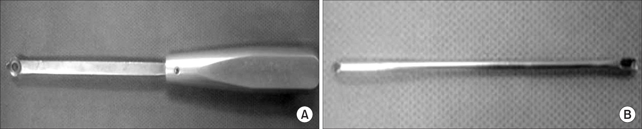
Figure 2
Preoperative marking of incision was made over the area of the rupture in mini-incision percutaneous repair (A) and open repair (B). Cited from the article of Kong et al. (J Korean Foot Ankle Soc. 2012;16:181-9).10)
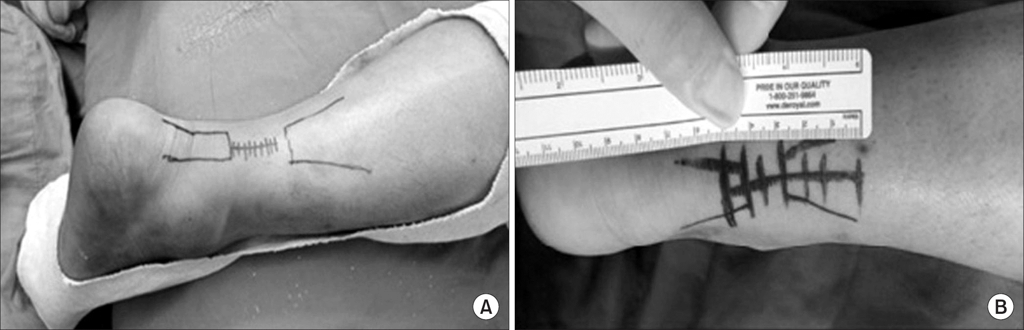
Figure 3
The paratenon and skin were closed in mini-incision percutaneous repair (A) and open repair (B). Cited from the article of Kong et al. (J Korean Foot Ankle Soc. 2012;16:181-9).10)
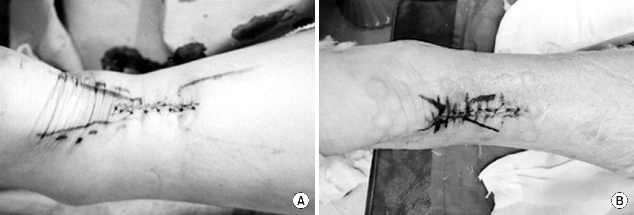
Figure 4
One leg heel raise test after 6 months from surgery in mini incision percutaneous repair (A) and open repair (B). Cited from the article of Kong et al. (J Korean Foot Ankle Soc. 2012;16:181-9).10)
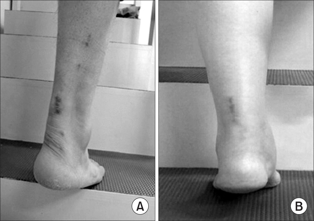
Table 1
Configuration of Demographics in Both Groups
Table 2
Configuration of Results in Both Groups
 XML Download