

 PDF
PDF ePub
ePub Citation
 Print
Print


Abstract
Paraspinal or extremity foreign-body reactions are a rare disease, which could be caused by foreign bodies to prevent intraoperative bleeding and can result in severe complications or death. However, they are often neglected. We report three cases of paraspinal and parapelvic gossypiboma mimicking a soft tissue tumor on magnetic resonance imaging, which were diagnosed and treated by surgical excision.
Figures and Tables
Figure 1
(A) Sagittal T1-weighted magnetic resonance image (MRI) shows hypointense, ellipsoid lesion with a central hypointense nidus in the left paravertebral soft tissue at the level of L4-5. (B) Sagittal T2-weighted MRI with contrast medium demonstrates a ring-enhanced, hyperintense lesion.
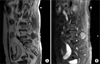
Figure 2
The excised specimen was cut. Retained sponge material was found within the thick capsule of the paraspinal mass.
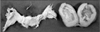
Figure 3
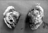
Histological findings (H&E staining). Submitted as a grayish white partially calcified fat tissue. Foreign body (gauze) granuloma (×200).
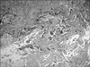
Figure 4
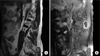
(A) Coronal T1 weighted magnetic resonance image (MRI) with contrast medium shows well-encapsulated inhomogenous mass with peripheral rim enhancement. (B) Coronal and (C) Axial T2 weighted MRI show a well-encapsulated complex mass containing hyper and hypointense regions. Central low and high signal intensity, peripheral low signal rim.
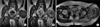
Figure 5
(A) Submitted subcutaneous mass, measuring 16×14×18 cm with a weight of 450 g. The cut surface of the section shows a cystic space, containing purulent exudates and (B) gauze.
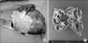
Figure 6
Histological findings. Cystic mass with a thick fibrous hyallinized wall (H&E stain, ×12.5) with central cystic degeneration. Foreign body (gauze) granuloma.
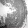
Figure 7
(A) Sagittal T2 weighted magnetic resonance image (MRI) shows the well-defined paravertebral soft tissue mass. Central low and high signal intensity, with a peripheral low signal rim. (B) Sagittal T1 weighted MRI with contrast medium shows the defined paravertebral soft tissue mass with peripheral rim enhancement.
References
1. Park JS, Park DI. Gossypiboma (Textiloma) due to retained surgical gauze. Korean J Gastroenterol. 2006. 48:143–144.
2. Rajković Z, Altarac S, Papeš D. An unusual cause of chronic lumbar back pain: retained surgical gauze discovered aft er 40 years. Pain Med. 2010. 11:1777–1779.
3. Ramirez LF, Thisted R. Complications and demographic characteristics of patients undergoing lumbar discectomy in community hospitals. Neurosurgery. 1989. 25:226–230.

4. Nabors MW, McCrary ME, Clemente RJ, et al. Identification of a retained surgical sponge using magnetic resonance imaging. Neurosurgery. 1986. 18:496–498.
5. Matsuki M, Matsuo M, Okada N. Case report: MR findings of a retained surgical sponge. Radiat Med. 1998. 16:65–67.
6. Okten AI, Adam M, Gezercan Y. Textiloma: a case of foreign body mimicking a spinal mass. Eur Spine J. 2006. 15:626–629.
7. Massie JB, Heller JG, Abitbol JJ, McPherson D, Garfin SR. Postoperative posterior spinal wound infections. Clin Orthop Relat Res. 1992. 284:99–108.
8. Marquardt G, Rettig J, Lang J, Seifert V. Retained surgical sponges, a denied neurosurgical reality? Cautionary note. Neurosurg Rev. 2001. 24:41–43.
9. Manzella A, Filho PB, Albuquerque E, Farias F, Kaercher J. Imaging of gossypibomas: pictorial review. AJR Am J Roentgenol. 2009. 193:S94–S101.
10. Turgut M, Akyüz O, Ozsunar Y, Kacar F. Sponge-induced granuloma ("gauzoma") as a complication of posterior lumbar surgery. Neurol Med Chir (Tokyo). 2005. 45:209–211.
 XML Download