

 PDF
PDF ePub
ePub Citation
 Print
Print


Abstract
Developmental process of the clavicle was studied on 35 cases of Korean fetuses of 9 to 300mm crown-rump length by histological observation and radiological study following silver impregnation, with the following results.
1. Primordia of the clavicle of the 14mm fetus were blastemal tissue, while that of the 110 to 130mm showed definite evidence of ossification center at the sternal and acromial ends as cartilagenous mass.
2. The membranous ossification center, which could be recognized in the 16 to 18mm fetuses, were two in number.
3. In the 24mm specimen the centers were shown to unite each other, forming a long single mass of bone and in the 35mm the clavicle resumed growth as a long cartilagenous mass.
4. The development of the acromio-clavicuiar joint began in the 50mm specimen as a cavity without the three-layered interzone which could commonly be recognized in developing joint of other long bone.
5. That the recognition of the ossification center was first possible in the 45mm specimen by radiological study showed that this method was inferior to histological observation for detecting the ossification center.
REFERENCES
1. Andersen H. Histochemistry and development of human shoulder and acromioclavicular joints with perticular REFERENCEs to the early development of the clavicle. Acta Anat. 55:124–165. 1963.
3. . The development and ossification of the human clavicle. J. Anat. (Lond). 47:225–234. 1913.
4. Gardner E.Bourne G. H., editorOsteogenesis in the human embryo and fetus, in “The Biochemistry and Physiology of Bone”. New York, Academic Press: 1956.
5. Gardner E., Gray D.J.Prenatal development of the human shoulder and acromioclayiclar joint. Amer, J. Anat. 92:219–226. 1953.
6. Gibson D.A., Caroll M.Congenital psedarthrosis of the clavicle. J. Bone Joint Surg. Vol. 52-B(No. 4):629. 1970.
7. Grav D.J., Cardner E.Prenatal development of the human elbow joint, Amer. J. Anat. 88:429–470. 1951.
8. Koch A.R.Die Fruhentwicklung der Clavicula beim Menchen. Acta Anat. 42:177–212. 1953.
9. Mall F.P.On ossification centers in human embryos less than one hundred days old. Amer. J. Anat. 5:433–458. 1906.

10. Morris H.Morris’ human anatomy. 12 Ed.245. McGraw-Hill, N. Y.;1966.
11. Noback C.R., Robertson C.G.Sequence of appearance of ossifecation centers in the human skeleton during the five prenatal months. Amer. J. Anat. 89:1–28. 1951.
12. O'Rahiley R., Meyer D.B.. Roentgenographic investigation of the human skeleton during early fetal life. Amer. J. Roentgenol. & Rad. Therapy. 7:455–468. 1956.
13. Robert C.Congenital pseudarthrosis of the clavicle. J. Bone Joint Surg. Vol. 52-B(No. 4):644. 1970.
14. Streeter G.L.Developmental horizons in human embryos: Age group 11 to 23. Embryology Reprint. Vol. 2:Carnegie Institute of Washington D.C.;1951.
1. Brower T.D., Wilde A.H.(1966); Femoral Neuropathy in Hemophilia. J. B. J. S. 48-A:487.
2. Calverley J.R., Mulder D.W.(1960); Femoral Neuropathy. Neurology. 10:963.
3. Douglas A.S., McAlpine S.G.(1956); Neurological Complication of Haemophilia and Christmas Disease. Scottish Medical Journal. 1:270.
4. Fearn C.B.(1968); Iliacus Hematoma Syndrome as a Complication of Anticoagulant Therapy. British Medical Journal. 4:97.
5. Gertzbein S.D.Femoral Nerve Neuropathy Complicating Iliopsoas Haemorrhage in patient's without Hemophilia. J.B.J.S. 54-B:149. 1972.
6. Green J.P.Proximal Avulsion of Iliacus with Paralysis of the Femoral Nerve. J.B.J.S. 54-B:154. 1972.
7. Groch S.N., Hurwitz L.J., McDevitt E., Wright I.S.(1959); Problems of Anticoagulant in Cerebrovascular disease. Neurology. 9:786.
8. Goodfellow J., Fearn C.B.D'A., Matthew's J.M.(1967); Iliacus Hematoma. J.B.J.S. 49-B:748.
9. Joseph G., Chusid M.D.(1973); Correlative Neuroanatomy & Functional Neurology. 15th edition. pp. 121–123.
10. Kettlekamp D.B., Power's S.R.(1969); Femoral Compression Neuropathy in Hemorrhagic disorders. Archives of Surgery. 98:367.
11. Kubacz G.J.(1971); Femoral and Sciatic Compression Neuropathy. British Journal of Surgery. 58:580.
12. Patten B.M.(1969); Neuropathy induced by Hemorrhage. Archives of Neurology (Chicago). 21:381.
13. Seddon J.H.Three types of Nerve Injury, Brain. 66:237, 1943. The American Academy of Orthopedic Surgeons Instructional Course Lectures. Vol. XIX:1970; pp. 41–54.
14. Sigler L., Raut P.S., Vollman R.W.(1970); Ilacus muscle Hematoma. Angiology. 21:114.
15. Strandell G.(1942); Subcutaneous rupture of the Iliopsoas muscle. Acta Chirurgica Scandinavica. 86:149.
16. Susens G.P., Hendrickson C.G., Mulder M.J., Sams B.(1968); Femoral Nerve Entrapment Secondary to a Heparin Hematoma. Annals of INternal Medicine. 69:575.
17. Licht S.Electrodiagnosis and Electromyelography. New, Haven, Conn.: 1956; Elizabeth Licht, Publisher;p. 317.
18. Tallroth A.(1939); Hemophilia with Spontaneous Hemorrhage in the Iliopsoas Muscle Followed by Injury to the Femoral Nerve. Acta Chirurgica Scandinavica. 82:1.
19. Tallroth A.(1941); Hemorrhage in the Iliopsoas Muscle Causing Injuries to the Femoral Nerve. Acta Chirurgica Scandinavica. 84:124.
20. Toshima Hiroshi & Yamahiro Kenzo. (1974); Clinical Orthopedic Surgery. Vol. 9(No. 1):Jan. 94.
Fig. 1
Photomicrigraph to show horizontal section of the clavicle of 16mm fetus. There are two centers, a smaller lateral one on the left merging into a cellular mass and a larger medial one on the right continuous with the condensation directed toward the ventral midline. H-E stain. X 100.
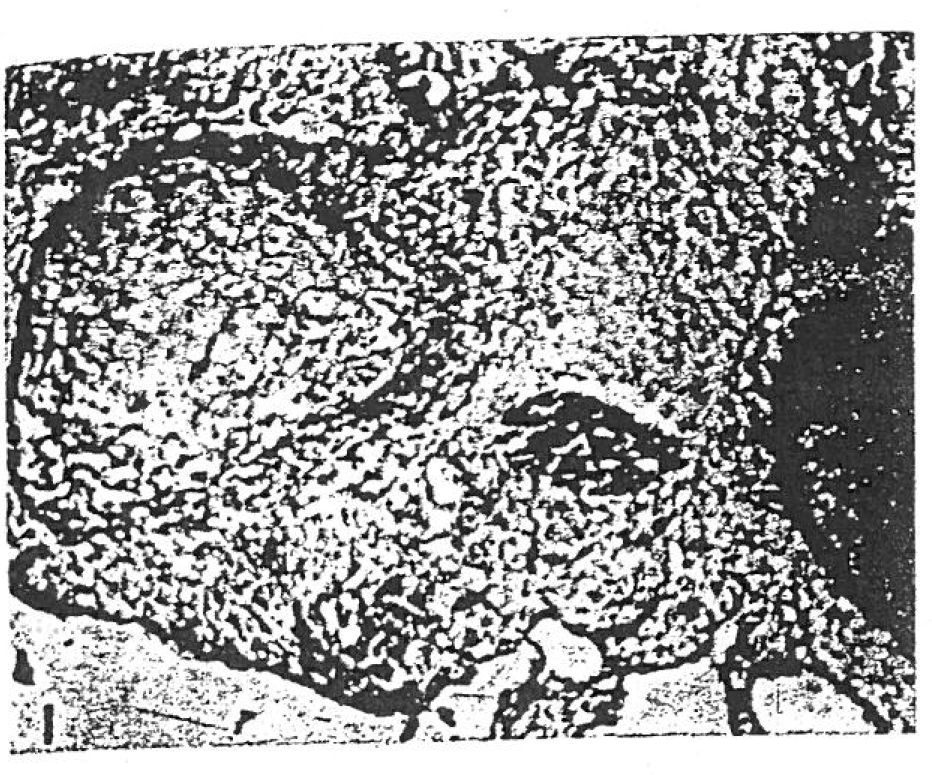
Fig. 2
Photomicrograph of specimen from 18 mm fetus showing the clavicle with early membranous bone formation. H-E stain, X 100.
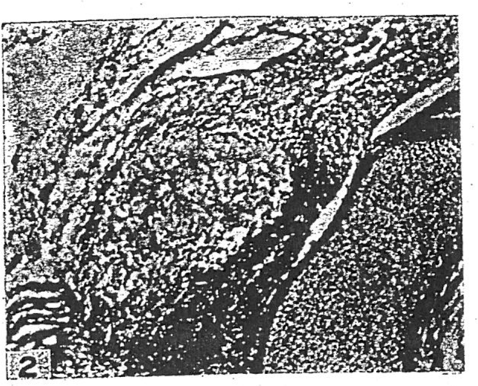
Fig. 3
Photomicrograp h of specimen from 130 mm fetus showing the cartilagenous acromial end. H-E stain, X 100.
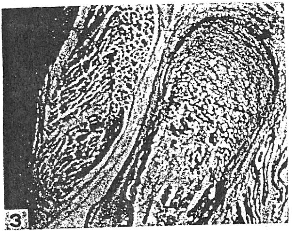
Fig. 4
Photomicrograph to show frontal section of the sternal end of 24mm fetus. A cavity is present. H-E stain, X 100.
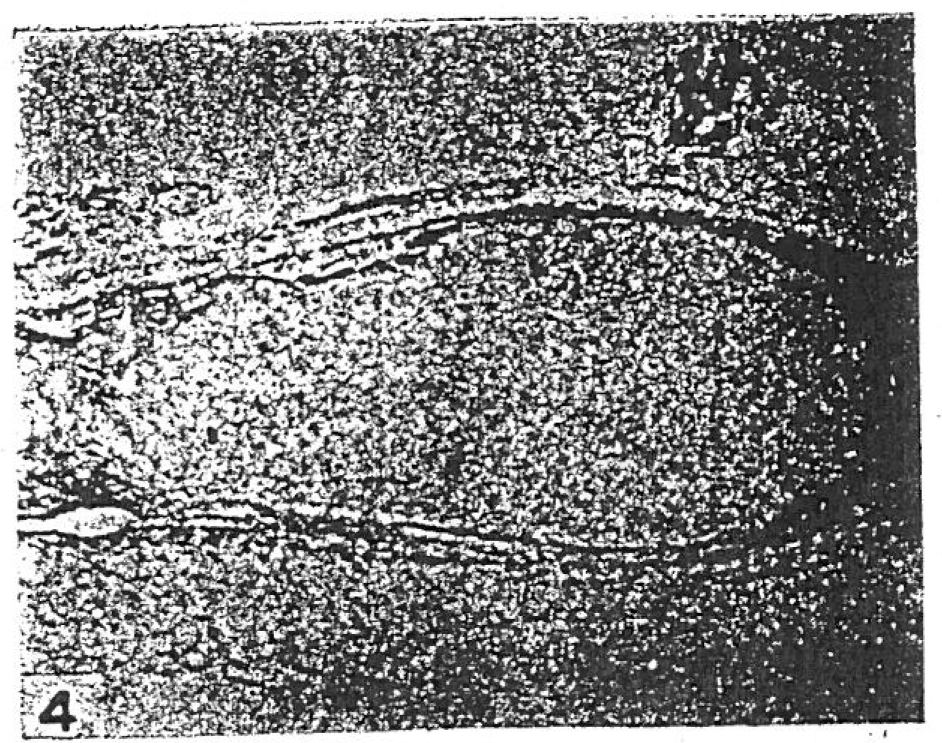
Fig. 5
Photomicrograph to show horizontal section of clavicle of 40 mm fetus. Ossification is spreading from the center of the shaft toward both ends. H-E stain, X 140.
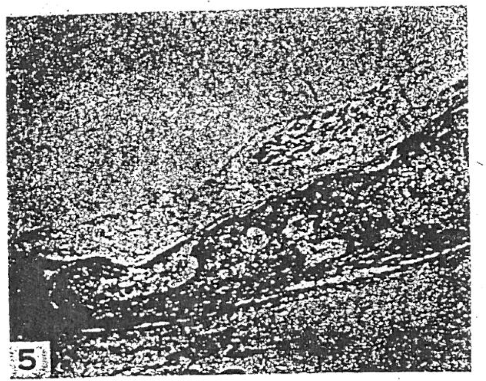
Fig. 6
Photomicrograph of specimen from 70 mm fetus showing well-advanced ossification with a marrow cavity. H-E stain, X 40.
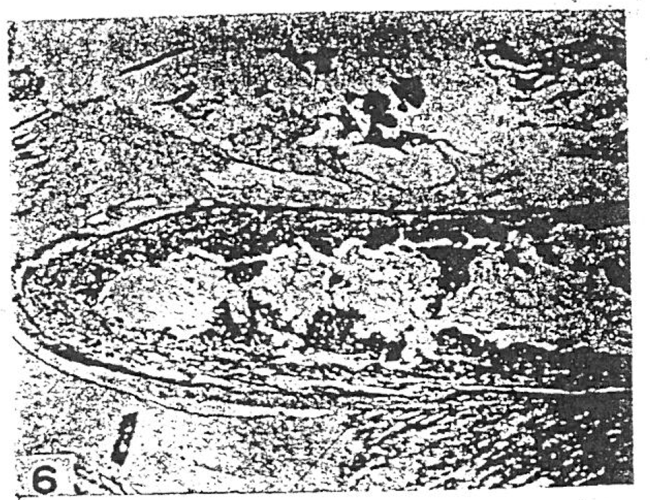
Fig. 7
Photo micrograph of the clavicle of 125mm fetus showing the sternal end which is largely made of well formed cartilage. H-E stain, X 40.
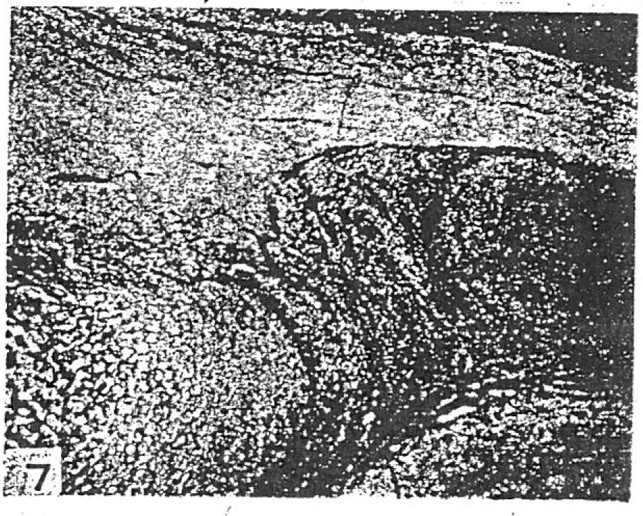
Fig. 8
Photomicrograph to show horizontal section of shoulder region of 50mm fetus. The acromio-clavicular joint cavity appears. H-E stain, X 40.
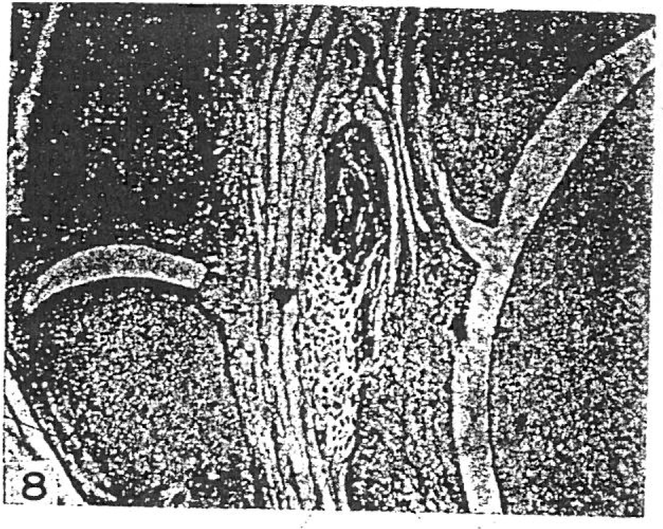
Fig. 9
Photomicrograph to show frontal section of shoulder region of 70mm fetus. The acromioclavicular joint cavity is complete. H-E stain, X 40.
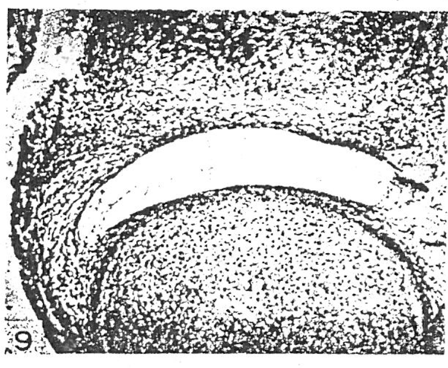
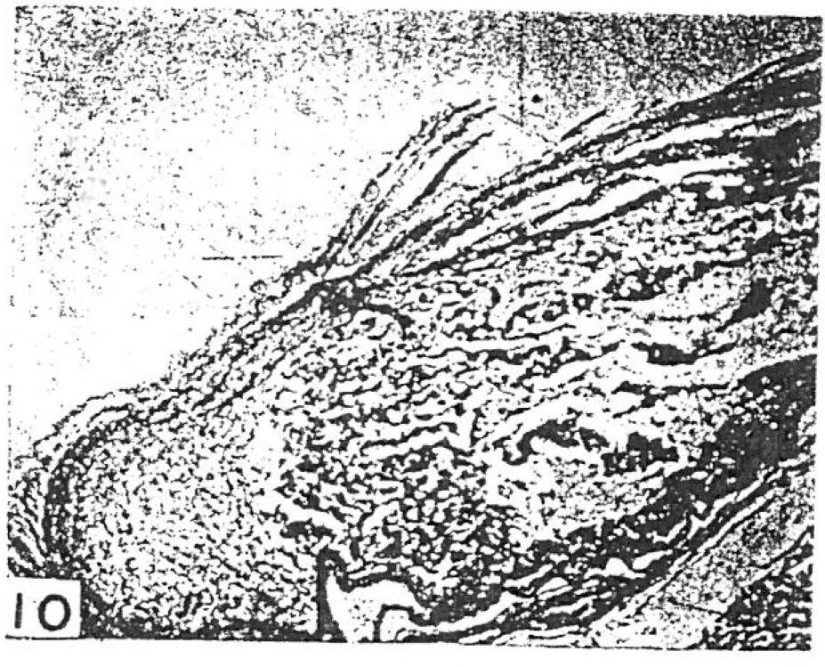
Fig. 10
Photomicrograph to show horizontal section of the clavicle of 170mm fetus. The clavicle is consisted mostly of bone. H-E stain, X 40.
 XML Download