

 PDF
PDF ePub
ePub Citation
 Print
Print


INTRODUCTION
Breast cancer is the most common cause of cancer-related deaths in women globally and the second most common malignancy in Korean women, accounting for 14.8% of all malignancies newly diagnosed in 2011 [1]. The incidence of invasive breast cancer (IBC) has increased continuously over time, reaching 58 cases per 100,000 women in 2011 [1]. Although early cancer detection and current standard treatments specific for individual tumor characteristics have increased the survival of patients with breast cancer, there remains a subset of patients displaying unresponsiveness to standard therapy. The selection and control of these patients are problems awaiting solutions in oncology.
Epithelial-mesenchymal transition (EMT) of epithelial cells is defined as the loss of epithelial characteristics and acquisition of a mesenchymal phenotype. Tumor cells undergoing EMT acquire migratory potential, stem cell-like features, and chemoresistance to conventional and targeted therapeutics [234]. A variety of biomarkers, including transcription factors (zinc finger factor SNAIL [Snail], snail2 [slug], zinc finger E-box-binding homeobox 1 [ZEB1], twist, and forkhead box protein 2 [FOXC2]), extracellular matrix proteins (fibronectin and laminin), cell surface proteins (E-cadherin and N-cadherin), and cytoskeletal proteins (vimentin and α-smooth muscle actin), have been used to define EMT phenotypes in cancer tissue; however, no biomarker has been accepted for EMT signatures [5].
E-cadherin is a transmembrane glycoprotein that has an important role in epithelial cell adhesion. Loss of E-cadherin expression has been considered a hallmark of EMT [4]. Fibronectin is a large heterodimeric glycoprotein that can exist in an insoluble, cellular form or soluble form in plasma. It plays a role in cell-matrix and cell-cell adhesion, cell migration, differentiation, morphogenesis, and oncogenic transformation [6]. A variety of benign and malignant epithelial and mesenchymal cells can produce fibronectin, and upregulation of fibronectin occurs during the EMT process of epithelial tumors [7]. Fibronectin has been used to detect EMT phenotypes in human cancers [489].
Earlier studies reported that the EMT phenotype defined by patterns of EMT-related protein expression was an independent prognostic factor in gastrointestinal cancers involving the esophagus, stomach, or small intestine [8910]. However, little is known about the clinical significance of EMT phenotypes in IBC. In this study, investigated whether EMT phenotypes could be used to select a distinct breast cancer subgroup with a worse clinical outcome, as demonstrated for other tumors [789]. For this purpose, we assessed the expression of E-cadherin and fibronectin in tumor samples from patients with IBC and defined EMT phenotypes based on the combined expression patterns of these markers. The correlation of EMT phenotypes with clinicopathological factors and patient survival was analyzed to evaluate of the clinical significance of EMT phenotypes in patients with IBC.
METHODS
Tissue samples and tissue microarray construction
Two sets of tissue microarrays (TMAs) of IBCs were used [1112]. Samples for a total of 1,596 patients with primary IBC who underwent surgical resection between January 1995 and December 2007 were collected retrospectively from the archives of the Department of Pathology at Yeungnam University Hospital, Daegu, Korea. Patients who could tolerate treatment received standard radiotherapy or adjuvant systemic therapy (hormone therapy or chemotherapy) after surgery according to a medical insurance program administered by the Ministry of Health and Welfare of Korea. For TMA construction, we reviewed hematoxylin and eosin-stained slides of all patients and selected a representative tumor block for each patient. The first set consisted of 13 TMA blocks containing 594 samples (obtained between 1995 and 2003). The method for TMA construction was described in our previous study [11]. The second set comprised 43 TMA blocks constructed with 1,002 samples (obtained between 2004 and 2007) using a Quick-Ray® Manual Tissue Microarrayer (Unitma, Seoul, Korea) and Quick-Ray® recipient blocks of 2 mm cores (Unitma) [12].
Patient and tumor characteristics, including age at the time of the initial diagnosis, pathologic tumor stage (pT), pathologic lymph node stage (pN), histological grade [13], the presence or absence of lymphovascular invasion, and follow-up information, were obtained from the pathology reports and patients' medical records. Information on the presence and cause of death of each patient was also obtained using the microdata service system provided by Statistics Korea (http://mdss.kostat.go.kr). Overall survival (OS) was defined as the interval between the date of surgical resection and the date of disease-specific death or the last follow-up. Disease-free survival (DFS) was expressed as the number of months from the date of surgical resection to the date of documented relapse, including locoregional recurrence and distant metastasis. This study was approved by the Institutional Review Board of Yeungnam University Hospital (YUH-12-0344-O20), and the requirement for informed consent was waived.
Immunohistochemistry and scoring
For molecular classification of the patients, because the staining conditions and interpretation criteria changed over the study period (1995-2007), we repeated immunohistochemistry for estrogen receptor (ER), progesterone receptor (PR), and human epidermal growth factor receptor 2 (HER2) and interpreted the results according to the current guidelines for ER/PR and HER2 testing [1415].
TMA sections measuring 4 µm in thickness were used for immunohistochemistry. Immunohistochemical staining for ER (SP1, prediluted; Ventana Medical Systems, Tucson, USA), PR (1E2, prediluted; Ventana Medical Systems), HER2 (CONFIRM™ anti-HER2/neu (4B5) rabbit monoclonal; Ventana Medical Systems), E-cadherin (clone 4A2C7, 1:200 dilution; Invitrogen, Carlsbad, USA), and fibronectin (clone F14, prediluted; Biogenex, San Ramon, USA) was performed in the automated Benchmark® platform (Ventana Medical Systems) and labeled using an UltraView™ universal DAB detection kit (Ventana Medical Systems), as described in previous studies [916]. Silver-enhanced in situ hybridization for HER2 using an INFORM® HER2 DNA probe (Ventana Medical Systems) was performed for equivocal cases in HER2 staining [12].
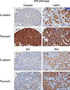
Regarding the interpretation of E-cadherin and fibronectin, staining intensity and the proportion of tumor cells were evaluated as described previously [9]. Staining intensity was classified as follows: 1, weak; 2, moderate; and 3, strong. Positive cells were quantified as a percentage of the total number of tumor cells, and the value was categorized as follows: 0, ≤5%; 1, >5% and ≤25%; 2, >25% and ≤50%; 3, >50% and ≤75%; 4, >75%. The sum of the tumor area of two consecutive tumor cores was regarded as the total tumor area (100%) instead of scoring the immunostaining results in each tumor core separately. The percentage of epithelial cell positivity and staining intensity were multiplied to generate an immunoreactivity score (IS) for each sample, which ranged from 0 to 12. The Kaplan-Meier method with the log-rank test was used to select a cutoff point for designating immunopositivity for each marker that was most meaningful with respect to prognosis [8]. Using this method, samples with an E-cadherin IS ≥8 were considered positive for E-cadherin, and those with a fibronectin IS ≥2 were regarded as positive for fibronectin.
IBCs were classified according to the following types of EMT by modifying the proposal provided by Sung et al. [8]: complete type (E-cadherin-negative/fibronectin-positive), incomplete type (hybrid type, E-cadherin-positive/fibronectin-positive; null type, E-cadherin-negative/fibronectin-negative), and wild-type E-cadherin-positive/fibronectin-negative).
Based on their hormone receptor (HR, ER or PR) and HER2 statuses, the IBC samples were divided into four molecular subtypes: HR+/HER2-, HR+/HER2+, HR-/HER2+, and HR-/HER2-.
Statistical analysis
Statistical analysis was performed using SPSS version 21.0 for Windows (IBM Corp., Armonk, USA). The chi-square test was used to evaluate the significance of correlations between EMT phenotypes and patient characteristics. Univariate and multivariate analyses were performed to assess the effect of EMT phenotypes on patient survival (OS and DFS). Survival curves were plotted using the Kaplan-Meier method and the log-rank test was used to test the significance of survival differences. Significant variables identified in univariate analyses were further analyzed using a Cox regression proportional hazard model. Adjusted hazard ratios and associated 95% confidence intervals (CIs) were estimated for each variable. A p-value of <0.05 was considered statistically significant.
RESULTS
Patient demographics and tumor characteristics
Of the 1,596 patients, 101 were excluded from the analysis because of a failure to obtain immunohistochemical results as a result of noninformative cores (no invasive tumor) or a loss of cores while performing immunohistochemistry. Therefore, 1,495 patients were included in this study. Patients ranged in age from 20 to 86 years (median, 46 years; mean, 48 years). Among these patients, 878 (58.7%) underwent mastectomy, and 617 (41.3%) underwent breast-conserving surgery. Sentinel node biopsy or axillary dissection was performed in 1,492 patients. Concerning adjuvant chemotherapy, 1,010 patients (67.6%) received anthracycline-based chemotherapy, 259 patients (17.3%) received nonanthracycline chemotherapy, and the remaining 226 patients (15.1%) did not receive chemotherapy. A total of 1,025 patients (68.6%) received hormone therapy. The median follow-up period was 62 months for OS and 60 months for DFS (range, 1-202 months).
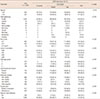
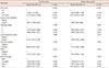
Of the 1,495 patient samples, 1,008 (67.4%) were positive for ER, and 871 (58.3%) were positive for PR. Protein overexpression or amplification of HER2 was observed in 293 samples (19.6%). Regarding molecular subtypes, 893 tumors (59.7%) were HR+/HER2-, 131 (8.8%) were HR+/HER2+, 162 (10.8%) were HR-/HER2+, and 309 (20.7%) were triple-negative. Positive expression for E-cadherin and fibronectin was observed in 1,193 (79.8%) and 354 (23.7%) samples, respectively. The characteristics of the tumors are shown in Table 1.
Classification of EMT phenotype and its correlation with clinicopathological features and patient survival
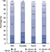
Classification of the 1,495 patients with IBC according to EMT phenotypes resulted in the identification of 64 patients (4.3%) with the complete EMT type, 903 patients (60.4%) with the wild-type, and 528 patients (35.3%) with the incomplete EMT type (hybrid type, 290 cases; null type, 238 cases) (Figure 1). The EMT phenotype displayed significant associations with age (p=0.015), pT (p<0.001), pN (p<0.001), histological grade (p<0.001), lymphovascular invasion (p=0.035), and molecular phenotype (p<0.001) (Table 1). The complete EMT phenotype was observed more frequently in patients with a younger age (<50 years) (Figure 2), higher pT and pN stages, histologic grade 3, or triple negativity. Lymphovascular invasion was observed more frequently in patients with complete or hybrid phenotypes than in those with null or wild-type phenotypes. In total, 52 (81.3%), nine (14.1%), and three (4.7%) tumors with the complete EMT phenotype were invasive (ductal) carcinoma, no special type, invasive lobular carcinoma, and mixed type, respectively. Concerning other histologic subtypes, including invasive micropapillary, mucinous, tubular, medullary, metaplastic, and invasive papillary carcinomas, none of the tumors displayed the complete EMT phenotype (p<0.001). Patients who underwent mastectomy had higher rates of complete and hybrid EMT phenotypes than those who underwent breast-conserving surgery (p<0.001). Patients with the complete EMT phenotype received adjuvant chemotherapy more frequently compared to those with the wild-type phenotype (p<0.001).
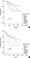
In the comparison of survival differences according to the EMT phenotype of patients with IBC, there was a significant survival difference among the four EMT subgroups (OS, p=0.001; DFS, p<0.001) (Figure 3). Survival differences were observed between the different EMT phenotypes (complete vs. wild-type, OS, p=0.004, DFS, p<0.001; hybrid vs. wildtype, OS, p=0.002, DFS, p=0.001; hybrid vs. null, OS, p=0.026, DFS, p=0.018; complete vs. null, OS, p=0.014, DFS, p=0.003), but not between wild-type and the null type (OS, p=0.896; DFS, p=0.956) or between hybrid and complete types (OS, p=0.407; DFS, p=0.203).
In addition to the EMT phenotype, pT (p<0.001 and p<0.001), lymph node status (p<0.001 and p<0.001), histologic grade (p<0.001 and p=0.001), lymphovascular invasion (p<0.001 and p<0.001), hormone receptor (p=0.019 and p=0.892) and HER2 status (p=0.02 and p=0.014) predicted OS and DFS based on univariate analysis. In multivariate analysis, advanced pT stage (p<0.001), the presence of lymph node metastasis (p=0.014), and the presence of lymphovascular invasion (p=0.006) were independent prognostic factors for poor OS in patients with IBC. For DFS, advanced pT stage (p=0.004), the presence of lymph node metastasis (p=0.001), histologic grade 3 (p=0.031), and the presence of lymphovascular invasion (p=0.015) were independent poor prognostic factors. Among EMT phenotypes, patients with the hybrid phenotype exhibited a 1.45-fold (95% CI, 1.03-2.04; p=0.032) higher risk of disease recurrence than those with the wild-type phenotype (Table 2).
DISCUSSION
We investigated the aberrant expression of the EMT-related proteins E-cadherin and fibronectin in the cancer cells of 1,495 patients with IBC using immunohistochemistry. The EMT phenotype defined by the combined expression pattern of E-cadherin and fibronectin displayed significant correlations with clinicopathological factors indicating aggressive biological behavior, including advanced pT and pN stages, high histologic grade, the presence of lymphovascular invasion, and triple negativity. In addition, patients with complete and hybrid EMT phenotypes exhibited poorer OS and DFS than those with the wild-type phenotype, and the hybrid EMT phenotype was an independent prognostic factor in patients with IBC.
EMT is a dynamic and reversible process induced by a variety of signaling pathways including Wnt, tumor necrosis factor α/nuclear factor κB, Notch, MAPK/PI3K, and transforming growth factor β pathways [17]. These EMT signaling pathways are also known to be involved in the generation of breast cancer stem cells (BCSCs) [1819]. Sarrió et al. [20] reported an association of EMT with the basal-like phenotype of breast cancer, and they suggested that EMT is related to high aggressiveness and the metastatic spread of basal-like breast cancer. Prat et al. [21] recently identified a molecular subtype of triple-negative breast cancer, the claudin-low subtype, in which markers linked to EMT and BCSCs are expressed concurrently. Breast cancer cells expressing these overlapping molecular features (EMT, BCSC, and claudin-low) are expected to have migratory potential, metastatic growth, and chemoresistance [212223]. There have been reports demonstrating that residual breast cancer cells remaining after conventional therapy displayed both BCSC and mesenchymal features [2425]. These basic and fundamental findings support our results that the EMT phenotype was significantly correlated with advanced stage, lymphovascular invasion, high histologic grade, triple-negativity, and poor clinical outcome.
The EMT phenomenon represents an interaction of complex EMT-related markers that are affected by each other [2]. Although EMT is characterized by a lack of epithelial features and attainment of the mesenchymal features of epithelial cells, a wide spectrum of EMT phenotypes have been reported. Sung et al. [8], proposed four specific types of EMT, including wild-type (epithelial), complete (mesenchymal), and intermediate (hybrid and null) phenotypes, based on the combined expression patterns of epithelial (E-cadherin) and mesenchymal (fibronectin, smooth muscle actin, and vimentin) markers. They found that the EMT phenotype has significant prognostic value in esophageal squamous cell carcinoma. In their study, OS and DFS were worst in the complete EMT type group, better in the incomplete type group, and best in the wild type group. In addition, the EMT phenotype displayed significant associations with tumor size, histological differentiation and invasion depth. Ryu et al. [10] investigated the expression status of five EMT-related markers (E-cadherin, vimentin, snail1, ZEB1, and β-catenin) and CD44 in gastric cancers to determine the roles of EMT-related proteins in gastric cancer progression. Although the loss of E-cadherin expression and aberrant expression of vimentin were associated with poor patient survival, altered expression of snail1, ZEB1, CD44, and β-catenin did not have a significant effect on patient survival. However, when they selected four markers, E-cadherin, snail1, vimentin, and CD44, tumors with altered expression of three or more proteins displayed highly aggressive clinical features and less favorable outcomes than those with altered expression of two or less proteins. Their results suggest that combined analysis of the expression of EMT-related proteins may provide more information about the biological behavior of tumors than the alteration of any single EMT-related marker.
In our previous study [9], we used three markers (E-cadherin, vimentin, and fibronectin) to define the EMT phenotype of small intestinal adenocarcinomas because none of the patients exhibited positivity for smooth muscle actin in their tumor cells, and the results obtained were similar to those reported by Sung et al. [8]. The complete EMT phenotype displayed significant correlations with undifferentiated histology and poor survival in patients with small intestinal adenocarcinoma and a trend toward an association with advanced pT classification. In this study, we also observed the expression of other mesenchymal markers including vimentin and smooth muscle actin in these patients. Unlike previous studies [89], vimentin and smooth muscle actin did not exhibit significant prognostic value in breast cancer in our study (data not shown). Therefore, only E-cadherin and fibronectin were used to define EMT phenotypes in this study because we intended to stratify IBC according to EMT phenotypes with prognostic significance. These previous studies and our present research suggest that EMT phenotype defined by specific epithelial and mesenchymal markers may provide useful information for predicting patient outcome regardless of the location (esophagus, small intestine, and breast) and histology (squamous cell carcinoma and adenocarcinoma) of the tumors.
Logullo et al. [26] studied the concomitant expression of EMT-related markers (E-cadherin, β-catenin, Snail, transforming growth factor β1 [TGF-β1], TGFβ type II receptor [TBRII], and the HGF receptor [c-met]) in both ductal carcinoma in situ (DCIS, n=95) and invasive ductal carcinoma (IDC, n=55) using a TMA. Excluding c-met and TGF-β1, EMT markers were not associated with differences in positivity rates between DCIS and IDC. In addition, none of the EMT markers was correlated with patient survival. Choi et al. [27] recently reported that the expression of EMT markers (vimentin, smooth muscle actin, osteonectin, N-cadherin, E-cadherin, and β-catenin) and CD146 was significantly higher in invasive carcinoma than in DCIS of the basal-like subtype. They suggested an important role of EMT in the progression from in situ to invasive basal-like breast cancer. To the best of our knowledge, this is the first study to perform survival analysis according to the combined expression patterns of EMT-related proteins rather than the expression of a single EMT-related marker in patients with IBC.
There are several limitations in our study. First, the EMT process occurs primarily in the infiltrative tumor border [28]; however, our study was performed using TMAs. While constructing TMA blocks, we attempted to remove tumor cores from the representative tumor area while avoiding areas tumor necrosis and central fibrous scars. Although some tumor cores may correspond to the peripheral portion of the tumor, most of tumor cores do not exactly reflect the infiltrative margin of the tumor. Second, unlike other studies that used three or more epithelial and mesenchymal markers to define EMT phenotypes [8910], only one epithelial marker and one mesenchymal marker were selected for EMT phenotype analysis in the present study. In addition to these epithelial and mesenchymal markers, there are many EMT-related markers, including transcription factors, which are more biologically significant in terms of the activation of signaling pathways. Therefore, additional research using other EMT-related markers is necessary to validate clinical significance of EMT phenotypes in cancer samples.
In conclusion, we investigated the expression patterns of EMT-related epithelial (E-cadherin) and mesenchymal (fibronectin) markers in 1,495 patients with IBC. The EMT phenotypes defined by the combination of expression patterns for both proteins exhibited significant associations with clinicopathological factors, and they could further stratify patients with IBC into subgroups with prognostic significance.
 XML Download