

 PDF
PDF ePub
ePub Citation
 Print
Print


INTRODUCTION
Resin cements are used for cementation of crowns, porcelain veneers, inlays, onlays, and inlay-retained fixed partial dentures. Resin cements have many advantages, such as improved bond strength, low microleakage, low solubility, good mechanical properties, and good esthetics.1234 Conventional resin luting depends on prior etching of the tooth structure and subsequent penetration of the adhesive system, which bonds to demineralized enamel or dentin by forming a ‘hybrid layer’ – i.e. an interdiffusion zone with exposed collagen fibrils – that is responsible for retention.5678 By contrast, self-etching resin luting cements require no acid etching, washing, or drying of the tooth surface since the system's acidic primer demineralizes the tooth surface by modifying the smear layer without removing it completely.7910 Long-term success of restorations depends upon the durability and strength of the bonds between restoration, cement, and dental substrate.10111213 Cementation failure of restorations is frequently observed in clinical practice.14 Clinical studies have reported restoration failure rates of 7% for porcelain laminate veneers; 8 - 24% for all-ceramic, full-coverage crowns, inlays, and onlays; and 12 - 60% for resin-retained fixed partial dentures in 8 to 15 years follow-ups.15161718 Adhesive failure of porcelain restorations is related to debonding, fracture, and/or leakage.1418 In cases of adhesive cementation failure, re-bonding of the failed restorations or bonding of new restorations to previously bonded enamel or dentin can be required.1419
Some studies have examined the re-bond strength to enamel of re-bonded resin-bonded fixed partial dentures and brackets.202122 However, limited information is available on how enamel and dentin bonding is affected by repeated bonding with different resin luting cements. Therefore, the initial and repeated bond strengths of different resin cements to enamel and dentin surfaces were compared in this study. The tested hypotheses were: (1) there are differences between the initial and repeated bond strengths of resin cement to both enamel and dentin; (2) the type of resin cement affects shear bond strength values.
MATERIALS AND METHODS
The protocol for this study was approved by the Ethics Committee of Kirikkale University (Approval no: 25/05, 27.10.2014). In this study, composite resin cylinders were luted to intact and/or debonded tooth surfaces in order to evaluate the initial bond, and the first and second bond strengths of 3 different resin cements [Variolink II (#M43314; IvoclarVivadent, Schaan, Liechtenstein), RelyX ARC (#20090609; 3M ESPE, St. Paul, MN, USA), and Panavia F (#41245; Kuraray, Osaka, Japan)] to enamel and dentin. A total of 180 bond strength values were compared. Failure modes were identified by stereomicroscopy and bonding interfaces were examined by scanning electron microscopy (SEM).
The study was conducted with 90 caries-free and restoration-free undamaged human maxillary central incisors extracted for periodontal reasons. Calculus, dental plaque, and periodontal fibers were removed from the teeth, and the teeth were stored in distilled water and used within 6 months of extraction.423 The roots were cut 3 mm below the cemento-enamel junction and the crowns were bisected longitudinally in the facial-palatal direction using a precision cutter (Micracut; Metkon Instruments, Bursa, Turkey). A total of 180 tooth halves were obtained. Each half was then placed in autopolymerizing acrylic resin (Vertex Orthoplast; #XU081P04, Vertex-Dental B.V., Zeist, Netherlands) with the buccal surface facing upwards.
A total of 360 composite resin (Filtek Ultimate, #N 236366; 3M ESPE) cylinders were prepared for bonding by filling composite resin into acrylic tubes 3 mm in height and 3 mm in diameter (Mediset, Bicakcilar, Istanbul, Turkey).24 Composite resin cylinders were placed on the central third of the labial surface of each tooth specimen to ensure smooth bonding area. The cylinders were then light polymerized (Elipar S10; 3M ESPE) with a light intensity of 1200 mW/cm2 for 30 seconds.
Using the 90 tooth halves, bond strengths to enamel were assessed. Enamel bonding surfaces were prepared by grinding the specimens with wet 400-and 600-grit silicon carbide abrasive paper (Atlas-Brand; London, UK) in order to create flat surfaces of enamel in the central third of each specimen.423 In order to remove only the superficial enamel and avoid dentin exposure, the sample thickness was measured by digital calipers (Mitutoyo digital caliper, Mitutoya Corp., Kawasaki, Japan) at regular intervals during enamel grinding; in this way 0.1 mm grinding depth was ensured.
The prepared 90 specimens were then divided into 3 equal groups (n = 30) according to the resin cement to be used. These groups are further divided into 3 equal groups (n = 10) for the initial bond strength to the enamel, and the first and second bond strengths when the restorations were re-bonded to the enamel. The following set was used for the initial bond strength: Group V (n = 10): Variolink II (Ivoclar Vivadent); Group R (n = 10): RelyX ARC (3M ESPE); Group P (n = 10): Panavia F (Kuraray). The initial bonding procedures are described in Table 1. The prepared composite cylinders were cemented to the prepared enamel bonding surfaces with resin cement. The surfaces were joined together using finger pressure242526 and excess cement was removed with a brush. The samples were then light polymerized (Elipa S10; 3M ESPE) with a light intensity of 1200 mW/cm2 for 30 seconds on the bonding area. All procedures were performed according to the manufacturers' recommendations, and by the same researcher. After the specimens were stored in distilled water at 37℃ for 24 hours, their shear bond strengths were measured using a universal testing machine (Lloyd-LRX; Lloyd Instruments, Fareham, UK) with a crosshead speed of 0.5 mm/min.
Same as the initial bonding, 10 specimens were used for measuring the bond strengths for the first re-bonding, corresponding to each of the three different resin cement types. The composite resin cylinders were luted on the enamel surfaces as the initial enamel bonding described in Table 1. After polymerization, the bonded composite resin cylinders were separated from the tooth surfaces using a portegue.2227 Before debonding, the margins of the composite cylinders were marked with an acetate pen in order to use the same surface for re-bonding. The adhesives that remained on the tooth surfaces after debonding were removed with a finishing carbide bur (Meisinger, #A34754; Neuss, Germany) until the enamel surfaces regained their glossy appearance, and the teeth were examined under a stereomicroscope to ensure that there were no minor cracks.2122 Bonding to enamel was repeated using new composite resin cylinders for each debonded tooth. Shear bond strength tests and failure identifications were performed and recorded for the first re-bonding of the restoration to the enamel.
For the second re-bonding, the procedures described above for the first re-bonding were repeated. The shear bond strength tests and failure identifications were performed after the second re-bonding.
The initial and repeated bond strengths of resin cements to dentin were measured using the new 90 tooth halves. 1.5 - 2 mm of buccal tooth structure (enamel and dentin) was removed by using a diamond bur (KG #4103/ KG Sorensen Ind. Com. Ltd., Cotia, SP, Brazil) to create flat surfaces of superficial dentin in the central third of the specimen (burs were replaced after every four preparations), and then dentin bonding surfaces were prepared by grinding the specimens using wet 400-and 600-grit silicon carbide abrasive paper.423 The prepared 90 specimens were then divided into 3 equal groups (n = 30) according to the resin cement to be used. These groups are further divided into 3 equal groups (n = 10) for the initial bond strength, and the first and second bond strengths. The following set was used for the initial bond strength: Group V (n = 10): Variolink II (IvoclarVivadent); Group R (n = 10): RelyX ARC (3M ESPE); Group P (n = 10): Panavia F (Kuraray). The initial dentin bonding was performed as described in Table 1. The new composite cylinders were cemented to the dentin surfaces using finger pressure242526 by the same researcher, and excess cement was removed with a brush. Samples were then light polymerized (Elipa S10; 3M ESPE) with a light intensity of 1200 mW/cm2 for 30 seconds on the bonding area. After the specimens were stored in distilled water at 37℃ for 24 hours, shear bond strength tests and failure mode identifications were performed.
Same as initial bonding, 10 specimens were used for measuring the first bond strengths corresponding to each of the three different resin cement types. The composite resin cylinders were luted to the dentin surfaces as describe before (Table 1). After polymerization, the composite resin cylinders were separated from the tooth surfaces using a portegue.2227 Before debonding, the margins of the composite cylinders were marked with an acetate pen in order to use the same surface for re-bonding. Residual adhesive was removed with a finishing carbide bur (Meisinger).2122 Bonding to dentin was repeated as described for the initial dentin bond (Table 1) with new cylinders, and the shear bond strength tests and failure identifications were performed.
For the second re-bonding to the dentin, the procedures described above for the first re-bonding to the dentin were repeated. The shear bond strength tests and failure identifications were performed after the second re-bonding.
The debonded specimens were examined under a stereomicroscope (Leica MZ 12; Leica Microsystems, Bensheim, Germany) at × 50 magnification by the same researcher. Bond failure was assessed as either (1) adhesive failure at the enamel/dentin-resin cement interface or the resin cement-composite resin interface; (2) cohesive failure within the enamel/dentin, resin cement, or composite resin; or (3) mixed failure (cohesive and adhesive failure on the tooth surface).122425
Eighteen specimens (initial bonding, and first and second re-bondings to the enamel as well as initial bonding, and first and second re-bondings to the dentin for each cement group) were prepared for SEM analysis of the bonding interfaces. Each specimen was bi-sectioned in the facial-palatal direction using a precision cutter (Micracut; Metkon Instruments, Bursa, Turkey) to examine the bonding interface. Following sectioning, the specimens were fixed in 10% neutral buffered formalin (#146260412040, Smyras, Izmir, Turkey) for 24 hours at 37℃, and then the surfaces were prepared using silicon carbide papers (240, 400, 600-grit) and etched with phosphoric acid (Total Etch, # M37173, IvoclarVivadent) for 3 - 5 seconds. The teeth were rinsed with water for 15 seconds, subjected to 5-min treatment with 5% sodium hypochlorite (Ace, Procter&Gamble, Istanbul, Turkey) solution, thoroughly rinsed again with distilled water, and then dried at room temperature.626 The specimens were sputter-coated with gold palladium alloy (Hummer VII; Anatech Ltd., Alexandria, VA, USA) and evaluated under a scanning electron microscope (JSM-5600; Serial Number: MP 17400041, JEOL Ltd., Tokyo, Japan) at × 1000 and × 1500 magnifications.
The differences in shear bond strengths between the groups were evaluated using One-way analysis of variance (ANOVA) and the Duncan test. The differences between the initial bond strength and the first and second bond strengths of resin cements to the enamel and dentin surfaces were assessed using repeated measures One-way ANOVA and the Bonferroni test with SPSS 22 (IBM SPSS Statistic 2013, Armonk, NY, USA). The significance level was set at P = .05.
RESULTS
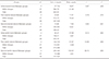
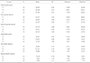
Table 2 presents the results of the One-way ANOVA. The mean shear bond strengths and standard deviations for each group are given in Table 3. The results of One-way ANOVA showed that the initial bond strength and the first and second bond strengths to enamel do not differ significantly. However, the mean initial shear bond strength to dentin was significantly lower in Group P (11.2 ± 1.6 MPa) when compared to those in Group V (15.3 ± 2.5 MPa) and Group R (14.6 ± 2.9 MPa) (P < .01, df = 2, F = 8.13). Group V showed significantly higher first (15.3 ± 2.2 MPa) (P <.0001, df = 2, F = 12.69) and second bond strengths (10.4 ± 2.2 MPa) (P < .0001, df = 2, F = 39.15) to dentin when compared to Groups P and R.
Repeated measures One-way ANOVA and the Bonferroni test found no significant differences in bond strength values among the initial enamel bonding, and the first and second enamel re-bondings or between the initial dentin bonding and the first dentin re-bonding for any of the cements tested. However, significant differences in bond strength values were found between the initial dentin bonding and the second dentin re-bonding and between the first and second dentin re-bondings (P < .0001, Sphericity Assumed sum of squares = 6154.85, Sphericity Assumed mean of squares = 1230.97, df = 5, F = 126) for all of the cements tested. Significant differences were also found between the mean enamel and mean dentin bond strengths for all of the groups (P < .0001).
Failure types for the initial bonding and the first and second re-bondings to enamel and dentin are given in Tables 4 and 5, respectively. The majority of all enamel bond failures (initial bonding as well as first and second re-bondings) were mixed failures (63 of 90), followed by cohesive failures within the cement (16 of 90). Failure in the dentin bonding differed by group; Group V exhibited mainly mixed failure (19 of 30); Groups R exhibited the same proportions of mixed (15 of 30) and adhesive (15 of 30) failure; and Groups P exhibited mainly adhesive failure (21 of 30). For the second dentin re-bonding, failures were primarily adhesive in both Group P (7 of 10) and Group R (6 of 10) and mixed (8 of 10) in Group V. No cohesive failure of enamel, dentin, or composite was observed in any of the specimens.
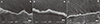
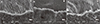
SEM images of the specimens from each group are shown in Figures 1, 2, 3, 4, 5, 6. No significant differences were observed in the resin penetration patterns of the specimens, whose enamel was etched with phosphoric acid (Groups V, R and P). Also, no significant differences were observed among the initial bonding, and the first and second enamel re-bondings (Fig. 1, Fig. 2, and Fig. 3). The specimens from all groups showed long resin tags on the dentin surfaces and a homogeneous hybrid layer at both the initial bonding and the first re-bonding, although more and longer tags were observed in Group V as compared to Groups P and R (Fig. 4, Fig. 5 and Fig. 6). At the second re-bonding, the number of resin tags decreased, and gaps and discontinuity of the hybrid layer between the dentin and the resin cement were observed in all groups (Fig. 4C, Fig. 5C, and Fig. 6C).
DISCUSSION
Prosthodontic practice frequently requires re-bonding of the failed restorations as well as the bonding of new restorations to previously bonded enamel and/or dentin. This is especially true for the cemented restorations with resin cement such as inlay-retained fixed partial dentures, porcelain inlays/onlays, crowns, and laminate veneers.14151618192024 However, limited data is available about the bond strength of re-bonded resin cements to enamel and dentin surfaces.
The findings of the present study partially confirmed the first hypothesis that differences would be found between the initial and repeated bond strengths of resin cements on both enamel and dentin surfaces. Whereas no significant differences were observed among the bond strengths of the initial bonding, and the first and second re-bondings to enamel, the bond strength of all resin cements to dentin decreased significantly after the first re-bonding procedure. These results may be explained by differences in the morphological characteristics of enamel and dentin. Enamel is comprised of 92% hydroxyapatite crystals within an inorganic structure, compared to only 45% hydroxyapatite in dentin. Dentin also has a much more complex histological structure than enamel, with numerous fluid-filled tubules that run from the pulp to the dentino-enamel junction. As a result, enamel bonding is more durable, and dentin bonding is more sensitive, requiring demineralization of the intertubular dentin.47 Dentin depth may also effect the decreasing strength of resin cements to dentin observed with rebonding.56 In line with clinical practice, the current study used a carbide bur to remove residual luting cement prior to re-bonding, which also exposed a deeper layer of dentin surface.21
Studies have shown that dentin depth is an important parameter in bond strength, and bond strength decreases with dentin depth.56 This may be attributed to the higher water content in deep dentin as compared to superficial dentin - a result of the larger diameters of the tubules and their greater numbers per unit area in deep dentin.6 Finally, the lower bond strength to dentin observed in re-bonding may be due to residual resin tags, which may obstruct formation of new resin tags, thereby decreasing bond strength.
The findings of the present study also partially confirmed the second hypothesis that the type of resin cement used would affect the shear bond strength values. The cements used in the study included 2 total-etch (Variolink II and RelyX ARC) and 1 self-etch (Panavia F) dual-cure resin cements frequently used in prosthodontic practice.123482425 In line with the manufacturers' recommendations, prior to cement application, enamel was pre-etched with phosphoric acid at Group P, which is known to significantly improve micro-retention and enhance bonding effectiveness.91023 In this study, no significant differences in enamel bond strength were observed between the total-etch and the selfetch resin cements. This finding is in line with a previous in vitro study, showing pre-etching of enamel increased the bond strength of a self-etch adhesive system.910 However, pre-etching of dentin is controversial before application of a self-etching primer.928 Some previous studies have shown that pre-etched dentin with phosphoric acid exposes collagen to a depth of several micrometers and has a beneficial effect on bonding effectiveness2328 In the present study, the initial bond strengths to dentin were higher for Variolink II and RelyX ARC resin cements (total-etch resin cement), with pre-etching of dentin, than for Panavia F resin cement (self-etch resin cement), without pre-etching. The results from the present study are consistent with those of the previous studies, showing that total-etch adhesive resin cement resulted in higher bond strengths than self-etch adhesive resin cement to dentin.829
In terms of dentin bond strength in re-bonding, Variolink II resin cement exhibited the highest shear bond strength values at both the first and second re-bondings to dentin. This may be attributed to the composition of the Variolink II resin cement, which includes urethane dimethacrylate-UDMA, maleic acid, and glutaraldehyde in the dentin primer, and the adhesives that condition the tooth surface in order to improve adhesion to dentin. By contrast, RelyX ARC relies on ethanol contained in the adhesive for conditioning, and Panavia contains an acidic monomer (10-methacryloyloydecyl dihydrogen phosphate-MDP) in the self-etching primer for conditioning dentin and enamel. The variations in bond strengths found in this study are in line with the previous studies, showing that adhesive type and composition might influence their dentin bond strength.12 Altintas et al.2 found that the shear bond strength to dentin of Variolink II resin cement after 24 hours (5.3 ± 2.2 MPa) was higher than that of Panavia F (4 ± 0.8). Öztürk et al.4 also found that Variolink II resin cement had a higher shear bond strength to dentin (13.8 ± 8.8) than RelyX Veneer (5.4 ± 6.6); the authors also found that tooth substrates (enamel vs dentin) had an effect on the bond strengths of resin cements.
Bond strengths of resin cements are best evaluated through clinical trials; however, clinical trials are difficult and time consuming, making in vitro studies essential for identifying potentially superior materials and methods prior to clinical trials. Shear, tensile, microtensile, and microshear tests are tests of mechanical properties of resin bonding to dental structure.91012242530 Among these the shear bond test is the most used test.2101222242529
A shear bond-strength value of 20 MPa is considered as the minimum value needed to provide an adequate bond to enamel.11 In the present study, all of the cements tested exhibited values close to or above 20 MPa in their initial bonding as well as re-bonding to enamel. However, the mean initial bonding and re-bonding shear bond strength values to dentin of all of the resin cements tested were below 17 MPa, which is considered as the minimum value for clinically adequate bond strength to dentin.41113 Previous in vitro studies have reported that the resin cement shear bond strengths to dentin ranged from 5.4 ± 2.3 MPa to 13.78 ± 8.8 MPa for Variolink II, 4.0 ± 0.8 MPa for Panavia F, and 5.42 ± 6.6 MPa for RelyX Veneer resin cements, which are in line with the values obtained in this study.24 A large range of variations (7 to 40 MPa) in the bond strengths of different bonding and resin luting agents were also found.1 The relatively low bond strengths reported by this study and previous studies may be explained by microstructural variations in tooth structure, tooth storage conditions, time, temperature, and the dimensions of the adhesive surface.1 Given the high rate of cohesive failures reported, shear tests have been criticized as inappropriate for adhesive bond-strength testing.2 However, in this study, high rates of adhesive failure between dentin and resin cement were observed, especially for re-bonding in Group P (80% at the first re-bonding, 70% at the second re-bonding) and Group R (50% at the first re-bonding, 60% at the second re-bonding). The literature suggests a relationship between bond strength values and failures modes.224 In this study, all 3 resin cements exhibited high bond strength to enamel, and the majority of failures at the enamel bonding and the re-bondings were either mixed failures or cohesive failures within the resin cement (Table 4). However, in terms of dentin bonding, Groups P and R, which exhibited comparatively low bond strengths, had higher rates of adhesive failures at the dentin/cement interface than Group V, which exhibited comparatively high bond strength. Group V had a higher rate of mixed failures (Table 5).
SEM views of the specimens, whose enamel was etched with phosphoric acid prior to bonding showed similar resin-penetration patterns for the initial enamel bonding, and the first and second re-bondings. However, SEM showed differences in the bonding and re-bonding of resin cements to dentin surfaces. When compared to Groups P and R, Group V produced more and longer resin tags at both the initial bonding and the first re-bonding to dentin. Moreover, when compared to the initial bonding and the first re-bonding to dentin, the second re-bonding to dentin resulted in fewer and shorter resin tags, as well as gaps between the dentin and cement in all groups.
In clinical situation, the results of this study suggests that the bond strength of re-bonded resin cement to dentin decreases especially after the first re-bonding, therefore the choice of resin cement type and adhesive cementation failure must be considered. In addition, microleakage can occur and mechanical properties of restoration may be adversely affected by the low bond strength of resin cement. In this study, the composite resin cylinders were cemented to natural tooth rather than ceramics because the aim of the present study was to evaluate only the bond strength of resin cements to tooth.24
Resin bond strength to enamel and dentin in clinical situations can be affected by cement composition and polymerization type.124 In vitro bond strength testing is also affected by thermal cycling and long-term storage of specimens.224 The present study is limited by its lack of evaluation of these factors. In addition, the number of the resin cement (2 total-etch and 1 self-etch) used in this study could be increased. Thus, future studies should examine the effects of different cement composition, long-term storage, polymerization type, and thermal cycling on repeated bond strength of adhesives.
CONCLUSION
Considering the limitations of this in vitro study: In clinical situation, when re-bonding of restorations is needed, Variolink II, RelyX ARC, and Panavia F resin cements have similar initial and repeated bond strengths to enamel. However, there are differences in the initial and repeated bond strengths of these resin cement to dentin. Repeated bonding to dentin was found to reduce the bond strength of resin cements after the first re-bonding. Variolink II, total-etch resin cement showed the highest bond strength to dentin for re-bonding (P < .0001).


 XML Download