Journal List > Tuberc Respir Dis > v.82(2) > 1119331

Jeon: Infection Source and Epidemiology of Nontuberculous Mycobacterial Lung Disease

Abstract

Nontuberculous mycobacteria (NTM) are ubiquitous organisms that are generally found not only in the natural environment but also in the human engineered environment, including water, soil, and dust. These organisms can form biofilms and can be readily aerosolized because they are hydrophobic owing to the presence of the lipid-rich outer membrane. Aerosolization and subsequent inhalation were the major route of NTM lung disease. Water distribution systems and household plumbing are ideal habit for NTM and the main transmission route from natural water to household. NTM have been isolated from drinking water, faucets, pipelines, and water tanks. Studies that used genotyping have shown that NTM isolates from patients are identical to those in the environment, that is, from shower water, showerheads, tap water, and gardening soil. Humans are likely to be exposed to NTM in their homes through simple and daily activities, such as drinking, showering, or gardening. In addition to environmental factors, host factors play an important role in the development of NTM lung disease. The incidence and prevalence of NTM lung disease are increasing worldwide, and this disease is rapidly becoming a major public health problem. NTM lung disease is associated with substantially impaired quality of life, increased morbidity and mortality, and high medical costs. A more comprehensive understanding of the infection source and epidemiology of NTM is essential for the development of new strategies that can prevent and control NTM infection.

Introduction

The term nontuberculous mycobacteria (NTM) generally refers to mycobacteria species other than the Mycobacterium tuberculosis complex and M. leprae12. Although, NTM were first identified in the late 19th century, NTM were not widely recognized as a cause of human disease until the late 1950s3. They have been called by various names including “atypical mycobacteria,” “environmental mycobacteria,” and “mycobacteria other than tuberculosis.” As these names imply, NTM disease have received less attention than tuberculosis (TB) and were considered as a minor disease.
Currently, NTM, which are evidently different from M. tuberculosis, have emerged as important pathogens worldwide45. The incidence and prevalence of NTM diseases are increasing, and their impact on human health has been a topic of interest. The diagnosis and treatment of NTM lung disease remains challenging for clinicians6. Microbiological eradication using the currently available dugs is difficult7. Furthermore, microbiological recurrence is common even after a successful treatment, with a substantially high reinfection rate8910. Therefore, to appropriately prevent and treat NTM diseases, the infection sources and epidemiology of NTM infection should be understood.

Infection Source

1. Environmental source of infection

1) Natural environment

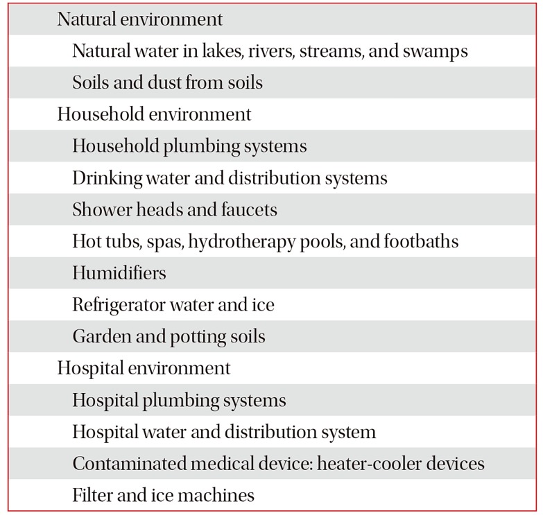
NTM are naturally occurring environmental pathogens. These organisms have been isolated from natural water, including rivers, lakes, swamps, and streams, as well as soil (Table 1)1112. NTM have a lipid-rich outer membrane13, which is a key factor in its survival and proliferation in the natural environment11. As an effect of the lipid-rich outer membrane, these organisms are resistant to acid14, antibiotics15, disinfectants16, and high temperature17. NTM can survive within amoebae18. These pathogens are quite hydrophobic due to their lipid-rich outer membrane and prefer to attach to surfaces and subsequently form biofilms19. NTM are readily aerosolized from natural water and soils due to its surface hydrophobicity20. Naturally occurring aerosolization and subsequent inhalation were the major route of NTM lung disease.
Epidemiology studies have supported the claims that environmental factors could affect the risk for NTM lung disease. In the United States, eight of 55 states were identified as hot spots, with a high incidence of NTM lung disease21. The eight states had a higher mean daily potential evapotranspiration levels and percentages covered by surface water and were more likely to have greater copper and sodium levels in the soil21. Another study on a registry of patients with cystic fibrosis has reported that high saturated vapor pressure is associated with the increased prevalence of NTM lung disease22.

2) Household environment

NTM are widely distributed in human engineered and household environment. Water distribution systems are thought to be a main transmission route from natural surface water reservoirs to the household. NTM have been isolated from drinking water pipelines2324 and water tanks25. The number of NTM increases by 2-fold in a drinking water distribution system, that is, from the treatment plant to the end user26. Cell surface hydrophobicity is a major determinant of the survival and proliferation of NTM in the water distribution systems. These organisms can attach to pipe surfaces and form biofilms, which prevents them from being flushed from the water distribution system. Furthermore, NTM are more resistant to common water disinfectants, such as chlorine, than other microorganisms. Therefore, the use of disinfects kills off competitors and selects NTM.
Household and building plumbing systems provide a stable, nutrient-limited, disinfectant-containing habitat that is ideal for NTM growth and persistence. As NTM are relatively resistant to high temperature, these organisms can survive in water heaters and hot water pipes. Households with water heater temperatures <50°C were more likely to yield NTM (17/20, 85%) than those with water temperature >55°C (6/15, 40%)27. Numerous studies have shown that showerheads and tap water as well as the end points of drinking water distribution systems are the reservoirs of NTM in the household28. Studies that used genotyping have shown that NTM isolates from patients are identical to those in the environment, that is, from shower water2930, showerheads31, and household tap water32.
NTM species have been isolated from house dust33 and soils34. Dry potting soil is a problem because the soil particles can be aerosolized during gardening, and thus, an individual can inhale these particles. DNA fingerprinting methods have shown that NTM isolates from a patient and those from the patient's dried potting and garden soil were identical34.

3) Hospital environment

NTM are also found in hospital environment. Hospital water distribution system may serve as a reservoir of NTM. These organisms contaminate hospital materials and can cause nosocomial outbreaks and pseudo-outbreaks35. Recently, several outbreaks due to M. chimaera have occurred from heatercooler devices used during open-heart surgery3637.

2. Transmission

NTM are generally acquired from the environment via ingestion, inhalation, and dermal contact12. These organisms are everywhere in the human environment and can be readily aerosolized. NTM in household plumbing systems are readily aerosolized from taps, showerheads, and hot tubs. Home humidifiers filled with household water could be a potential generator of a high number of NTM in aerosols12. Use of indoor swimming pools has been associated with NTM lung disease among individuals with cystic fibrosis38. Dusts generated during potting of plants or gardening are also likely sources of NTM infection. Humans are likely to be exposed to NTM in their homes through simple daily activities, such as drinking, showering, or gardening39. The most common routes for pulmonary NTM infection involve the inhalation of aerosols generated by hot tubs and showerheads. Indeed, the global increase in NTM infections may reflect the use of showers rather than bathing40.

1) Human to human transmission

Traditionally, it was believed that human-to-human NTM transmission is not possible. Whole genome sequencing, a new molecular technique, has provided the first genetic evidence of human-to-human transmission of M. abscessus complex among patients with cystic fibrosis4142. Bryant et al.42 have performed a whole genome sequencing analysis of 1,080 clinical isolates of M. abscessus complex obtained from 517 patients from cystic fibrosis centers in seven countries. Results have shown that most M. abscessus complex infections were acquired via human-to-human transmission, potentially via fomites and aerosols.

3. Preventive measure

Strategies that are effective in preventing NTM lung disease are still limited. Vaccines and prophylaxis medications are not available. Unlike TB, preventive therapy is only recommended in patients with advanced human immunodeficiency virus (HIV) disease. A more comprehensive understanding of NTM transmission can help in establishing targeted prophylactic measures. Some preventive measures are recommended to avoid NTM exposure and reduce the number of NTM in the household12. Although the efficacy of these recommendations has not yet been validated via clinical trials, these are still helpful for vulnerable individuals with a high risk for infection.
Preventive measures recommended by experts are the following12: (1) raise hot water temperature to 55°C; (2) use bacteriologic filters (pore size <0.45 mm) on taps and showerheads; (3) utilize well water rather than piped utility supply; (4) clean and disinfect showerheads regularly to remove the biofilm; (5) use showerheads with large holes to reduce mist formation; (6) avoid spas and hot tubs or any water with an aerator; (7) increase bathroom exhaust rate; (8) boil drinking water for 10 minutes to kill NTM; (9) wear mask when gardening; and (10) moisten garden and potting soils.

Epidemiology

1. Incidence and prevalence of NTM

The identification of the epidemiology of NTM lung disease has been challenging because reporting of cases is not mandatory in most countries. Furthermore, diagnosis is difficult because simple isolation of the organism does not necessarily indicate the actual lung disease. Various epidemiological methodologies have been used, including surveillance, population-based studies, analysis of large linked datasets, studies that used laboratory-based convenience samples, and combinations of these approaches43.
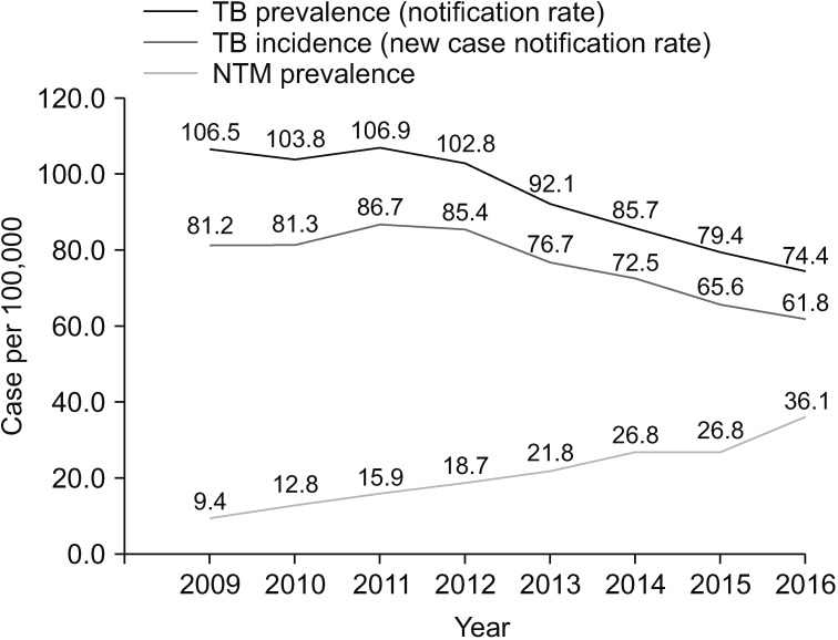
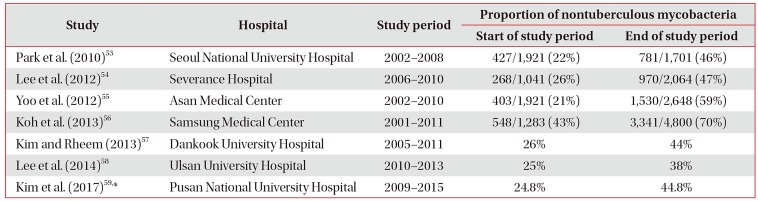
Numerous studies have shown that the incidence and prevalence of NTM lung disease are increasing worldwide44. Population-based data from the United States45, Canada46, the United Kingdom47, and Australia48 have shown that the prevalence of NTM lung disease has been increasing. Large tertiary care facility-based studies in East Asian countries, including Japan49, Taiwan50, and South Korea5152, have also suggested an increasing prevalence of NTM lung disease. In South Korea, no population-based study on the epidemiology of NTM lung disease was conducted. Most studies were conducted in a single institution using laboratory-based convenience samples. Those studies have consistently shown that the proportion of NTM among positive mycobacterial cultures is increasing (Table 2)53545556575859. In a 10-year observational study56, the proportion of NTM among all positive mycobacterial cultures increased from 43% in 2001 to 70% in 2011. Furthermore, the recovery rate of NTM isolates from acid-fast bacilli smear-positive specimens increased from 9% in 2001 to 64% in 2011. This finding has suggested that early differentiation between pulmonary TB and NTM lung disease has become a clinically important issue in South Korea due to the increasing incidence and prevalence of NTM lung disease. Recently, Yoon et al.60 have reported a nationwide increasing trend in the prevalence of NTM disease from 2009 to 2016 in South Korea based on the claims data of the Health Insurance Review and Assessment Service (Figure 1). Interestingly, the increase in the proportion of NTM lung disease may be associated with a simultaneous decrease in the incidence of TB in most countries61, including South Korea60.

2. Species distribution

The distribution of NTM species varies by region and country. This geographical diversity can provide important clues to identify factors associated with the acquisition of NTM infection, such as geographic factors, population density, or host factors. In 2008, the NTM-Network European Trials Group provided species identification data for 20,182 patients from 62 laboratories in 30 countries62. This study has shown that the Mycobacterium avium complex (MAC) predominated in most countries. However, there were important differences in the geographical distribution of M. xenopi, M. kansasii, and other rapid-growing mycobacteria. M. xenopi and M. malmoense were extremely rare in East Asia. However, they are predominantly found in Europe.
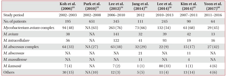
In South Korea, the MAC is the most common organism causing NTM lung disease (Table 3)53545860636465, which is similar to other countries. M. abscessus complex is the second most common organism, whereas M. kansasii does not commonly cause the disease. However, M. kansasii was the second most common NTM species in Ulsan58.

3. Risk factor

Exposure to NTM is quite common. However, NTM disease is relatively rare. This observation suggests that host susceptibility likely plays a key role in the development of NTM lung disease. Patients who develop NTM lung disease usually have some degree of host impairment that make them vulnerable to these infections. NTM lung disease is more prevalent in patients with structural lung disease, such as cystic fibrosis, non-cystic fibrosis bronchiectasis, primary ciliary dyskinesia, chronic obstructive pulmonary disease (COPD), previous TB, and pneumoconiosis66. An immunosuppressed status, which is related to HIV infection, transplantation, or the use of tumor necrosis factor α inhibitor, is also associated with NTM disease66.
However, patients can develop NTM lung disease without any obvious underlying cause. These patients are typically postmenopausal women who have a unique body morphotype6768. Recently, a whole-exome sequencing study has begun to identify genetic predispositions for those patients and found an increased prevalence of genetic mutations that control the function of the immune system, ciliary body, and connective tissues69. This suggests that NTM lung disease is a multigenic disease, which is characterized by a combination of variants across gene categories and environmental exposures that increase susceptibility to infection.

4. Impact on public health

The impact of NTM infection on public health has been underestimated. Generally, NTM have been considered to have a low pathogenicity, and it was believed that these organisms cannot be transmitted from person to person. However, NTM lung disease is associated with a substantially impaired quality of life70, increased morbidity and mortality71, and high medical costs43. Huang et al.72 have shown that patients with COPD who have NTM isolates had a greater decline in lung function and more frequent exacerbation than those with no isolate. Park et al.73 have shown that treatment failure was associated with a substantial decline in lung function in individuals with NTM lung disease. A recent study in Germany have revealed that a higher mortality, direct costs, and indirect costs were observed among patients with NTM lung disease than in matched controls74. Considering the increasing burden of NTM lung disease globally, this disease may soon become a significant health and economic burden.

Conclusion

NTM are generally found not only in the natural but also in the human engineered environment. Remarkable changes in the human environment and lifestyle habits were observed during the past several decades. These changes have made humans more comfortable but also resulted in a more favorable environment for NTM growth. In addition, with the development of medicine, an aging population with multiple comorbidities who are vulnerable to NTM infection is growing rapidly. As a result, the incidence and prevalence of NTM lung disease are increasing worldwide, and this disease is rapidly becoming a major public health problem. NTM lung disease is more difficult to treat and prevent than TB. The elimination of NTM from environmental sources and the destruction of the transmission route are critical issues that must be urgently addressed. Further comprehensive works to investigate the source, transmission route, and epidemiology of NTM are needed to develop new strategies that can prevent and control NTM infection.

Notes

Conflicts of Interest: No potential conflict of interest relevant to this article was reported.

References

1. Griffith DE, Aksamit T, Brown-Elliott BA, Catanzaro A, Daley C, Gordin F, et al. An official ATS/IDSA statement: diagnosis, treatment, and prevention of nontuberculous mycobacterial diseases. Am J Respir Crit Care Med. 2007; 175:367–416. PMID: 17277290.
crossref
2. Haworth CS, Banks J, Capstick T, Fisher AJ, Gorsuch T, Laurenson IF, et al. British Thoracic Society guidelines for the management of non-tuberculous mycobacterial pulmonary disease (NTM-PD). Thorax. 2017; 72(Suppl 2):ii1–ii64.
crossref
3. Runyon EH. Anonymous mycobacteria in pulmonary disease. Med Clin North Am. 1959; 43:273–290. PMID: 13612432.
crossref
4. Griffith DE, Aksamit TR. Understanding nontuberculous mycobacterial lung disease: it's been a long time coming. F1000Res. 2016; 5:2797. PMID: 27990278.
crossref
5. Koh WJ. Nontuberculous mycobacteria: overview. Microbiol Spectr. 2017; 1. [Epub]. DOI: 10.1128/microbiolspec.TNMI7-0024-2016.
6. Ryu YJ, Koh WJ, Daley CL. Diagnosis and treatment of nontuberculous mycobacterial lung disease: clinicians' perspectives. Tuberc Respir Dis. 2016; 79:74–84.
crossref
7. Stout JE, Koh WJ, Yew WW. Update on pulmonary disease due to non-tuberculous mycobacteria. Int J Infect Dis. 2016; 45:123–134. PMID: 26976549.
crossref
8. Wallace RJ Jr, Zhang Y, Brown-Elliott BA, Yakrus MA, Wilson RW, Mann L, et al. Repeat positive cultures in Mycobacterium intracellulare lung disease after macrolide therapy represent new infections in patients with nodular bronchiectasis. J Infect Dis. 2002; 186:266–273. PMID: 12134265.
9. Wallace RJ Jr, Brown-Elliott BA, McNulty S, Philley JV, Killingley J, Wilson RW, et al. Macrolide/Azalide therapy for nodular/bronchiectatic mycobacterium avium complex lung disease. Chest. 2014; 146:276–282. PMID: 24457542.
crossref
10. Koh WJ, Moon SM, Kim SY, Woo MA, Kim S, Jhun BW, et al. Outcomes of Mycobacterium avium complex lung disease based on clinical phenotype. Eur Respir J. 2017; 50:1602503. PMID: 28954780.
11. Falkinham JO 3rd. Surrounded by mycobacteria: nontuberculous mycobacteria in the human environment. J Appl Microbiol. 2009; 107:356–367. PMID: 19228258.
12. Falkinham JO 3rd. Environmental sources of nontuberculous mycobacteria. Clin Chest Med. 2015; 36:35–41. PMID: 25676517.
crossref
13. Brennan PJ, Nikaido H. The envelope of mycobacteria. Annu Rev Biochem. 1995; 64:29–63. PMID: 7574484.
crossref
14. Bodmer T, Miltner E, Bermudez LE. Mycobacterium avium resists exposure to the acidic conditions of the stomach. FEMS Microbiol Lett. 2000; 182:45–49. PMID: 10612729.
15. Rastogi N, Frehel C, Ryter A, Ohayon H, Lesourd M, David HL. Multiple drug resistance in Mycobacterium avium: is the wall architecture responsible for exclusion of antimicrobial agents? Antimicrob Agents Chemother. 1981; 20:666–677. PMID: 6798925.
crossref
16. Taylor RH, Falkinham JO 3rd, Norton CD, LeChevallier MW. Chlorine, chloramine, chlorine dioxide, and ozone susceptibility of Mycobacterium avium. Appl Environ Microbiol. 2000; 66:1702–1705. PMID: 10742264.
17. Schulze-Robbecke R, Buchholtz K. Heat susceptibility of aquatic mycobacteria. Appl Environ Microbiol. 1992; 58:1869–1873. PMID: 1622262.
crossref
18. Cirillo JD, Falkow S, Tompkins LS, Bermudez LE. Interaction of Mycobacterium avium with environmental amoebae enhances virulence. Infect Immun. 1997; 65:3759–3767. PMID: 9284149.
19. Faria S, Joao I, Jordao L. General overview on nontuberculous mycobacteria, biofilms, and human infection. J Pathog. 2015; 2015:809014. PMID: 26618006.
crossref
20. Parker BC, Ford MA, Gruft H, Falkinham JO 3rd. Epidemiology of infection by nontuberculous mycobacteria. IV. Preferential aerosolization of Mycobacterium intracellulare from natural waters. Am Rev Respir Dis. 1983; 128:652–656. PMID: 6354024.
21. Adjemian J, Olivier KN, Seitz AE, Falkinham JO 3rd, Holland SM, Prevots DR. Spatial clusters of nontuberculous mycobacterial lung disease in the United States. Am J Respir Crit Care Med. 2012; 186:553–558. PMID: 22773732.
crossref
22. Adjemian J, Olivier KN, Prevots DR. Nontuberculous mycobacteria among patients with cystic fibrosis in the United States: screening practices and environmental risk. Am J Respir Crit Care Med. 2014; 190:581–586. PMID: 25068291.
crossref
23. Nishiuchi Y, Iwamoto T, Maruyama F. Infection sources of a common non-tuberculous mycobacterial pathogen, Mycobacterium avium complex. Front Med (Lausanne). 2017; 4:27. PMID: 28326308.
crossref
24. Thomson R, Tolson C, Sidjabat H, Huygens F, Hargreaves M. Mycobacterium abscessus isolated from municipal water: a potential source of human infection. BMC Infect Dis. 2013; 13:241. PMID: 23705674.
crossref
25. Torvinen E, Suomalainen S, Lehtola MJ, Miettinen IT, Zacheus O, Paulin L, et al. Mycobacteria in water and loose deposits of drinking water distribution systems in Finland. Appl Environ Microbiol. 2004; 70:1973–1981. PMID: 15066787.
crossref
26. Tuffley RE, Holbeche JD. Isolation of the Mycobacterium avium-M. intracellulare-M. scrofulaceum complex from tank water in Queensland, Australia. Appl Environ Microbiol. 1980; 39:48–53. PMID: 7356321.
27. Falkinham JO 3rd, Norton CD, LeChevallier MW. Factors influencing numbers of Mycobacterium avium, Mycobacterium intracellulare, and other mycobacteria in drinking water distribution systems. Appl Environ Microbiol. 2001; 67:1225–1231. PMID: 11229914.
28. Falkinham JO 3rd. Nontuberculous mycobacteria from household plumbing of patients with nontuberculous mycobacteria disease. Emerg Infect Dis. 2011; 17:419–424. PMID: 21392432.
crossref
29. Thomson R, Tolson C, Carter R, Coulter C, Huygens F, Hargreaves M. Isolation of nontuberculous mycobacteria (NTM) from household water and shower aerosols in patients with pulmonary disease caused by NTM. J Clin Microbiol. 2013; 51:3006–3011. PMID: 23843489.
crossref
30. Falkinham JO 3rd, Iseman MD, de Haas P, van Soolingen D. Mycobacterium avium in a shower linked to pulmonary disease. J Water Health. 2008; 6:209–213. PMID: 18209283.
31. Feazel LM, Baumgartner LK, Peterson KL, Frank DN, Harris JK, Pace NR. Opportunistic pathogens enriched in showerhead biofilms. Proc Natl Acad Sci U S A. 2009; 106:16393–16399. PMID: 19805310.
crossref
32. von Reyn CF, Maslow JN, Barber TW, Falkinham JO 3rd, Arbeit RD. Persistent colonisation of potable water as a source of Mycobacterium avium infection in AIDS. Lancet. 1994; 343:1137–1141. PMID: 7910236.
33. Reznikov M, Dawson DJ. Serological investigation of strains of Mycobacterium intracellulare (“battey” bacillus) isolated from house-dusts. Med J Aust. 1971; 1:682–683. PMID: 5553815.
34. De Groote MA, Pace NR, Fulton K, Falkinham JO 3rd. Relationships between Mycobacterium isolates from patients with pulmonary mycobacterial infection and potting soils. Appl Environ Microbiol. 2006; 72:7602–7606. PMID: 17056679.
35. Sood G, Parrish N. Outbreaks of nontuberculous mycobacteria. Curr Opin Infect Dis. 2017; 30:404–409. PMID: 28548990.
crossref
36. Allen KB, Yuh DD, Schwartz SB, Lange RA, Hopkins R, Bauer K, et al. Nontuberculous Mycobacterium infections associated with heater-cooler devices. Ann Thorac Surg. 2017; 104:1237–1242. PMID: 28821331.
37. Williamson D, Howden B, Stinear T. Mycobacterium chimaera spread from heating and cooling units in heart surgery. N Engl J Med. 2017; 376:600–602. PMID: 28177865.
38. Prevots DR, Adjemian J, Fernandez AG, Knowles MR, Olivier KN. Environmental risks for nontuberculous mycobacteria: individual exposures and climatic factors in the cystic fibrosis population. Ann Am Thorac Soc. 2014; 11:1032–1038. PMID: 25068620.
crossref
39. Halstrom S, Price P, Thomson R. Review: environmental mycobacteria as a cause of human infection. Int J Mycobacteriol. 2015; 4:81–91. PMID: 26972876.
crossref
40. O'Brien DP, Currie BJ, Krause VL. Nontuberculous mycobacterial disease in northern Australia: a case series and review of the literature. Clin Infect Dis. 2000; 31:958–967. PMID: 11049777.
41. Bryant JM, Grogono DM, Greaves D, Foweraker J, Roddick I, Inns T, et al. Whole-genome sequencing to identify transmission of Mycobacterium abscessus between patients with cystic fibrosis: a retrospective cohort study. Lancet. 2013; 381:1551–1560. PMID: 23541540.
42. Bryant JM, Grogono DM, Rodriguez-Rincon D, Everall I, Brown KP, Moreno P, et al. Emergence and spread of a human-transmissible multidrug-resistant nontuberculous mycobacterium. Science. 2016; 354:751–757. PMID: 27846606.
43. Prevots DR, Loddenkemper R, Sotgiu G, Migliori GB. Nontuberculous mycobacterial pulmonary disease: an increasing burden with substantial costs. Eur Respir J. 2017; 49:1700374. PMID: 28446563.
crossref
44. Prevots DR, Marras TK. Epidemiology of human pulmonary infection with nontuberculous mycobacteria: a review. Clin Chest Med. 2015; 36:13–34. PMID: 25676516.
45. Adjemian J, Olivier KN, Seitz AE, Holland SM, Prevots DR. Prevalence of nontuberculous mycobacterial lung disease in U.S. Medicare beneficiaries. Am J Respir Crit Care Med. 2012; 185:881–886. PMID: 22312016.
crossref
46. Marras TK, Mendelson D, Marchand-Austin A, May K, Jamieson FB. Pulmonary nontuberculous mycobacterial disease, Ontario, Canada, 1998–2010. Emerg Infect Dis. 2013; 19:1889–1891. PMID: 24210012.
crossref
47. Moore JE, Kruijshaar ME, Ormerod LP, Drobniewski F, Abubakar I. Increasing reports of non-tuberculous mycobacteria in England, Wales and Northern Ireland, 1995-2006. BMC Public Health. 2010; 10:612. PMID: 20950421.
crossref
48. Thomson RM. NTM working group at Queensland TB Control Centre and Queensland Mycobacterial Reference Laboratory. Changing epidemiology of pulmonary nontuberculous mycobacteria infections. Emerg Infect Dis. 2010; 16:1576–1583. PMID: 20875283.
crossref
49. Ide S, Nakamura S, Yamamoto Y, Kohno Y, Fukuda Y, Ikeda H, et al. Epidemiology and clinical features of pulmonary nontuberculous mycobacteriosis in Nagasaki, Japan. PLoS One. 2015; 10:e0128304. PMID: 26020948.
crossref
50. Lai CC, Tan CK, Chou CH, Hsu HL, Liao CH, Huang YT, et al. Increasing incidence of nontuberculous mycobacteria, Taiwan, 2000–2008. Emerg Infect Dis. 2010; 16:294–296. PMID: 20113563.
crossref
51. Kwon YS, Koh WJ. Diagnosis and treatment of nontuberculous mycobacterial lung disease. J Korean Med Sci. 2016; 31:649–659. PMID: 27134484.
crossref
52. Ko RE, Moon SM, Ahn S, Jhun BW, Jeon K, Kwon OJ, et al. Changing epidemiology of nontuberculous mycobacterial lung diseases in a tertiary referral hospital in Korea between 2001 and 2015. J Korean Med Sci. 2018; 33:e65. PMID: 29441757.
crossref
53. Park YS, Lee CH, Lee SM, Yang SC, Yoo CG, Kim YW, et al. Rapid increase of non-tuberculous mycobacterial lung diseases at a tertiary referral hospital in South Korea. Int J Tuberc Lung Dis. 2010; 14:1069–1071. PMID: 20626955.
54. Lee SK, Lee EJ, Kim SK, Chang J, Jeong SH, Kang YA. Changing epidemiology of nontuberculous mycobacterial lung disease in South Korea. Scand J Infect Dis. 2012; 44:733–738. PMID: 22720876.
crossref
55. Yoo JW, Jo KW, Kim MN, Lee SD, Kim WS, Kim DS, et al. Increasing trend of isolation of non-tuberculous mycobacteria in a tertiary university hospital in South Korea. Tuberc Respir Dis. 2012; 72:409–415.
crossref
56. Koh WJ, Chang B, Jeong BH, Jeon K, Kim SY, Lee NY, et al. Increasing recovery of nontuberculous mycobacteria from respiratory specimens over a 10-year period in a tertiary referral hospital in South Korea. Tuberc Respir Dis. 2013; 75:199–204.
crossref
57. Kim JK, Rheem I. Identification and distribution of nontuberculous mycobacteria from 2005 to 2011 in Cheonan, Korea. Tuberc Respir Dis. 2013; 74:215–221.
crossref
58. Lee MY, Lee T, Kim MH, Byun SS, Ko MK, Hong JM, et al. Regional differences of nontuberculous mycobacteria species in Ulsan, Korea. J Thorac Dis. 2014; 6:965–970. PMID: 25093094.
59. Kim N, Yi J, Chang CL. Recovery rates of non-tuberculous mycobacteria from clinical specimens are increasing in Korean tertiary-care hospitals. J Korean Med Sci. 2017; 32:1263–1267. PMID: 28665061.
crossref
60. Yoon HJ, Choi HY, Ki M. Nontuberculosis mycobacterial infections at a specialized tuberculosis treatment centre in the Republic of Korea. BMC Infect Dis. 2017; 17:432. PMID: 28619015.
crossref
61. Brode SK, Daley CL, Marras TK. The epidemiologic relationship between tuberculosis and non-tuberculous mycobacterial disease: a systematic review. Int J Tuberc Lung Dis. 2014; 18:1370–1377. PMID: 25299873.
crossref
62. Hoefsloot W, van Ingen J, Andrejak C, Angeby K, Bauriaud R, Bemer P, et al. The geographic diversity of nontuberculous mycobacteria isolated from pulmonary samples: an NTM-NET collaborative study. Eur Respir J. 2013; 42:1604–1613. PMID: 23598956.
crossref
63. Koh WJ, Kwon OJ, Jeon K, Kim TS, Lee KS, Park YK, et al. Clinical significance of nontuberculous mycobacteria isolated from respiratory specimens in Korea. Chest. 2006; 129:341–348. PMID: 16478850.
crossref
64. Jang MA, Koh WJ, Huh HJ, Kim SY, Jeon K, Ki CS, et al. Distribution of nontuberculous mycobacteria by multigene sequence-based typing and clinical significance of isolated strains. J Clin Microbiol. 2014; 52:1207–1212. PMID: 24501029.
crossref
65. Kim HS, Lee Y, Lee S, Kim YA, Sun YK. Recent trends in clinically significant nontuberculous mycobacteria isolates at a Korean general hospital. Ann Lab Med. 2014; 34:56–59. PMID: 24422197.
crossref
66. Honda JR, Knight V, Chan ED. Pathogenesis and risk factors for nontuberculous mycobacterial lung disease. Clin Chest Med. 2015; 36:1–11. PMID: 25676515.
crossref
67. Kartalija M, Ovrutsky AR, Bryan CL, Pott GB, Fantuzzi G, Thomas J, et al. Patients with nontuberculous mycobacterial lung disease exhibit unique body and immune phenotypes. Am J Respir Crit Care Med. 2013; 187:197–205. PMID: 23144328.
crossref
68. Kim RD, Greenberg DE, Ehrmantraut ME, Guide SV, Ding L, Shea Y, et al. Pulmonary nontuberculous mycobacterial disease: prospective study of a distinct preexisting syndrome. Am J Respir Crit Care Med. 2008; 178:1066–1074. PMID: 18703788.
69. Szymanski EP, Leung JM, Fowler CJ, Haney C, Hsu AP, Chen F, et al. Pulmonary nontuberculous mycobacterial infection: a multisystem, multigenic disease. Am J Respir Crit Care Med. 2015; 192:618–628. PMID: 26038974.
crossref
70. Yeung MW, Khoo E, Brode SK, Jamieson FB, Kamiya H, Kwong JC, et al. Health-related quality of life, comorbidities and mortality in pulmonary nontuberculous mycobacterial infections: a systematic review. Respirology. 2016; 21:1015–1025. PMID: 27009804.
crossref
71. Novosad SA, Henkle E, Schafer S, Hedberg K, Ku J, Siegel SA, et al. Mortality after respiratory isolation of nontuberculous mycobacteria: a comparison of patients who did and did not meet disease criteria. Ann Am Thorac Soc. 2017; 14:1112–1119. PMID: 28387532.
crossref
72. Huang CT, Tsai YJ, Wu HD, Wang JY, Yu CJ, Lee LN, et al. Impact of non-tuberculous mycobacteria on pulmonary function decline in chronic obstructive pulmonary disease. Int J Tuberc Lung Dis. 2012; 16:539–545. PMID: 22325332.
crossref
73. Park HY, Jeong BH, Chon HR, Jeon K, Daley CL, Koh WJ. Lung function decline according to clinical course in nontuberculous mycobacterial lung disease. Chest. 2016; 150:1222–1232. PMID: 27298072.
crossref
74. Diel R, Jacob J, Lampenius N, Loebinger M, Nienhaus A, Rabe KF, et al. Burden of non-tuberculous mycobacterial pulmonary disease in Germany. Eur Respir J. 2017; 49:1602109. PMID: 28446559.
crossref
Figure 1

Trend in the prevalence of tuberculosis (TB) and nontuberculous disease from 2009 to 2016 in South Korea. NTM: nontuberculous mycobacteria. Adopted from Yoon et al. BMC infect Dis 2017;17:432, according to Creative Commons license60.

trd-82-94-g001
Table 1

Source of nontuberculous mycobacteria

trd-82-94-i001
Natural environment
 Natural water in lakes, rivers, streams, and swamps
 Soils and dust from soils
Household environment
 Household plumbing systems
 Drinking water and distribution systems
 Shower heads and faucets
 Hot tubs, spas, hydrotherapy pools, and footbaths
 Humidifiers
 Refrigerator water and ice
 Garden and potting soils
Hospital environment
 Hospital plumbing systems
 Hospital water and distribution system
 Contaminated medical device: heater-cooler devices
 Filter and ice machines
Table 2

Changes in the proportions of nontuberculous mycobacteria among mycobacterial isolates from clinical respiratory specimens in South Korea

trd-82-94-i002
Study Hospital Study period Proportion of nontuberculous mycobacteria
Start of study period End of study period
Park et al. (2010)53 Seoul National University Hospital 2002–2008 427/1,921 (22%) 781/1,701 (46%)
Lee et al. (2012)54 Severance Hospital 2006–2010 268/1,041 (26%) 970/2,064 (47%)
Yoo et al. (2012)55 Asan Medical Center 2002–2010 403/1,921 (21%) 1,530/2,648 (59%)
Koh et al. (2013)56 Samsung Medical Center 2001–2011 548/1,283 (43%) 3,341/4,800 (70%)
Kim and Rheem (2013)57 Dankook University Hospital 2005–2011 26% 44%
Lee et al. (2014)58 Ulsan University Hospital 2010–2013 25% 38%
Kim et al. (2017)59,* Pusan National University Hospital 2009–2015 24.8% 44.8%

*This study included both respiratory and non-respiratory specimens.

Table 3

Etiologic species of nontuberculous mycobacterial lung disease in South Korea

trd-82-94-i003
Koh et al. (2006)63 Park et al. (2010)53 Lee et al. (2012)54 Jang et al. (2014)64 Lee et al. (2014)58 Kim et al. (2014)65 Yoon et al. (2017)60
Study period 2002–2003 2002–2008 2006–2010 2012 2010–2013 2007–2011 2011–2016
No. of patients 195 651 345 111 245 90 64
Mycobacterium avium complex 94 (48) NA (63) 263 (76) 73 (66) 132 (54) 61 (68) 29 (45)
M. avium 38 NA 141 32 39 42 13
M. intracellulare 56 NA 122 41 93 19 16
M. abscessus complex 64 (33) NA (27) 63 (18) 32 (29) 22 (9) 15 (17) 27 (42)
M. abscessus NA NA NA 21 NA 11 NA
M. massiliense NA NA NA 11 NA 4 NA
M. kansasii 7 (4) NA 7 (2) 1 (1) 80 (33) 1 (1) 4 (6)
Others 30 (15) NA (10) 12 (3) 5 (5) 11 (4) 13 (14) 4 (6)

Values are presented as number (%).

NA: not available.

TOOLS
Similar articles