

 PDF
PDF ePub
ePub Citation
 Print
Print


Introduction
Extrapulmonary tuberculosis (EPTB) is defined as tuberculosis (TB) involving organs other than the lungs12. Of 5.4 million new TB cases in 2014, 0.8 million were classified and notified as EPTB according to the revised World Health Organization (WHO) definition2. Among EPTB, tuberculous pleural effusion (TBPE) is one of the most common forms13.
The definitive diagnosis of TBPE is detection or isolation of Mycobacterium tuberculosis (MTB) in respiratory specimens, pleural fluid, or pleural biopsy specimens, or histological demonstration of caseating granuloma in the pleura145. But, in actual clinical practice of areas with intermediate-to-high prevalence of TB, the diagnosis of TBPE is frequently established on the basis of a lymphocyte-predominant exudate and a high adenosine deaminase (ADA) level in the pleural fluid1467. However, it should be noted that because drug-resistant TB is more common in areas with an intermediate-to-high burden28, it is necessary to isolate and identify MTB even in a patient with TBPE to allow definitive diagnosis
Recent advances in culture techniques, including the use of liquid media, have improved sensitivity1491011, while from a clinical point of view, several studies have attempted to identify predictors for cultivation of MTB from TBPE10111213.
Loculation of pleural effusion is generally the result of intense intra-pleural inflammation and organization, and can occur in association with various clinical conditions including parapneumonic effusion, empyema, malignant pleural effusion (PE), hemothorax, and TBPE1415.
In TBPE, the pathogenesis is now considered as a result of direct pleural infection of MTB and followed with an immunologic response such as delayed hypersensitivity according to recent advance of culture technique14. Assuming that pleural inflammation is associated with the amount of MTB into the pleural space, the presence of loculation may be an important predictor of a high probability of cultivation of MTB from a TBPE. However, there are no data concerning the clinical role of loculated TBPE as a predictor of the cultivation of MTB from TBPE.
In this study, we hypothesized that loculation is an important predictor of a positive MTB culture in TBPE. We undertook this study to compare the clinical, radiological, serological, and pleural fluid characteristics of patients with positive and negative TBPE cultures to determine the predictors of a positive MTB culture in TBPE.
Materials and Methods
1. Study population and design
We retrospectively reviewed the records of all consecutive patients diagnosed with TBPE who underwent diagnostic thoracentesis between January 2011 and July 2015 at Hallym University Kangdong Sacred Heart Hospital (Seoul, Korea), which is situated in an area of intermediate TB burden with a reported estimated prevalence of 143/100,000 persons in 20132. Patients who had received any anti-TB treatment before diagnostic thoracentesis, or patients who had been previously treated for pulmonary tuberculosis (PTB) or TBPE, were excluded because these conditions can result in pleural adhesions that mask the loculation of TBPE. The protocol for this study was approved by the Institutional Review Board of Hallym University Kangdong Sacred Heart Hospital (IRB 2015-12-013). Informed consent was waived because of the retrospective nature of the study.
2. Diagnosis of TBPE
A diagnosis of TBPE was made based on the following criteria: (1) positive culture for MTB in pleural fluid or pleural tissue; (2) granulomatous inflammation in biopsy tissue of parietal pleura; (3) positive culture for MTB in a respiratory specimen such as sputum or lower respiratory tract specimen obtained via bronchoscopy, and a PE that resolved with anti-TB treatment; (4) lymphocytic exudates from the first or subsequent thoracentesis, high ADA levels (>40 U/L) in pleural fluid, negative cytological results, and effusions that resolved in response to anti-TB treatment116.
3. Definition of loculation and classification of the amount of TBPE
Loculated TBPE was defined as PE that did not shift on decubitus film and/or loculation on chest computer tomography and/or real-time chest ultrasonography1718. The absence or presence of loculation of TBPE was classified based on radiologic data before pleural aspiration. The radiographic finding of loculated TBPE are shown in Figure 1.
The amount of TBPE was classified as small, moderate, or large scale based on the chest radiographs before pleural tapping: (1) small: a level of TBPE that blunted the costophrenic angle but did not obscure the entire diaphragm; (2) moderate: a level that obscured the entire diaphragm but was below the hilum; and (3) large: a level up to and above the hilum1920. Radiologic studies were reviewed independently by a pulmonologist and a radiologist. If a discrepancy was noted between their interpretations, the image was reviewed further by another pulmonologist blinded to the results.
4. Microbiologic examination of respiratory and pleural fluid specimens
Pleural fluid specimen were obtained during thoracentesis and transported to the laboratory. It was concentrated but not decontamination then inoculated for MTB culture in the laboratory as recommended21. The acid-fast bacilli (AFB) smears were examined after auramine–rhodamine fluorescence staining. All specimens including pleural fluid were simultaneously cultured on both solid and liquid media, 3% Ogawa medium (Eiken Chemical, Tokyo, Japan) using the mycobacteria growth indicator tube 960 system (Becton Dickinson, Mountainview, CA, USA). TB polymerase chain reaction was performed using the AdvanSure TB/NTM RT-PCR kit (LG Life Sciences, Seoul, Korea) according to the manufacturer’s protocol.
5. Statistical analysis
The data are presented as medians and interquartile range (IQR) for continuous variables and number (percentage) for categorical variables. The data were compared using the Mann-Whitney U test for continuous variables and Pearson’s chi-square test or Fisher exact test for categorical variables. Multiple logistic regression analysis was used to identify independent predictors of cultivation of MTB, as measured by the estimated odds ratios (OR) with 95% confidence intervals (CI), including variables with a p-value <0.2 on univariate analysis22. To reduce the risk of multicollinearity, one closely correlated variable was a candidate for inclusion in the final model. All tests were two-sided, and a p-value of less than 0.05 was considered to indicate statistical significance. Data were analyzed using IBM SPSS Statistics version 19 (IBM Corp., Armonk, NY, USA).
Results
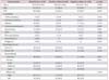
During the study period, 232 consecutive patients diagnosed with TBPE were screened for this study. Of these, 13 were excluded: nine because they received anti-TB therapy before thoracentesis and four because of a previous history of PTB; thus 219 were finally analyzed. The demographic and radiologic characteristics of the 219 patients with TBPE are summarized in Table 1. There were 141 males (64.4%) with a median age of 51.0 (IQR, 32.0–69.0) years. Of these, 117 patients (53.4%) had only TBPE based on radiologic studies and 102 had PTB with concurrent TBPE. Loculated TBPE was identified in 86 cases (39.3%).
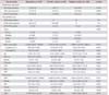
Table 2 shows the microbiological and laboratory data for these patients, of whom 69 (31.5%) were culture positive for MTB in the pleural fluid and 150 were culture negative. There were no case with TB empyema, defined as the presence of frank pus or smear positive for AFB on pleural aspiration. Of the 150 patients with negative pleural culture for MTB, 50 had a positive culture from a respiratory specimen and had PE that resolved with anti-TB treatment, and 100 were clinically diagnosed based on lymphocytic exudates, high ADA levels in the pleural fluid, negative cytological results, and effusions that resolved in response to anti-TB treatment.
Among the 146 TBPE patients in whom MTB was not isolated from a respiratory specimen, it was possible to cultivate MTB in the pleural fluid of 46 patients. The combination of both respiratory specimen and pleural fluid culture showed a diagnostic sensitivity of 54.3% (119/219). There were nine patients (median neutrophil %, 70.5; IQR, 55.0–85.0) with neutrophil-predominant TBPE, all of whom were culture positive for MTB, two of whom were positive for MTB by polymerase chain reaction of TBPE, and six of whom had loculated TBPE.
1. Comparison of clinical, radiological, microbiological, and laboratory findings
Univariate comparisons of the clinical, radiological, microbiological, and laboratory characteristics of MTB culturepositive and culture-negative patients are presented in Tables 1 and 2. There were no significant differences between the groups of patients who were culture-positive and culture-negative for MTB in the pleural fluid with regard to age, sex, underlying disease, frequency of TBPE combined with PTB, and radiological findings, except for loculation. Compared with the patients with TBPE that was culture-negative for MTB, the patients with TBPE that was culture-positive for MTB had a lower lymphocyte percentage, pH, and glucose level in their TBPE. In addition, the culture-positive group had a higher neutrophil percentage, higher protein and lactate dehydrogenase (LDH) levels in their TBPE and a higher C-reactive protein (CRP) level in their blood than the culture-negative group.
2. Predictors of MTB culture positivity in patients diagnosed with TBPE
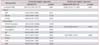
To identify clinical predictors suggestive of culture positivity for MTB in TBPE, eight variables that were significantly different between the two groups in univariate analysis were further analyzed using multivariate logistic regression. After adjusting for potential confounding factors, the loculation of TBPE was independently associated with culture positivity for MTB in TBPE (adjusted OR, 40.062; 95% CI, 9.355–171.556; p<0.001). In contrast, the lymphocyte percentage of TBPE (adjusted OR, 0.934; 95% CI, 0.899–0.971; p=0.001) was inversely associated with culture positivity for MTB in TBPE (Table 3).
3. Comparison of clinical, radiological, microbiological, and laboratory parameters in patients with loculated and free-flowing TBPE
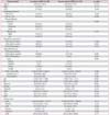
Table 4 compares the characteristics of patients with loculated and free-flowing TBPE. The median values of the white blood cell count, lymphocyte percentage, pH, and glucose level in patients with loculated TBPE were lower than their median values in patients with free-floating TBPE. However, the neutrophil percentage, LDH levels in TBPE, and blood levels of CRP were higher in patients with loculated TBPE.
Discussion
To our knowledge, this is the first study to evaluate the clinical role of loculated TBPE as a predictor of culture positivity for MTB in TBPE. This study aimed to identify clinically useful factors that predict cultivation of MTB in patients with TBPE. The results showed that loculation of TBPE was an independent positive predictor of culture positivity for MTB in TBPE, while a high lymphocyte percentage in TBPE was inversely associated with MTB culture positivity in TBPE.
It is known that concurrent PTB and TBPE is not as uncommon as previously believed, and PTB can be diagnosed by induced sputum in approximately 52% of cases of TBPE232425. However, in the remaining cases of TBPE, it is necessary to cultivate the MTB in the TBPE rather than in respiratory specimens to identify it as the pathogen. Thus, cultivation of MTB from TBPE should not be overlooked in the diagnostic process. Furthermore, it can allow targeted therapy according to the DST14.
Although previous few studies reported that drug-resistant TB occurs less frequently in EPTB than in PTB2627, there is no theoretical reason for a difference in drug-resistance between PTB and EPTB. The reason is that TB is an infectious disease and the proportion of drug-resistant strains of MTB in a community similarly affects both PTB and EPTB2829. Moreover, the proportion of EPTB is increasing among new cases according to the WHO global TB report230. Therefore, it is necessary to isolate the MTB even in EPTB and verify whether actually drug-resistant or not. In our study, MTB was isolated only in the TBPE (46/146, 31.5%), not in respiratory specimen. Furthermore, among these 46 cases identified only by TBPE, two had multidrug-resistant TB, one was resistant to isoniazid and para-aminosalicylic acid, and one was resistant to streptomycin.
Until a recent day, the pathogenesis of TBPE were considered largely as delayed hypersensitivity response of tuberculous protein into the pleural space for most, according to experimental study of guinea pigs and negative cultivation of MTB in patients with TBPE1431. However, the TBPE is now believed to be the consequence of direct infection of the pleural space by paucibacillary MTB related to serial immunologic responses by various pro-inflammatory cytokines based on recent studies131323334. After inoculation of MTB into pleural space, TBPE is initially developed as a result of rapid neutrophilic inflammatory reaction and followed by a CD4+-lymphocyte driven immunologic response of delayed hypersensitivity response over time131. Actually, automated liquid culture systems, now widely used, provide a higher yield of positive cultures, with up to 75% in human immunodeficiency virus (HIV)–positive TBPE patients and up to 60% in HIV-negative TBPE patients, unlike solid media culture at past.
Based on the mechanism of TBPE, from a clinical point of view, it is necessary to identify possible predictors of positive mycobacterial culture in TBPE. Previous studies focused on this point and demonstrated that a low lymphocyte percentage, a high neutrophil percentage, a low protein level, a low glucose level, low pH and high LDH level of TBPE, cancer as an underlying disease, and lack of a radiologically detectable lung infiltrate may be predictors for positive mycobacterial culture10111213. Unfortunately, these studies did not demonstrate any predictors that were markedly superior compared with others.
However, there were interesting finings observed in previous studies. It is that higher neutrophil percentage and lower lymphocyte percentage is associated with a high frequency of positive MTB cultures10111213. It can be explained that higher neutrophil percentage and lower lymphocyte percentage indicate more severe intra-pleural inflammation such as TB empyema or early phase of mycobacterial infection before immunologic response of anti-mycobacterial activity1011. The current study cohort also demonstrated a similarly high culture frequency, but the proportion with a high neutrophil percentage in the TBPE was relatively low, with only 4.1% in this study cohort compared with 11.0%–17.0% in previous studies11133536.
Loculation of pleural fluid is not an uncommon feature of inflammatory exudates including TBPE and parapneumonic PE, and is considered to be an intense intra-pleural inflammation resulting in fibrin deposit and subsequent adhesions in the pleural space1533. In TBPE, this may be caused by elevation of the release of pro-inflammatory cytokines such as tumor necrosis factor α, interleukin 1β, transforming growth factor β1, and vascular endothelial growth factor in response to pleural infection with MTB3337. Since it was demonstrated that loculated TBPE causes pulmonary impairment by residual pleural thickening, most studies have concentrated on reducing the sequelae of TBPE.
However, we focused on the predictive power of loculation of TBPE for cultivation of MTB based on the pathogenesis. The analysis of our study cohort demonstrated that compared with other factors, the loculation of TBPE is potent predictor for positive mycobacterial culture. It has been suggested that loculation of TBPE indicates intense intra-pleural inflammation caused by pleural invasion of more bacillary, with not yet effective immune-clearance system14101131. In our study, loculated TBPE group has higher neutrophil percentage and higher LDH level of pleural fluid, while it has lower lymphocyte percentage, lower glucose and pH level (Table 4). Moreover, loculation of TBPE is not uncommon and easily identifiable by the clinician. Features of loculation are shown by 22.4%–68.8% of TBPE patients, with 39.3% in this study cohort13172038.
There are several limitations to this study. First, it was a retrospective hospital patient-based study. Second, the rate of culture-positive TBPE in this study cohort, despite using both solid and liquid media, was relatively low compared with that in previous studies (31.5% vs. 41.3%–63.1%)101139. Third, this study was conducted in an area with intermediate TB and low HIV infection burden, and there were no HIV-infected patients in this study cohort. Fourth, the discrepancy of classification of loculated TBPE may have impacted result of this study. Among 219 cases, 177 case were underwent chest computed tomography. Of these, there were no discrepancy between interpretations. Other 42 cases were only underwent chest plain X-ray without thoracic sonography and two cases were classified as loculation group by one pulmonologist but not radiologist. However, that cases finally were classified as non-loculated group by other pulmonologist. Thus, the effect of loculation of TBPE as a predictor of positive mycobacterial culture may not be generalizable to other clinical situations.
In conclusion, our study demonstrates that loculation of TBPE is clinically useful predictor of MTB culture positivity in TBPE. The radiological identification of loculation in TBPE is easy, reliable to measure in existing practice, not uncommon and may be used in clinical practice to help to predict the possibility of positive mycobacterial culture.

 XML Download