

 PDF
PDF ePub
ePub Citation
 Print
Print


Abstract
The number of immunocompromised patients has increased over the past decades due to HIV infection, solid and stem cell transplantation, intensified chemotherapy and treatment of autoimmune disease. Pneumonia is a major cause of both morbidity and mortality in immunocompromised patients. Clinical management of pneumonia is difficult, since differential diagnosis in this setting is broad and includes both infectious and noninfectious processes. Because the development of pneumonia in immunocompromised patients is frequently life threatening, early therapeutic and diagnostic intervention is essential to obtain better outcomes.
Figures and Tables
Figure 1
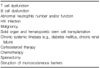
Summary of three major classes of immunosuppression with the infections most commonly associated with each class. Patients can develop other infections, but these represent infections associated with particular immunosuppression 4. This figure is reprinted from Baughman RP. The lung in the immunocompromised patient. Infectious complications Part 1. Respiration 1999;66:95-109.
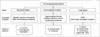
Figure 2
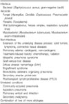
Time line for pathogens in pneumonia after SOT. Sizes of bars express the relative frequency and importance of a group of pathogens1. Bacterial infections in the first month are most often nosocomially acquired but can also be imported from the donors, particularly in lung transplant recipients2. Most often a consequence of ischemia-reperfusion injury5. CARVs: community-acquired respiratory viruses; CMV: cytomegalovirus; EGD: early graft dysfunction; HSV: herpes simplex virus; RSV: respiratory syncytial virus. This figure is reprinted from Chakinala MM, Trulock EP. Pneumonia in the solid organ transplant patient. Clin Chest Med 2005;26:113-21.
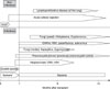
Figure 3
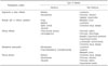
The timeline of pulmonary complications following hematopoietic stem cell transplantation1. HSV: herpes simplex virus; HZV: herpes zoster virus; CMV: cytomegalovirus; RSV: respiratory syncytial virus; CHF: congestive heart failure; ES: engraftment syndrome; VOD: veno-occlusive disease; DAH: diffuse alveolar hemorrhage; IPS: idiopathic pneumonia syndrome; BOOP: bronchiolitis obliterans organizing pneumonia; BO: bronchiolitis obliterans; PTLPD: post- transplant lymphoproliferative disorder. This figure is reprinted from Baughman RP. The lung in the immunocompromised patient. Infectious complications Part 1. Respiration 1999;66:95-109.
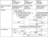
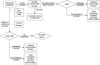
Figure 4
Diagnostic approach with FOB1. Target initial antibiotic therapy for previous isolates from patient. For lung transplant patients with obliterative bronchiolitis (chronic rejection), gram-negative coverage, especially for Pseudomonas aeruginosa, is advised2. FOB with transbronchial biopsy and bronchoalveolar lavage (±protected specimen brushing). If suspected on clinical grounds or suggested by routine hemotoxylin and eosin staining, additional stains for CMV (immunoperoxidase), fungi (Gomori methenamine silver), and mycobacteria (acid-fast) should also be performed3. If CMV pneumonitis is a consideration, begin intravenous ganciclovir (5 mg/kg twice a day, dose adjusted for level of renal function)4. SLBx: surgical lung biopsy5; FOB: fiberoptic bronchoscopy; CMV: cytomegalovirus. This figure is reprinted from Jain P, Sandur S, Meli Y, Arroliga AC, Stoller JK, Mehta AC. Role of flexible bronchoscopy in immunocompromised patients with lung infiltrates. Chest 2004;125:712-22.
References
1. Safadi AR, Soubani AO. Diagnostic approach of pulmonary disease in the HIV negative immunocompromised host. Eur J Intern Med. 2009. 20:268–279.
2. Cunha BA. Pneumonias in the compromised host. Infect Dis Clin North Am. 2001. 15:591–612.
3. Gasink LB, Blumberg EA. Bacterial and mycobacterial pneumonia in transplant recipients. Clin Chest Med. 2005. 26:647–659.
4. Baughman RP. The lung in the immunocompromised patient. Infectious complications Part 1. Respiration. 1999. 66:95–109.
5. Chakinala MM, Trulock EP. Pneumonia in the solid organ transplant patient. Clin Chest Med. 2005. 26:113–121.
6. Jain P, Sandur S, Meli Y, Arroliga AC, Stoller JK, Mehta AC. Role of flexible bronchoscopy in immunocompromised patients with lung infiltrates. Chest. 2004. 125:712–722.
7. Hayden RT. Diagnostic microbiology of the immunocompromised host. 2009. Washington, D.C: ASM Press.
8. Corti M, Palmero D, Eiguchi K. Respiratory infections in immunocompromised patients. Curr Opin Pulm Med. 2009. 15:209–217.
 XML Download