

 PDF
PDF ePub
ePub Citation
 Print
Print


Introduction
Desmoid tumors are uncommon tumors accounting for less than 0.03% of all neoplasms. They show infiltrating growth of well-differentiated fibroblasts or myofibroblasts, and tend to recur locally without metastasis1. Alternatively, the term "aggressive fibromatosis" has often been employed to emphasize the frequently aggressive behavior of these tumors. Desmoid tumors are most commonly intraabdominal, and the most common extraabdominal sites include the chest wall, shoulder girdle, thigh, and head and neck. True intrathoracic desmoid tumors are rare with most cases actually representing intrathoracic extension of chest wall tumors. Out of the reported intrathoracic desmoid tumors, of which a few extended into abdominal cavities2. Twelve cases occurred in the pleura, including one Korean case3-11.
Here, we report on a case of intrathoracic desmoid tumor protruding into the pleural cavity, and discuss its differential diagnoses along with a review of the literature.
Case Report
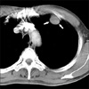
A 40-year-old male presented with progressive and marked weight loss of 13 kg over one year and dizziness for three weeks. The patient appeared to be chronically ill with a weight of 50 kg and height of 170 cm. He had 30 pack years (1.5 packs per day for 20 years) and had worked in printing and handled chemicals for six years. He showed no remarkable laboratory profiles, including a normal blood glucose level. Chest computed tomography (CT) revealed an oval mass of 2.0 cm in size in the left upper anterior field (Figure 1). At the base, the tumor appeared to invade into the left chest wall. Solitary fibrous tumor (SFT), primary or metastatic pleural tumor was radiologically suspected. Thoracoscopic surgery using three ports was performed. The main mass, measuring 0.5 cm, protruded into the pleural cavity with a short stalk, having a base in the left second intercostal muscle. No pleural adhesion, effusion, or tumor seeding was found. Thoracoscopic near total excision of the protruded mass was performed. During post-operative follow up, the patient's marked weight loss was proved to be ascribed to hyperthyroidism. No additional treatment was administered. Neither recurrence, nor metastasis was found during 6 years of follow up.
1. Pathologic findings
A needle biopsy taken from the mass by ultrasound-guidance revealed paucicellular fibrous tissue admixed with a few small capillaries. Spindle cells in the paucicellular fibrous tissue were immunoreactive for vimentin (V9; Dako, Glostrup, Denmark, prediluted), and negative for pancytokeratin (AE1/AE3; Dako, prediluted), and CD34 (QBEnd10; Dako, prediluted).
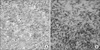
Grossly, the excised mass measured 2.5×2.0 cm. The external surface of the mass was smooth and glistening, covered by parietal pleura. The gray-tan, trabeculated cut surface was homogeneously solid without hemorrhage or necrosis. Microscopically, the mass was composed of a hypocellular arrangement of spindle-shaped cells in a massive collagenous background (Figure 2A). Spindle cells had small indistinct nucleoli and fine nuclear chromatin, and weakly eosinophilic cytoplasm. Mitosis or necrosis was not observed. Small capillary-sized vessels were contained in some areas. A hemangiopericytomatous vascular arrangement was not observed. The resection margin of the stalk was focally involved by the spindle cells. Immunohistochemistry was performed using an avidin-biotin peroxidase complex method. The cytoplasm of the spindle cells was strongly positive for vimentin (Figure 2B), while totally negative for CD34, pancytokeratin, S-100 protein (polyclonal; Dako, prediluted), CD68 (PG-M1; Dako, prediluted), muscle specific actin (HHF35; Dako, prediluted), desmin (D33; Dako, prediluted), progesterone receptor (PgR636; Dako, prediluted), estrogen receptor (SP1; Dako, prediluted), and p53 (DO-7; Dako, prediluted). The immunohistochemical stainability of Ki-67 (MIB-1; Dako, prediluted) was investigated using a quantitative manual counting method i-Solution 7.5 (IMT i-Solution company, Coquitlam, Canada). Maximum Ki-67 labeling index was less than 0.1%.
Discussion
The cause of desmoid tumor is uncertain. For pathogenesis, previous trauma including surgical scars, hormonal factors, and genetic factors such as Gardner's syndrome or familial adenomatous polyposis have been postulated. History of trauma can be considered first due to the fact that cases of desmoid tumor in operative scars of mastectomy and thoracotomy have been reported1. Endocrinologic hormonal etiology has been suggested due to the fact that desmoid tumor most commonly appears in young women during or after pregnancy, and may regress after menopause, or after tamoxifen treatment12. Molecular studies have revealed a high incidence of adenomatous polyposis coli (APC) and β-catenin gene mutations in desmoid tumors, resulting in nuclear accumulation of β-catenin proetin13. Nuclear staining patterns for β-catenin in desmoid tumors are shared with those of pleural SFT, suggesting that pleuropulmonary desmoid tumor and SFT might have somewhat overlapping pathogenetic pathways.
In pathologic differential diagnoses of intrathoracic desmoid tumor, pleural SFT is a primary consideration. Pleural SFT is an uncommon but increasingly recognized neoplasm derived from mesenchymal cells located in the submesothelial lining of the pleural space, and usually forming a pedunculated or non-pedunculated tumor from the parietal or visceral pleurae1. Typical pathology of pleural SFT shows a disorderly configuration of bipolar spindle cells and collagen fibers in a pattern-less arrangement, with abundant stag-horn shaped vessels that are hemangiopericytomatous in appearance. Desmoid tumor is composed of bland-looking spindle shaped nuclei and keloid-like connective tissue, with absence of hemangiopericytomatous vessels. Spindle cells of desmoid tumor show immunonegativitity for CD34 and immunopositivity for vimentin, variable reactions for smooth muscle actin, S-100 protein, or desmin, whereas SFT shows characteristic dual immunoreactivity for vimentin and CD34. Other pathologic differential diagnoses include inflammatory pseudotumor (inflammatory myofibroblastic tumor), low-grade fibromyxoid sarcoma, malignant fibrous histiocytoma and malignant mesothelioma after needle biopsy. Inflammatory pseudotumor is a tumor-like mass composed of fibrous spindle cells or granulation tissue infiltrated by inflammatory cells. Malignant fibrous histiocytoma shows a stroriform arrangement of atypical spindle cells with occasional pleomorphic stromal cells, for which a definable line of differentiation has not been found. Immnohistochemically, these spindle cells are usually reactive for vimentin, α1-antitrypsin, α1-antichymotrypsin, and CD68, and vary in their reactivity for actin, desmin, and lysozyme. Malignant mesothelioma commonly appears as a diffuse extension along the pleural surface, but occasionally shows a localized growth pattern. Localized type mesothelioma shows epithelioid features in small areas, even in the case of a sarcomatous type of malignant mesothelioma. Immunohistochemically, tumor cells showing positivity for low molecular weight cytokeratin and calretinin confirm malignant mesothelioma. Low-grade fibromyxoid sarcoma is rare, and benign looking, but local recurrences are frequent. Microscopically, low-grade fibromyxoid sarcoma displays a mixture of hypocellular, collagen-rich areas and more cellular, myxoid areas. Whorling growth patterns of spindle cells are helpful in the distinction from desmoid tumor.
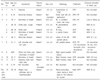
Review of the previously reported thirteen cases of intrathoracic pleural desmoid tumors including the present case showed a preponderance of females (8 females vs. 5 males)3-11. Age ranged 5 to 72 years. The size of intrathoracic pleural desmoid tumors ranged from 2.0 cm up to 16.0 cm. The most common presentation was dyspnea or shortness of breath (6/13, 46.2%), followed by chest pain (4/13, 30.8%), abnormal chest shadow (4/13, 30.8%) and cough (2/13, 15.4%), flank or back pain (2/13, 15.4%), shoulder and arm pain (1/13, 7.7%). Radiologic findings show desmoid tumor variability; high signal intensity on T2-weighted images and low to isointensity on T1-weighted images with respect to muscle14. These nonspecific clinicoradiologic features shared with those of the more common pleural SFT. The present case showed severe weight loss and dizziness, but were caused by incidentally accompanying hyperthyroidism. Clinicoradiologic summaries are shown in Table 1.
Treatment of intrathoracic desmoid tumor is challenging because this disease does not respect the usual surgical rules relating to resection and recurrence. Complete excision with clear margins is the mainstay of treatment. Due to the high local recurrence rate, adjuvant therapy may be conducted. Radiotherapy has been traditionally directed at known positive margins and in inoperable cases. Antiestrogen hormone therapy and cytotoxic chemotherapy have also been used for unresectable or recurrent disease, however, such experiences have been limited, and results have been inconclusive2. Like extraabdominal desmoid tumor of other sites, the natural course of intrathoracic desmoid tumor is widely variable, ranging from spontaneous remission to multiple recurrences, regardless of treatment modality12. Adjuvant radiotherapy can be reserved for the minority with a high risk of recurrence, particularly those with residual bulk disease or multiple previous recurrences. Despite controversies over adjuvant therapy, a desmoid tumor in an unusual location, preventing wide excision margins, may favor early consideration of adjuvant therapy, particularly in premenopausal women. Albeit rare, malignant transformation of extra-abdominal desmoid tumor may occur15. To date, no malignant transformation, including that of pleural mass, has been described among intrathoracic cases. The present case was focally involved by the tumor with no recurrence during the follow up period, suggesting pleural desmoid tumors might take more favorable prognosis than do non-pleural desmoid tumors. Due to lack of accumulated experience, intrathoracic desmoid tumors, including pleural masses, should be under long term follow-up.
 XML Download