

 PDF
PDF ePub
ePub Citation
 Print
Print


INTRODUCTION
Over the years, orthodontists have used various methods to accelerate orthodontic tooth movement. Some of these methods are corticotomy,1 corticision,23 micro-osteoperforations/piezopuncture,45 and piezocision. 67 A modification of corticotomy with or without alveolar bone grafting has been referred to as “accelerated osteogenic orthodontics” and more recently to as “periodontally accelerated osteogenic orthodontics”. Corticotomy acceleration of tooth movement has been reported for several types of tooth movements, including canine retraction,89 anterior teeth retraction,101112 decrowding,13 extrusion of impacted canine,14 and 3rd molar uprighting.15
Although corticotomy procedures are quite effective in assisting acceleration of orthodontic tooth movement, they are inherently invasive because of the requirement for significant flap elevations, which potentially result in postsurgical discomfort and complications that deter patients from undergoing such procedures. Some studies have also reported that procedures involving full-thickness flap elevation could cause periodontal problems, and increase tooth mobility and bone dehiscence.11617
Another minimally invasive procedure “Piezocision™” was introduced in 2009. This flapless technique used an ultrasonic piezosurgical knife to make micro-incisions in the gingiva and cortical alveolar bone.18 It combined piezoelectric bone decortication with selective tunneling that allowed for hard- or soft-tissue grafting. This novel approach led to a shorter treatment time, minimized discomfort, and greater patient acceptance.19
Both corticotomy and piezocision have already been implemented in conjunction with orthodontics worldwide. Nonetheless, most studies reporting such applications were case reports or cases series. Only a few clinical studies have compared the effectiveness of these two methods. Therefore, our research focused on evaluating the effectiveness of corticotomy and piezocision in accelerating orthodontic tooth movement. The objective of this systematic review was to evaluate the rate of canine retraction by using either corticotomy or piezocision in comparison with the conventional method and assessing their associated complications.
MATERIALS AND METHODS
Protocol registration
The study protocol was registered on the PROSPERO International prospective register of systematic reviews, 2017 number: CRD42017070359.
Search strategy
The search terms used were designed according to the PICO principle (Patient/Problem/Population; Intervention/Exposure; Comparison and Outcomes). The key terms were as follows: patients undergoing orthodontic treatments; and the intervention terms were corticotomy, piezocision, corticotomy-facilitated orthodontics, Wilckodontics, periodontally accelerated osteogenic orthodontics, corticision, interdental osteotomy, micro-osteoperforation alveolar decortication, decortication orthodontics, or regional acceleratory phenomenon procedure (RAP). The outcomes were tooth movement and rate of tooth movement. The details of the search terms with Boolean operators, classified according to the five electronic databases, are shown in Supplementary Table 1 (available at http://www.e-kjo.org only).
This systematic review was conducted according to the guidelines of the Preferred Reporting Items for Systematic Reviews and Meta-analyses (PRISMA). Abstracts were searched for on five principal electronic databases: PubMed, SCOPUS, Web of Science, Embase, and Cochrane Central Register of Controlled Trials (CENTRAL), dated up to July 2017. No restrictions (limits) were imposed on language, year published, or malocclusion type. Details of the search strategy, classified according to the five electronic databases are shown in Supplementary Table 1. The search results were exported to the Endnote program (version X7; Clarivate Analytics, Philadelphia, PA, USA), and duplicates were removed using the “remove duplicates” command.
Eligibility criteria
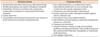
Abstract screening was conducted by two authors (SD and NV), according to the inclusion and exclusion criteria shown in Table 1. The authors independently selected the studies focusing on canine retraction. The included studies had to be randomized controlled clinical trials (RCTs), with a split-mouth design. The retrieved studies had to present primary outcomes in terms of accumulative moved distance, movement rate, velocity of tooth movement, or the duration of treatment. Exclusion criteria were laboratory studies, animal studies, descriptive studies, case reports, case series, review articles, systematic reviews, and meta-analysis, distraction, osteotomy, etc. The percentage of the two authors' agreement was 97.6%. Any disagreements regarding the selection of studies were resolved through discussion. Thereafter, the full texts of selected articles were retrieved and examined for eligibility by using a pilot data screening form. The lists of references in the retrieved articles were further manually searched for other pertinent publications.
Data extraction and quality assessment
Data were extracted independently by two authors using a data extraction form. Information from each study was then organized into tables that examined the characteristics of the participants, interventions, comparators, outcomes, and study design (PICOS). The results of the studies were collected. The primary outcome reported for canine retraction was either the amount of tooth movement, rate of tooth movement, or treatment time. The secondary outcome was complications, which could be periodontal parameters, pain, root resorption, or satisfaction.
Risk of bias
The risk of bias of the studies was independently assessed by two authors, using the Cochrane Collaboration's assessment tool.20 Seven domains of bias were evaluated; (1) random sequence generation, (2) allocation concealment, (3) blinding of participants and personnel, (4) blinding of outcome assessment, (5) incomplete outcome data, (6) selective reporting, and (7) others. The study was categorized as a) low risk if all domains were assessed as low risk of bias, b) as unclear risk if any domain was assessed as unclear risk of bias, and c) as high risk when any domain was judged as high risk of bias.20 Any disagreements between the authors were resolved through discussion.
RESULTS
Study selection
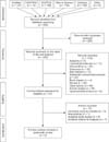
As shown in Figure 1, the database search retrieved 530 abstracts: 127 from PubMed, 12 from CENTRAL, 168 from Scopus, 114 from the Web of Science, 105 from Embase, and 4 from additional sources. Screening using the Endnote program helped exclude 290 duplicate abstracts, resulting in 240 abstracts for screening. After reviewing the abstracts, 218 records were removed according to the inclusion and exclusion criteria, leaving 22 full-text articles for examination. Details regarding the number of papers and reasons for exclusion at each step are depicted in Figure 1. Eventually, five full-text articles were included and evaluated in this systematic review.
Study characteristics
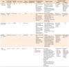
The characteristics of the five selected studies are presented in Table 2. All studies were RCTs, with a split-mouth design. The studies included measured the effectiveness either corticotomy or piezocision in comparison with conventional orthodontic treatment. Conventional canine retraction was compared with corticotomy in four studies,892122 and with piezocision in two studies.2123 The outcomes of interest were classified into primary outcomes (accumulative distance and velocity of tooth movement) and secondary outcomes (periodontal condition, root resorption, and pain).
Surgical interventions
Surgical details of corticotomy or piezocision are described in Table 2. Four studies had surgery performed immediately after the premolars were extracted, while one study by Al-Naoum et al.9 had surgery performed 4 weeks after extraction. Three corticotomy studies had only the buccal flap elevated,82122 while the remaining study had both the buccal and palatal flaps elevated.9 One corticotomy study used modified corticotomy procedures by drilling numerous holes into the cortical plate without vertical cuts,8 while the other three employed a combination of corticotomy cuts and perforating holes.92122 Piezocision studies were flapless, and used similar procedures by performing vertical interproximal incision under the interdental papilla by using piezosurgical devices.2123
Orthodontic interventions
According to Table 2, all five studies used conventional fixed orthodontic appliances and focused on canine retraction. Orthodontic appliances were placed before the surgical procedures in all studies.89212223 Force was applied immediately or 2 weeks after surgery. Three studies also used extra anchorages such as miniscrews822 or transpalatal arches.9
Rate of tooth movement
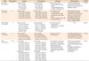
Table 3 shows the results of the outcomes from the five studies. The outcomes are briefly listed as follows.
Corticotomy vs. conventional
Three studies using corticotomy procedures demonstrated a statistical improvement in the rate of orthodontic tooth movement than did the conventional technique.
Aboul-Ela et al.8 found that the rate of canine retraction was twice as fast on the corticotomy side than on the control side during the first 2 months after surgery, followed by a decrease to 1.6 times and 1.06 times during the 3rd and the 4th months, respectively. Al-Naoum et al.9 reported 2 to 4 times faster rates on the corticotomy side. During the 1st and 2nd weeks after corticotomy, the rate was 4 times faster, and during the 2nd–4th and 8th–12th weeks, it was almost 3 times faster than that in the control group. Similarly, Abbas et al.21 and Jahanbakhshi et al.23 also reported 1.5 to 2 times faster rates in the corticotomy group.
Piezocision vs. conventional
The piezocision procedures also presented statistical progress in the rate of orthodontic tooth movement. Aksakalli et al.22 found that piezocision produced a 2 times faster rate of tooth movement than did the conventional approach. This was slightly higher than the 1.5 times faster rate described in the study by Abbas et al.21
Corticotomy vs. piezocision
The study by Abbas et al.21 was the only one in this systematic review that drew an indirect comparison between the effectiveness of corticotomy and piezocision on orthodontic tooth movement. In addition, they conducted a comparison between each surgical group and the conventional technique. Their results showed a higher rate of canine crown tip movement in both the corticotomy and piezocision groups as opposed to the conventional group. Moreover, the rate of canine crown tip movement in the corticotomy group was greater than that in the piezocision group.21
Risk of bias assessment
The quality of the studies was assessed as shown in Table 4. In general, the five studies included showed an overall high risk of bias. Allocation concealment was ranked as low because all five articles were RCTs with a split-mouth design. Blinding of participants and personnel in all studies was impossible because both the operators and patients were aware of the corticotomy/piezocision side. Blinding of outcome assessment was conducted only in one study.23 The details of the support for judgment are also listed in Table 4.
DISCUSSION
This systematic review indicated that both corticotomy and piezocision resulted in greater acceleration of tooth movement than did conventional techniques. The rate of orthodontic tooth movement in corticotomy varied from 1.5 to 4 times that of the conventional rate depending on the surgical methods used.892122 Similarly, piezocision also led to an effective tooth movement rate of 1.5 to 2 times faster than that of the conventional method.2123
A strength of our study was the homogeneity in the design of the included studies, which were all RCTs with a split-mouth design and using canine retraction. Although there were some other interesting RCTs using a) corticotomy for impacted canines14 or en-masse six-teeth anterior retraction10 or b) piezocision in non-extraction19 or 3rd molar impaction,15 they were not included in our study.
The results of our study were generally in agreement with those of Patterson et al.,24 who reported a systematic review on corticotomy, using a larger number of studies (six RCTs and eight controlled clinical trials). However, their study seemed to have more limitations. The quality of the body of evidence presented by Patterson et al.24 was regarded as low, owing to the presence of multiple methodological issues, high risks of bias, and heterogeneity of the included articles. For example, they included studies on impacted canine,14 canine retraction of the six upper anterior teeth,10 and micro-osteoperforations with a Propel device.4
Differential tooth movement rate
The three corticotomy papers in our study showed initially high acceleration of tooth movement within the first few months before a slow decrease in the rate of tooth movement over time.8922 Aksakalli et al.22 reported twice the cumulative canine movement in the piezocision group than in the conventional group. Because they did not report the rate of tooth movement, their results could not be compared with those in a study by Abbas et al.21
The plausible reason for this acceleration could be the RAP, which is a transient phenomenon that begins a few days after the surgery. It peaks between 1 and 2 months and then declines over time and lasts for approximately 4 months.172526 As a consequence, intentional bone damage by corticotomy and piezocision procedures can increase bone turnover, which results in rapid tooth movement that lasts only as long as the RAP is active. On the contrary, a study by Abbas et al.21 reported faster tooth movement rates over time up to 10 to 12 weeks after both corticotomy and piezocision, which did not comply with RAP phenomenon.
Surgical technique
In general, our study showed that the more aggressive the surgical technique, the greater the acceleration of tooth movement was. This phenomenon was shown in the study by Al-Naoum et al.,9 which used a combination of corticotomy cuts and perforations on both the buccal and palatal sides and reported that corticotomy could accelerate tooth movement up to 4 times during the first 2 weeks. In contrast, the studies by Aboul-Ela et al.8 (which used only perforations without vertical cuts), Jahanbakhshi et al.,23 and Abbas et al.21 (which used both corticotomy cuts and perforations only on the buccal side) revealed that corticotomy produced a rate of tooth movement only 1.5 to 2 times greater than that produced by the conventional techniques. Therefore, further study is needed to verify the effectiveness of corticotomy using perforations only and perforations with vertical cuts.
Lastly, corticotomy exhibited only slightly greater rates of canine movement than did piezocision. These differences might be attributable to the more extensive surgery required for corticotomy, which might have enhanced the RAP to a greater extent than in piezocision.21
Timing of force application
Many studies applied force before the surgical procedures, possibly because of a desire for convenient bonding prior to surgery. Thus, it would have allowed for safer force application as soon as surgery was completed.891921222324
In this study, the initial force application was conducted within 2 weeks after surgery, thus complying with the study by Abu-Hussein et al.27 who stated that orthodontic force application should be initiated within 2 weeks after surgery. Beyond that period, the full benefit of the RAP will not be realized. In addition, the orthodontic appliance should be adjusted more frequently every 2 weeks throughout the duration of treatment.28
Treatment time
Reduction in treatment time is a great benefit of surgical-assisted orthodontics. In the corticotomy study by Aboul-Ela et al.,8 a Class I canine relationship was established 2 or 3 months faster than the usual rate of 7 months required for the conventional technique.28
Likewise, the canine distalization phase in piezocision procedures was also completed in 3.5 months, which was faster than the 5.6 months required in the conventional treatment group.23 Charavet et al.,19 in a non-extraction study, showed that piezocision helped reduce the treatment time by 43%. They observed a reduction in the total treatment duration from bracket placement to bracket removal. These results demonstrated that piezocision techniques may assist in accelerating orthodontic tooth movement.
Periodontal parameters
The adverse effects of corticotomy on periodontal tissue are controversial. In the past, there were case reports about periodontal problems after corticotomy, such as interdental bone loss, decrease of the attached gingiva, and periodontal defects.129
In this study, the adverse effects on the periodontal tissue after corticotomy821 and piezocision192123 were not significantly different from those in the control group. The reasons could be attributed to the flap design. Aboul-Ela et al.8 used a special technique called submarginal Luebke-Ochsenbein flap to avoid intrasulcular and marginal bone incision, which resulted in the preservation of the periodontal condition after surgery. They also stated that the reason for the absence of any adverse effects on the periodontium after corticotomy could be the manner of bone removal, which were as follows; a) corticotomy was not performed as a true osteotomy (with a block of bone removed), and b) the procedure only perforated the bone, leaving the original bony architecture intact.8 This allowed the resorption-deposition cellular process to proceed in the existing bony architecture with fewer side effects.
Pain, discomfort, and satisfaction levels
There was only one study that examined the levels of pain and discomfort.9 Al-Naoum et al.9 who performed two-sided corticotomy indicated that 50% of patients experienced severe pain during meals on the day after corticotomy. However, the pain gradually receded within the next week, while approximately 60% to 70% of patients reported no pain or only mild discomfort. The findings of this report were in agreement with those of the studies by Cassetta et al.30 and Wilcko et al.,1 which showed that post-surgical pain completely disappeared within a 7- to 10-day period. Moreover, the pain reported in corticotomy studies was possibly due to the effects of flap operations, which could influence the patients' acceptance of the procedure.9
Root resorption and dehiscence
The study of Abbas et al.21 found greater canine resorption in the conventional group than in the corticotomy and piezocision groups. This was consistent with the findings of several papers, which reported that teeth retained their vitality without any evidence of resorption after corticotomy or piezocision.119 Less root resorption may be due to the increased osteoclastic activity and decreased bone density that were associated with the RAP. It may also decrease the likelihood of hyalinization necrosis and subsequent root resorption.
Similarly, Charavet et al.19 examined the effects of piezocision on root resorption and dehiscence, and demonstrated no significant differences between the piezocision and conventional groups. Anyhow, in the study by Abbas et al.,21 patients undergoing all procedures were observed during an equal 3-month period without mentioning the method used to evaluate root resorption.
Limitations
This study included a low number of previously published RCTs which is a limitation. The five selected studies showed an overall high risk of bias. This highlights the need for more primary research to be conducted in the future. In clinical studies, several confounding factors may account for the variability in acceleration rates, including patient characteristics, different study designs, patient numbers, operator skill, different surgical protocols, corticotomy/piezocision, and variations in orthodontic protocols (such as wire size, bracket type, force application, force magnitudes, types of tooth movement, different activation and reactivation regimes, and durations of force activation).24 Another limitation was the patients' age. In our study, two publications comprised subjects younger than 18 years old.2123 Age could have had an impact on this outcome analysis.
It should also be noted that in the study by Abbas et al.,21 tooth movement was greater between 10 and 12 weeks in the corticotomy and piezocision groups when compared with other time intervals. This finding was contradictory to those of previous reports citing RAP phenomenon which mostly showed that tooth movement was faster in the early period of the study. The difference between Abbas et al.'s study21 and other studies had raised questions about the validity of their report. Therefore, the results of our study related to piezocision should be interpreted with caution. These contradictions may be resolved by future primary RCTs on piezocision using similar protocols.
Implications
Both corticotomy and piezocision could be helpful in accelerating canine retraction. However, piezocision may be a better alternative to corticotomy for canine retraction into the immediate premolar extraction site. This was because the accelerated rate of piezocision was quite similar to that of corticotomy (with only buccal flap elevation). In addition, piezocision seemed to be a less traumatic technique with greater patient acceptance. Nevertheless, more RCTs are required to confirm the rate of acceleration, risk-benefit ratio, long-term follow-up, and relapse after piezocision.
CONCLUSION
1. This study confirmed that corticotomy and piezocision increased the rate of orthodontic canine retraction.
2. Corticotomy had the potential to generate 2 to 4 times greater canine retraction rate than that seen in the control.
3. Corticotomy with both buccal and palatal flap elevation could generate greater canine retraction rate than could corticotomy with only buccal flap elevation.
4. Piezocision resulted in a canine retraction rate 1.5 to 2 times faster than that seen in the control.
5. For canine retraction into the immediate premolar extraction site, the rate of canine retraction after piezocision was almost comparable to that of corticotomy (with only buccal flap elevation).
6. Corticotomy (with a flap design avoiding marginal bone incision) or flapless piezocision did not have an adverse impact on the periodontal status, including the plaque index, probing depth, attachment levels, gingival recession, mobility scores, and alveolar crest levels or root resorption.
 XML Download