

 PDF
PDF ePub
ePub Citation
 Print
Print


Abstract
Objective
The purpose of this study was to analyze the dental and basal arch forms in patients with normal occlusion using the computed tomography (CT) imaging method.
Methods
CT images were taken from 27 normal occlusion subjects (male, 15; female, 12) and these images were reconstructed into three-dimensional models. A 3D-coordinate system was formed by setting the middle of the facial axis (FA) point of the maxillary central incisors as the origin. The morphology of the maxilla and mandibular dental and basal arches were analyzed by sectioning parallel to the maxillary occlusal plane.
Results
There was no significant difference between A point and B point and between the maxillary 1st molars in both sides of the maxillary and mandibular basal bone. The dental arch was located more labially than the basal arch in the anterior portion. The bucco-lingual crossover of the dental arch and basal arch was formed at the molar region in the maxilla, and at the premolar region in the mandible.
Figures and Tables
Fig. 1
Three-dimensional coordinate system. A, Maxillary occlusal plane (XY plane) is the plane passing the FA point of #16, 26 and the center of #11, 21 FA point; B, midpalatal plane (YZ plane) is the plane passing ANS and PNS perpendicular to the maxillary occlusal plane; C, frontal plane (ZX plane) is the plane passing the center of #11, 21 FA point perpendicular to the maxillary occlusal plane and midpalatal plane.
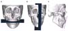
Fig. 2
Basal bone. A, A plane passing through A point parallel to XY plane; B, a plane passing through B point parallel to XY plane; C, five planes passing through the FA points of #16, #13, center of #11 and 21, #23, and #26 parallel to YZ plane; D, segmentation of 3D object; E, a plane passing through the FA points of #17, #27 perpendicular to XY plane; F, point on the basal bone arch (#17, #16, #13, A point, #23, #26, #27 area).
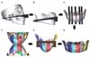
Fig. 3
Crown center. A, Crown center of the incisor shown as the midpoint of the incisal edge; B, crown center of the canine shown as the cusp tip of the canine; C, and D, crown center of premolar and molar, respectively, shown as the midpoint of the crown at the proximal contact point level.
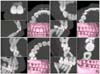
Fig. 4
Root center. A, B, C, The root apex for single-rooted teeth; D, the midpoint of the apical third of the root in three cross-sectional images for multiple-rooted teeth.
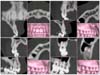
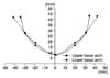
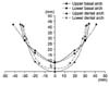
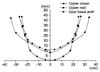
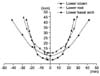
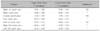
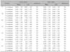
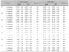
Table 5
Comparison of crown and root position between right and left sides (maxillary teeth, unit: mm)
References
1. Proffit WR, Fields HW, Sarver DM. Contemporary orthodontics. 2007. 4th ed. St Louis: Mosby;3–23.
2. Proffit WR. Muscle pressures and tooth position: North American whites and Australian aborigines. Angle Orthod. 1975. 45:1–11.
3. Tweed CH. The Frankfort-mandibular plane angle in orthodontic diagnosis, classification, treatment planning, and prognosis. Am J Orthod Oral Surg. 1946. 32:175–230.

4. Lundström AF. Malocclusion of the teeth regarded as a problem in connection with the apical base. Int J Orthod Oral Surg Radiogr. 1925. 11:1022–1042.
5. Betts NJ, Vanarsdall RL, Barber HD, Higgins-Barber K, Fonseca RJ. Diagnosis and treatment of transverse maxillary deficiency. Int J Adult Orthodon Orthognath Surg. 1995. 10:75–96.
6. Strang RH. The fallacy of denture expansion as a treatment procedure. Angle Orthod. 1949. 19:12–22.
7. Tweed CH. A philosophy of orthodontic treatment. Am J Orthod Oral Surg. 1945. 31:74–103.
8. Sergl HG, Kerr WJ, McColl JH. A method of measuring the apical base. Eur J Orthod. 1996. 18:479–483.
9. Ahn HS, Cha KS. A study on maxillary basal bone morphology in skeletal Class III malocclusion requiring orthognathic surgery. Korean J Orthod. 1995. 25:577–585.
10. Ko SD, Cha KS. A study on the labial & buccal surface contour in Korean permanent teeth using three-dimensional laser scanning. Korean J Orthod. 2002. 32:275–291.
11. Park HC, Lee JW. Study of horizontal skeletal pattern and dental arch in skeletal Class III malocclusion patients. Korean J Orthod. 2008. 38:358–370.
12. Baumrind S, Frantz RC. The reliability of head film measurements. 1. Landmark identification. Am J Orthod. 1971. 60:111–127.
13. Bergersen EO. Enlargement and distortion in cephalometric radiography: compensation tables for linear measurements. Angle Orthod. 1980. 50:230–244.
14. Jeon KJ, Park H, Lee HC, Kim KD, Park CS. Reproducibilities of cephalometric measurements of three-dimensional CT images reconstructed in the personal computer. Korean J Oral Maxillofac Radiol. 2003. 33:171–178.
15. Cavalcanti MG, Haller JW, Vannier MW. Three-dimensional computed tomography landmark measurement in craniofacial surgical planning: experimental validation in vitro. J Oral Maxillofac Surg. 1999. 57:690–694.
16. Lee SK, Kwon OW, Sung JH. A study on the dental arch characteristics of bialveolar protrusion patients using a three-dimensional digital model. Korean J Orthod. 2006. 36:45–54.
17. Kim NR, Kim YI, Park SB, Hwang DS. Three dimensional cone-beam CT study of upper airway change after mandibular setback surgery for skeletal Class III malocclusion patients. Korean J Orthod. 2010. 40:145–155.
18. Kim KD, Ruprecht A, Wang G, Lee JB, Dawson DV, Vannier MW. Accuracy of facial soft tissue thickness measurements in personal computer-based multiplanar reconstructed computed tomographic images. Forensic Sci Int. 2005. 155:28–34.
19. Steiner CC. Cephalometrics for you and me. Am J Orthod. 1953. 39:729–755.
20. Jacobson A. The "Wits" appraisal of jaw disharmony. Am J Orthod. 1975. 67:125–138.
21. Chan GK. A cephalometric appraisal of the Chinese (Cantonese). Am J Orthod. 1972. 61:279–285.
22. Yang HB. A study on the dental arch characteristic for bracket positioning of bicuspid extraction cases (thesis). 1996. Cheonan: Dankook University.
23. Yim JB. Three dimensional analysis of the dental arches in Korean adult with normal occlusion (thesis). 1999. Cheonan: Dankook University.
24. Lee DS. Three dimensional analysis of dental arch and basal structure in Korean adult with angle III malocclusion (thesis). 1999. Cheonan: Dankook University.
25. Biggerstaff RH. Three variations in dental arch form estimated by a quadratic equation. J Dent Res. 1972. 51:1509.
26. Pepe SH. Polynomial and catenary curve fits to human dental arches. J Dent Res. 1975. 54:1124–1132.
27. Han H. A study of the variances in pre- and post-treatment dental arch shapes in extraction and non-extraction cases (thesis). 1990. Cheonan: Dankook University.
28. Lim KH. A study of dental arch shape and dimensional change in Class II division 1 malocclusion treatment (thesis). 1997. Cheonan: Dankook University.
29. Lee DJ. Study on the arch form of normal occlusion after orthodontic treatment (thesis). 2002. Cheonan: Dankook University.
30. Shin MR. A study on the pre- and post dental arch shape changes in sectional and continuous arch technique (thesis). 1995. Cheonan: Dankook University.
 XML Download