

 PDF
PDF ePub
ePub Citation
 Print
Print


Abstract
Objective
The aim of this study was to investigate the 3-dimensional position of the center of resistance of the 4 maxillary anterior teeth, 6 maxillary anterior teeth, and the full maxillary dentition using 3-dimensional finite element analysis.
Methods
Finite element models included the whole upper dentition, periodontal ligament, and alveolar bone. The crowns of the teeth in each group were fixed with buccal and lingual arch wires and lingual splint wires to minimize individual tooth movement and to evenly disperse the forces to the teeth. A force of 100 g or 200 g was applied to the wire beam extended from the incisal edge of the upper central incisor, and displacement of teeth was evaluated. The center of resistance was defined as the point where the applied force induced parallel movement.
Results
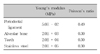
The results of study showed that the center of resistance of the 4 maxillary anterior teeth group, the 6 maxillary anterior teeth group, and the full maxillary dentition group were at 13.5 mm apical and 12.0 mm posterior, 13.5 mm apical and 14.0 mm posterior, and 11.0 mm apical and 26.5 mm posterior to the incisal edge of the upper central incisor, respectively.
Figures and Tables
Fig 1
Three-dimensional finite element mesh of tooth-periodontal ligament (PDL)-alveolar bone of the maxillary dentition. A and B, Frontal and lateral views of upper dentition and PDL; C and D, frontal and lateral views of tooth-PDL-alveolar bone.
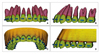
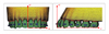
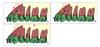
Fig 3
Finite element models of the teeth group. A, Four anterior teeth; B, six anterior teeth; C, full maxillary dentition. Blue wires on the buccal and lingual surface of the teeth are rigid and have no play with brackets, so the movement of the individual tooth is limited. Black wires cross-link left and right teeth, designed to distribute the applied force evenly on the dentition. D, vertical and horizontal force application.
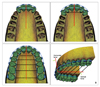
Fig 4
The sum of horizontal displacement (Δy) of four anterior teeth. A, Six anterior teeth; B, full maxillary dentition; C, varying on the position of force on the Z-axis.
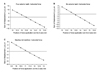
Fig 5
Contour plot of the full maxillary dentition according to the direction of horizontal retraction force. Original model (white mesh) and deformed model (color) in which the horizontal (Δy) displacement of teeth was magnified 500 times were superimposed. See the color scale bar for exact finite element analysis of analyzed horizontal teeth displacement. A, The line of force passing (z = 11 mm) through CR causes parallel tooth movement; B, the line of force passing (z = 0 mm) below CR causes counter-clockwise rotation of occlusal plane; C, the line of force passing (z = 16 mm) above CR causes clockwise rotation of the occlusal plane.
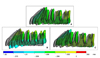
Fig 6
Standard deviation of vertical displacement (Δz) of four anterior teeth. A, Six anterior teeth; B, full maxillary dentition; C, varying on the position of force on the Y-axis.
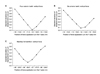
Fig 7
The contour plot of the full maxillary dentition according to the direction of vertical intrusion force. Original model (white mesh) and deformed model (color) in which the vertical (Δz) displacement of teeth was magnified 500 times were superimposed. See the color scale bar for exact finite element analysis of analyzed vertical teeth displacement. A, The line of force passing (y = -26.5 mm) through CR causes intrusion; B, the line of force passing (y = -17 mm) anteriorly to CR causes clockwise rotation of occlusal plane; C, the line of force passing (y = -35 mm) posteriorly to CR causes counter-clockwise rotation of the occlusal plane.
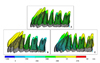
Fig 8
Position of the center of resistance. A, The center of resistance of four anterior teeth; B, the center of resistance of six anterior teeth; C, the center of resistance of the maxillary dentition.
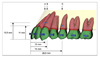
Fig 9
Comparison of the position of the center of resistance from other studies. A, 4 anterior teeth: present study (black dot); Vanden Bulcke et al11 (yellow); Pedersen et al6 (blue); Matsui et al12 (red); B, 6 anterior teeth: present study (black); Vanden Bulcke et al11 (yellow); Pedersen et al6 (blue); Choy et al7 (green); C, full maxillary dentition: present study (black); Billiet et al8 (violet).
References
1. Lee HA, Park YC. Treatment and posttreatment changes following intrusion of maxillary posterior teeth with miniscrew implants for open bite correction. Korean J Orthod. 2008. 38:31–40.

2. Kim SJ, Chun YS, Jung SH, Park SH. Three dimensional analysis of tooth movement using different types of maxillary molar distalization appliances. Korean J Orthod. 2008. 38:376–387.
3. Smith RJ, Burstone CJ. Mechanics of tooth movement. Am J Orthod. 1984. 85:294–307.
4. Reimann S, Keilig L, Jäger A, Bourauel C. Biomechanical finite-element investigation of the position of the centre of resistance of the upper incisors. Eur J Orthod. 2007. 29:219–224.
5. Lee HK, Chung KR. The vertical location of the center of resistance for maxillary six anterior teeth during retraction using three dimensional finite element analysis. Korean J Orthod. 2001. 31:425–438.
6. Pedersen E, Isidor F, Gjessing P, Andersen K. Location of centres of resistance for maxillary anterior teeth measured on human autopsy material. Eur J Orthod. 1991. 13:452–458.
7. Choy K, Kim KH, Burstone CJ. Initial changes of centres of rotation of the anterior segment in response to horizontal forces. Eur J Orthod. 2006. 28:471–474.
8. Billiet T, de Pauw G, Dermaut L. Location of the centre of resistance of the upper dentition and the nasomaxillary complex. An experimental study. Eur J Orthod. 2001. 23:263–273.
9. Woo JY, Park YC. Experimental study of the vertical location of the centers of resistance for maxillary anterior teeth during retraction using the laser reflection technique. Korean J Orthod. 1993. 23:375–390.
10. Park GH, Shon BW. The center of resistance of the maxillary anterior segment in the horizontal plane during intrusion by using laser reflection technique. Korean J Orthod. 1993. 23:619–632.
11. Vanden Bulcke MM, Burstone CJ, Sachdeva RC, Dermaut LR. Location of the center of resistance for anterior teeth during retraction using the laser reflection technique. Am J Orthod Dentofac Orthop. 1987. 91:375–384.
12. Matsui S, Caputo AA, Chaconas SJ, Kiyomura H. Center of resistance of anterior arch segment. Am J Orthod Dentofacial Orthop. 2000. 118:171–178.
13. Coolidge E. The thickness of the human periodontal membrane. J Am Dent Assoc. 1937. 24:1260–1267.
14. Kronfeld R. Histologic study of the influence of function on the human periodontal membrane. J Am Dent Assoc. 1931. 18:1942.
15. Block PL. Restorative margins and periodontal health: a new look at an old perspective. J Prosthet Dent. 1987. 57:683–689.
16. Tanne K, Sakuda M, Burstone CJ. Three-dimensional finite element analysis for stress in the periodontal tissue by orthodontic forces. Am J Orthod Dentofacial Orthop. 1987. 92:499–505.
17. Jeong HS, Moon YS, Cho YS, Lim SM, Sung SJ. Factors influencing the axes of anterior teeth during SWA en masse sliding retraction with orthodontic mini-implant anchorage: a finite element study. Korean J Orthod. 2006. 36:339–348.
18. Chung AJ, Cho JH, Kim SC, Kim US, Lee SH, Kang SS, et al. The pattern of movement and stress distribution during retraction of maxillary incisors using a 3-D finite element method. Korean J Orthod. 2007. 37:98–113.
19. Zeigler A, Keilig L, Kawarizadeh A, Jäger A, Bourauel C. Numerical simulation of the biomechanical behaviour of multi-root teeth. Eur J Orthod. 2005. 27:333–339.
20. Poppe M, Bourauel C, Jäger A. Determination of the elasticity parameters of the human periodontal ligament and the location of the center of resistance of single-rooted teeth a study of autopsy specimens and their conversion into finite element models. J Orofac Orthop. 2002. 63:358–370.
21. Andrews Lf. Straight wire, the concept and appliance. 1989. L.A.: Wells Co..
22. Germane N, Bentley BE Jr, Isaacson RJ. Three biologic variables modifying faciolingual tooth angulation by straight-wire appliannces. Am J Orthod Dentofac Orthop. 1989. 96:312–319.
23. Park CK, Yang WS. A three-dimentional finite element analysis on the location of center of resistance during intrusion of upper anterior teeth. Korean J Orthod. 1997. 27:259–272.
24. Chung KR, Oh MY, Ko SJ. Corticotomy-assisted orthodontics. J Clin Orthod. 2001. 35:331–339.
25. Sugawara J, Daimaruya T, Umemori M, Nagasaka H, Takahashi I, Kawamura H, Mitani H. Distal movement of mandibular molars in adult patients with the skeletal anchorage system. Am J Orthod Dentofacial Orthop. 2004. 125:130–138.
26. Park YC, Lee SY, Kim DH, Lee SH. Intrusion of posterior teeth using mini-screw implants. Am J Orthod Dentofacial Orthop. 2003. 123:690–694.
27. Chung KR, Kook YA, Kim SH, Mo SS, Jung JA. Class II malocclusion treatment by combining a lingual retractor and a palatal plate. Am J Orthod Dentofac Orthop. 2008. 133:112–123.
28. Chung KR, Kim SH, Kook YA. The C-orthodontic Micro-implant. J Clin Orthod. 2004. 38:478–486.
29. Min YG, Hwang CJ. A study about the change of locations of the center of resistance according to the decrease of alveolar bone heights and root lengths during anterior teeth retraction using the laser reflection technique. Korean J Orthod. 1998. 29:165–181.
30. Dykman JFP. Distribution of forces in orthodontic treatment. 1969. Nijmegan, HTe Netherlands: University of Nijmegam;[Thesis].
31. Türk T, Elekdag-Türk S, Dinçer M. Clinical evaluation of the centre of resistance of the upper incisors during retraction. Eur J Orthod. 2005. 27:196–201.
32. Lee HK, Chung KR. The vertical location of the center of resistance for maxillary six anterior theeth during retraction using three dimentional finite element analysis. Korean J Orthod. 2001. 31:425–438.
33. Bourauel C, Keilig L, Rahimi A, Reimann S, Ziegler A, Jäger A. Computer-aided analysis of the biomechanics of tooth movements. Int J Comput Dent. 2007. 10:25–40.
 XML Download