

 PDF
PDF ePub
ePub Citation
 Print
Print


Abstract
Objective:
The purpose of this study was to investigate the horizontal skeletal pattern and dental arch differences between Class III malocclusion patients and normal occlusion patients.
Methods:
Twenty skeletal Class III malocclusion patients and ten normal occlusion patients were selected and 3D facial CT were taken to analyze the horizontal skeletal differences between the two groups.
Results:
In the horizontal comparison of the maxilla, skeletal width and perimeter were significantly smaller in skeletal Class III patients on ANS and A point reference planes. The difference between maxillary width of ANS and A point reference planes showed that there was greater constriction of the first and second premolar in skeletal Class III patients. In the horizontal comparison of the mandible, the widths of the canine and premolar area were significantly larger in skeletal Class III patients on B point reference plane. The differences between width of the upper and lower jaws (comparison of A and B reference planes) were significantly large in the canine and premolar area.
REFERENCES
1.Proffit WR., Fields HW Jr., Moray LJ. Prevalence of malocclusion and orthodontic treatment need in the United States: estimates from the N-HANES III survey. Int J Adult Orthodon Orthognath Surg. 1998. 13:97–106.
2.Mills LF. Epidemiologic studies of occlusion. IV. The prevalence of malocclusion in a population of 1,455 school children. J Dent Res. 1966. 45:332–36.

3.Lee SJ., Suhr CH. Recognition of malocclusion and orthodontic treatment need of 7-18 year-old Korean adolescent. Korean J Orthod. 1994. 24:367–94.
4.Yoo YK., Kim NI., Lee HK. A study on the prevalence of malocclusion in 2,378 Yonsei university students. Korean J Orthod. 1971. 2:35–40.
5.Angle EH. Classification of malocclusion. Dental Cosmos. 1899. 41:248–64.
6.Broadbent BH. A new X-ray technique and its application to orthodontia. Angle Orthod. 1931. 1:45–66.
7.Park SJ., Ryu YK. A comparative study on craniofacial skeleton between Angle's class III malocclusion and normal occlusion. Korean J Orthod. 1987. 17:63–71.
8.Kim JH., Suhr CH. A roentgenocephalometric study on morphologic factors of normal occlusion and class III malocclusion. Korean J Orthod. 1987. 17:23–32.
9.Jacobson A., Evans WG., Preston CB., Sadowsky PL. Mandibular prognathism. Am J Orthod. 1974. 66:140–71.
10.Sanborn RT. Differences between the facial skeletal patterns of Class III malocclusion and normal occlusion. Angle Orthod. 1955. 25:208–22.
11.Sassouni V. A classification of skeletal facial types. Am J Orthod. 1969. 55:109–23.
12.Lee SJ., Baek SH., Kim SC., Kook YA. Size and forms of the mandibular dental arch in Korean malocclusion patients. Korean J Orthod. 2005. 35:15–22.
13.Lee HK., Son WS. A study on basal dental arch width in skeletal class III malocclusion. Korean J Orthod. 2002. 32:117–27.
14.Lee BD. Application of three-dimensional CT in dentistry. J Korean Dent Association. 2002. 40:853–9.
15.Kim KD., Ryu SK. Application of three-dimensional CT images using personal computer in dentistry. J Korean Dent Association. 2004. 42:197–208.
16.Jeon KJ., Park H., Lee HC., Kim KD., Park CS. Reproducibilities of cephalometric measurements of three-dimensional CT images reconstructed in the personal computer. Korean J Oral Maxillofac Radiol. 2003. 33:171–8.
17.Cavalcanti MG., Haller JW., Vannier MW. Three-dimensional computed tomography landmark measurement in craniofacial surgical planning: experimental validation in vitro. J Oral Maxillofac Surg. 1999. 57:690–4.
18.Williams DR. Maxillary growth velocity and variation in three dimensions during treatment of Class III malocclusion. Angle Orthod. 1973. 43:422–39.
19.Choi YS., Kim GT., Hwang EH. Basic principle of cone beam computed tomography. Korean J Oral Maxillofac Radiol. 2006. 36:123–9.
20.Chang HS., Baik HS. A proposal of landmarks for craniofacial analysis using three-dimensional CT imaging. Korean J Orthod. 2002. 32:313–25.
21.Kim WS., Lee KH., Hwang HS. Comparison of asymmetric degree between maxillofacial hard and soft tissue in facial asymmetric sugjects using three-dimensional computed tomography. Korean J Orthod. 2005. 35:163–73.
22.Kwon TG., Park HS., Ryoo HM., Lee SH. A comparison of craniofacial morphology in patients with and without facial asymmetry - a three dimensional analysis with computed tomography. Int J Oral Maxillofac Surg. 2006. 35:43–8.
23.Park JW., Kim NK., Chang YI. Formulation of a reference coordinate system of three-dimensional (3D) head and neck images: Part I. Reproducibility of 3D cephalometric landmarks. Korean J Orthod. 2005. 35:388–97.
24.Waitzman AA., Posnick JC., Armstrong DC., Pron GE. Craniofacial skeletal measurements based on computed tomography: Part II. Normal values and growth trends. Cleft Palate Craniofac J. 1992. 29:118–28.
25.Xia J., Wang D., Samman N., Yeung RW., Tideman H. Computer-assisted three-dimensional surgical planning and simulation: 3D color facial model generation. Int J Oral Maxillofac Surg. 2000. 29:2–10.
26.Xia J., Ip HH., Samman N., Wang D., Kot CS., Yeung RW, et al. Computer-assisted three-dimensional surgical planning and simulation: 3D virtual osteotomy. Int J Oral Maxillofac Surg. 2000. 29:11–7.
27.Xia J., Samman N., Yeung RW., Wang D., Shen SG., Ip HH, et al. Computer-assisted three-dimensional surgical planing and simulation. 3D soft tissue planning and prediction. Int J Oral Maxillofac Surg. 2000. 29:250–8.
28.Brodie AG. The apical base; zone of interaction between the intestitial and skeletal system. Angle Orthod. 1966. 36:131–5.
29.Lagerstrom LO., Brodie AG. A quantitative method for measuring change in the maxilla due to growth and orthodontic procedures. Angle Orthod. 1967. 37:241–50.
30.Lude JC. The technique for the determination of the size of the mandibular apical base; it's application to growth studies. Angle Orthod. 1967. 37:272–84.
31.Ahn HS., Cha KS. A study on maxillary basal bone morphology in skeletal class III malocclusion requiring orthognathic surgery. Korean J Orthod. 1995. 25:577–86.
32.Worms FW., Issacson RJ., Spedal TM. Surgical orthodontic treatment planning: profile analysis and mandibular surgery. Angle Orthod. 1976. 46:1–25.
Fig 1.
Reference plane. 1, Maxillary midsagittal plane (Na-ANS-Ba); 2, ANS horizontal plane (A plane passing ANS perpedicular to Maxillary midsagittal plane); 3, A point horizontal plane (A plane passing A point parallel to ANS horizontal plane); 4, B point horizontal plane (B point-Gonion Lt.-Gonion Rt.); 5, antegonion horizontal plane (A plane passing center of gonion parallel to B point horizontal plane).
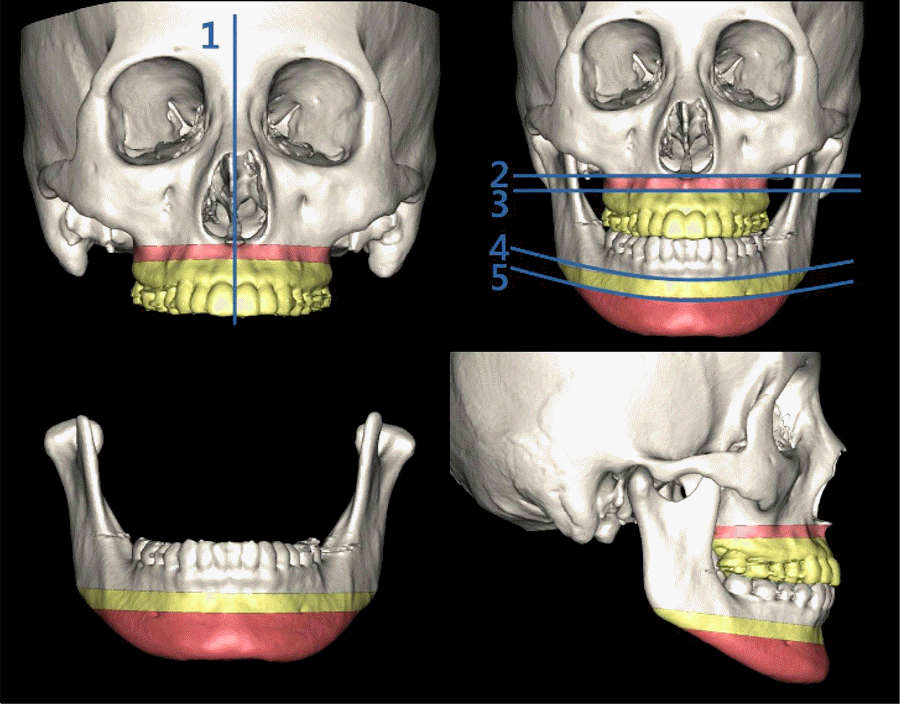
Fig 2.
Maxillary reference plane. 5 reference planes (A) perpendicular to ANS horizontal plane and reference points (B, C) for linear measurement.
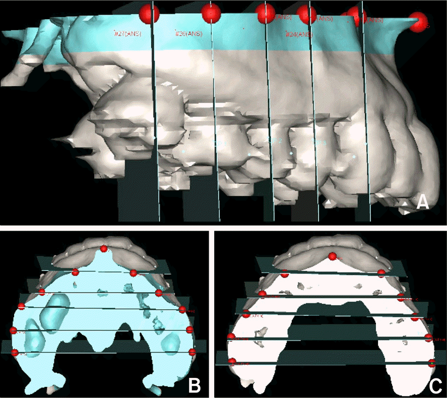
Fig 3.
Mandibular reference plane. 5 reference planes (A) perpendicular to B point horizontal plane and reference points (B, C) for linear measurement.
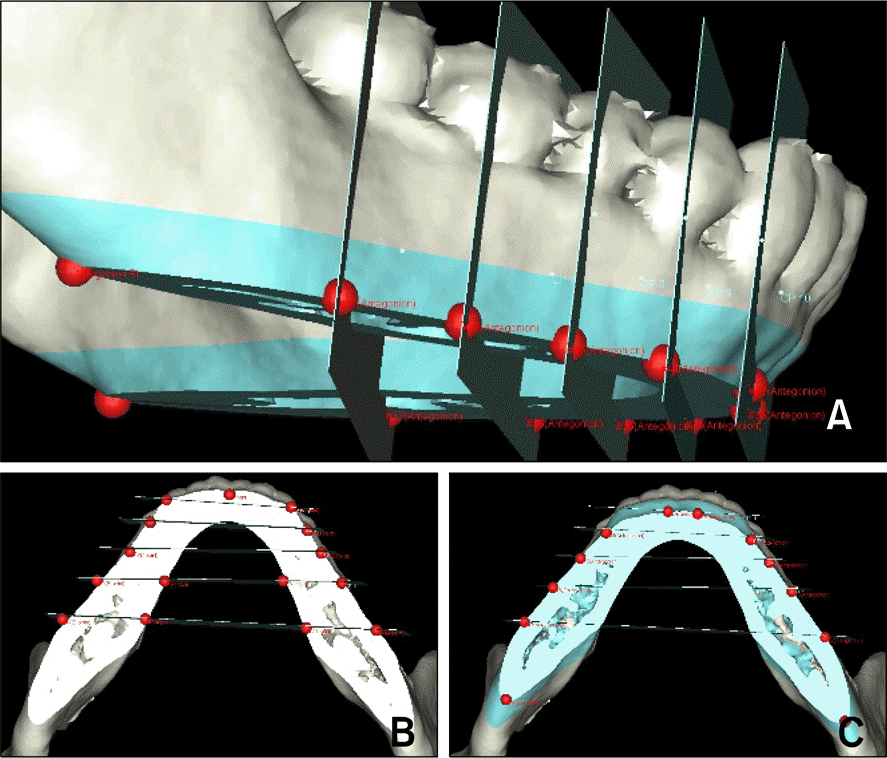
Fig 4.
ANS (A) and A point (B) horizontal plane and maxillary measurements. The landmarks are described in Table 1.
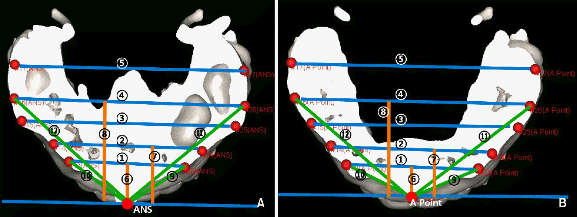
Fig 5.
B point (A) and antegonion (B) horizontal plane and mandibular measurements. The landmarks are described in Table 2.
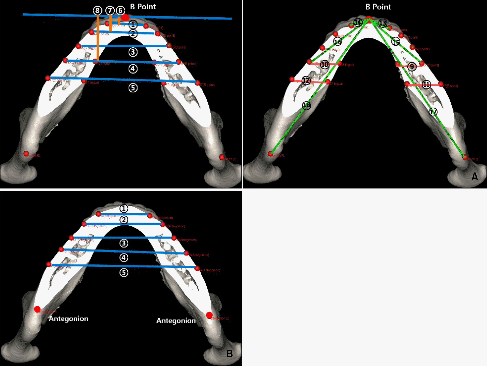
Fig 6.
The coordinates in A point reference plane (A) and superimposition (B), Class III group is red and normal group is blue.
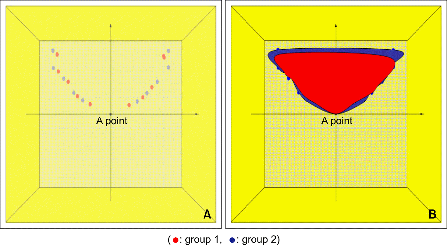
Fig 7.
The coordinates in B point reference plane (A) and superimposition (B), Class III group is red and normal group is blue.
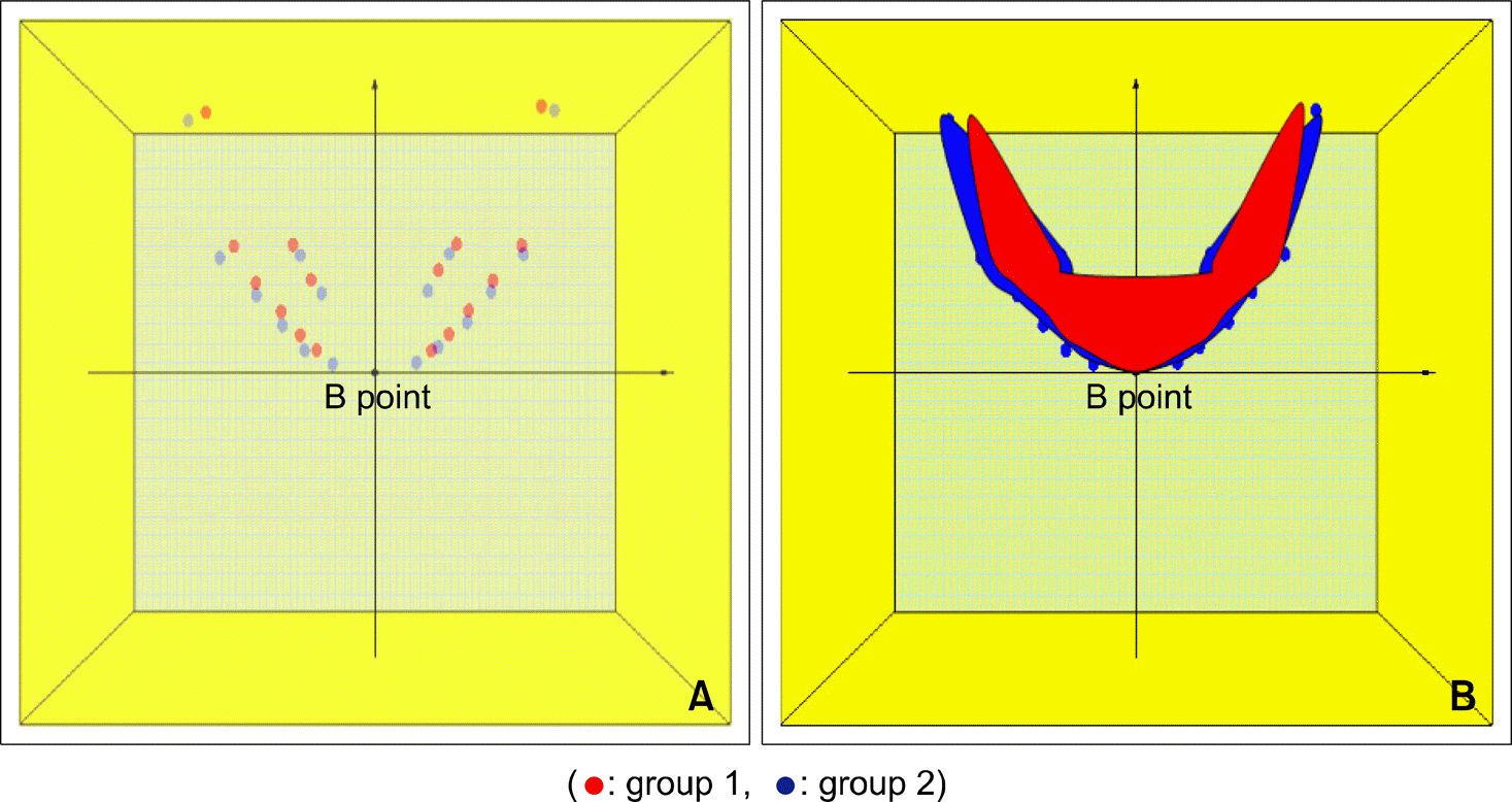
Table 1.
Maxillary measurements
Table 2.
Mandibular measurements
Table 3.
Transverse comparison of basal bone in ANS horizontal plane
Table 4.
Transverse comparison of basal bone in A point horizontal plane
Table 5.
Basal bone width difference between ANS, A point horizontal plane (bone width on ANS plane-bone width on A point plane)
Table 6.
Transverse comparison in B point horizontal plane
Table 7.
Transverse comparison in antegonion horizontal plane
Table 8.
Basal bone width difference between A point, B point horizontal plane (Bone width on A point plane-bone width on B point plane)
 XML Download