

 PDF
PDF ePub
ePub Citation
 Print
Print


Dear Editor,
Abducens nerve palsy is the most common type of ocular nerve palsy to occur in isolation, and it has several associated etiologies.1 It is important to identify the causative lesion to determine the etiology of abducens nerve palsy and optimize the treatment. In this context, magnetic resonance imaging (MRI) plays an important role in the management of abducens nerve palsy.2 In rare cases, pathology involving Dorello's canal, which is the osteofibrous canal in the skull base that includes the abducens nerve and the inferior petrosal sinus (IPS), can cause abducens nerve palsy; however, this is often overlooked.3 Here, we present a rare case of a left transverse-sigmoid sinus dural arteriovenous fistula (dAVF) with drainage into the left IPS that presented with isolated left abducens nerve palsy presumably caused by venous hypertension in the left IPS within Dorello's canal.
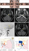
A previously healthy 64-year-old woman was referred to our hospital due to acute-onset painless diplopia without any associated symptoms, which exacerbated over the course of 1 week. A neurological examination revealed impaired abduction of the left eye with no other abnormal findings (Fig. 1A), which suggested isolated left abducens nerve palsy. Contrast-enhanced MRI did not reveal any causative tumor lesions or inflammatory lesions. However, time-of-flight magnetic resonance angiography (TOF-MRA) revealed venous enhancement of the left IPS at the left Dorello's canal, indicating the presence of rapid retrograde venous drainage into the left IPS (Fig. 1B and C). Subsequent cerebral angiography confirmed a left transverse-sigmoid sinus dAVF allowing retrograde venous drainage into the left IPS (Fig. 1D). Embolization of the dAVF alone resolved the enhancement of the left IPS at the left Dorello's canal in TOF-MRA (Fig. 1E) and, subsequently, the left abducens palsy.
Dorello's canal, which was first described by Wenzel Leopold Gruber in 1859 and later by Primo Dorello in 1905, is the osteofibrous canal in the skull base located under Gruber's ligament.4 Dorello's canal is bordered by Gruber's ligament superiorly, the dorsum sellae of the sphenoid bone medially, the clivus of the occipital bone inferiorly, and the petrous apex of the temporal bone laterally5 (Fig. 1F). Since the abducens nerve runs alongside the IPS within the canal4 (Fig. 1G), we should pay attention to Dorello's canal in cases of abducens nerve palsy. A dAVF is an abnormal arteriovenous shunt between the dural arteries and dural venous sinuses or meningeal veins, and presents with various symptoms caused by venous hypertension of the vein involved in the drainage.6 Based on the clinical course and radiographical findings in the present case, we believe that venous hypertension due to retrograde venous drainage into the IPS caused by the dAVF damaged the abducens nerve in Dorello's canal. Isolated abducens nerve palsy due to compression of the abducens nerve by the IPS in Dorello's canal is extremely rare, but has been reported in patients with IPS thrombosis6 and in those undergoing IPS sampling as part of a workup for Cushing's disease.7 Neurologists should pay careful attention to Dorello's canal when evaluating patients with isolated abducens nerve palsy, and investigating this canal using MRI with detailed anatomical knowledge of the canal is particularly important.
 XML Download