

 PDF
PDF ePub
ePub Citation
 Print
Print


Dear Editor,
Mutations of ANO3 have recently been found to cause isolated dystonia (DYT24, MIM 615034),1 predominantly involving the craniocervical area, with the age at onset ranging from childhood to the sixth decade of life.23 This report describes a Korean patient with segmental dystonia caused by a novel variant c.860G>A (p.Arg287Gln) in ANO3 (NM_031418.3 and NP_113606.2), with brain PET showing basal ganglia (BG) dysfunction.
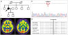
A 60-year-old left-handed woman presented with abnormal posture in her left arm and the neck that had first appeared 6 years previously. She had not taken any medication that can cause dystonia, and there was no family history of tremor or dystonia (Fig. 1A). A neurological examination showed an abnormal posture with slow and large-amplitude jerky tremors in her left arm. She also exhibited mild left torticollis combined with retrocollis arm (Supplementary Video 1 in the online-only Data Supplement). Brain MRI produced normal findings. Brain 18F-fluorodeoxyglucose (18F-FDG) PET showed increased uptake in the right putamen, with decreased uptake in the right subthalamic nucleus (STN) (Fig. 1B). Next-generation sequencing using a targeted panel of 93 genes for dystonia identified a novel heterozygous missense variant (a G to A substitution at position 860 of ANO3; transcript ID: ENST00000256737) that resulted in the replacement of an arginine residue by a glutamine residue at position 287 of the anoctamin 3 protein (p.Arg287Gln). This variant was confirmed by Sanger sequencing (Fig. 1C). The arginine residue at codon 276 is in the cytoplasmic domain of ANO3, which is highly conserved throughout evolution (Fig. 1D). The in silico prediction algorithms UMD-Predictor (http://umd-predictor.eu/) and MutationTaster2 (http://www.mutationtaster.org/) predicted that this missense mutation was pathogenic and disease-causing, respectively. The Combined Annotation-Dependent Depletion tool (CADD, version 1.3; http://cadd.gs.washington.edu/) produced a score of 34.0. No mutations were detected in other genes known to cause dystonia.
The guidelines of the American College of Medical Genetics and Genomics classify p.Arg287Gln of ANO3 as a variant of uncertain significance. The ANO3 variant in the present patient is possibly causative for DYT24 for the following reasons: First, the patient presented with segmental dystonia similar to findings in previous published dystonia cases with ANO3 mutations (Supplementary Table 1 in the online-only Data Supplement). Second, in silico analyses of this novel variant have predicted that this mutation is pathogenic. To date, 17 missense mutations and 1 in-frame insertion mutation in ANO3 have been reported as possible variants causing DYT24 (Supplementary Table 1 in the online-only Data Supplement). The CADD score of p.Arg276Gln is equal to the highest score of previously reported mutations. Third, this mutation has not been reported in patients with dystonia or in large public databases such as gnomAD, ExAC, and 1,000 Genomes Project.
18F-FDG-PET assessments of patients with hereditary dystonia have shown changes (increases or decreases) in glucose metabolism in the BG, cortices, and cerebellum.4 The altered glucose metabolism in the right putamen and STN in the present patient may reflect changes within the BG motor circuit. ANO3 encodes a calcium (Ca2+)-activated chloride channel that is highly expressed in the striatum, especially in the putamen, and is thought to play a role in modulating neuronal excitability.1 If abnormal calcium homeostasis is a disease mechanism in dystonia,5 then abnormal glucose metabolism in the BG of the present patient may explain the pathophysiological mechanisms underlying the development of dystonia in patients with ANO3 mutations.1 We did not detect any abnormality of glucose metabolism in the cortices and cerebellum in the present patient, because the 18F-FDG uptake was symmetrical in those regions. Additional studies using group comparisons such as network analysis are needed to confirm the exact topography of metabolic abnormalities revealed by 18F-FDG-PET in this type of dystonia.
We have described a Korean patient with segmental dystonia who had a novel ANO3 variant (c.860G>A) and abnormal glucose metabolism in the BG.

 XML Download