

 PDF
PDF ePub
ePub Citation
 Print
Print


Dear Editor,
Giant-cell arteritis (GCA), also known as temporal arteritis, is a primary systemic autoimmune disease characterized by infiltration of moderate-size and large blood vessels.1 GCA is the most common primary vasculitis in adults older than 50 years, and GCA patients commonly experience cranial or systemic symptoms, including unilateral or bilateral headache, scalp tenderness, weight loss, fatigue, and fever.2 An abrupt-onset headache is the main symptom in more than two-thirds of GCA patients.3 Severe headache often occurs on the side of the affected artery. Irreversible vision loss occurs in up to 20% of GCA patients, and this is considered a medical emergency.4 Arteritic anterior ischemic optic neuropathy (AAION) is the most serious ophthalmological complication of GCA.2 Here we report the case of a patient with unilateral headache attributed to GCA and contralateral AAION.
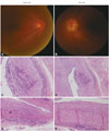
A 74-year-old man was referred with new-onset headache with a 1-month history in the right temporal area, with systemic manifestations including right jaw claudication, malaise, scalp tenderness, and loss of appetite. Laboratory findings revealed a white blood cell count of 11.67×10³/µL with a slight elevation of neutrophils and platelets at 320×103/µL. The erythrocyte sedimentation rate (ESR) and C-reactive protein (CRP) had increased to 49 mm/hr and 3.85 mg/dL, respectively. Ultrasonography showed thickening of the right superficial temporal artery, but brain MRI and angiography revealed no significant abnormalities. On the second day after the daily administration of 60 mg of oral prednisone, his left eye visual acuity deteriorated rapidly, although the headache was significantly improved. An ophthalmological examination indicated intact right eye visual acuity but significant impairments in the left eye, with only finger counting possible. A relative afferent pupillary defect was detected in the left eye. The intraocular pressure was 11 mm Hg in the right eye and 15 mm Hg in the left eye. A fundus examination confirmed the presence of papilledema (Frisen grade 2) in the left eye (Fig. 1A and B). Fluorescein angiography showed that the choroidal blood flow in the left eye was decreased. Intravenous methylprednisolone was administered at 1,000 mg daily for 5 days and both temporal arteries were biopsied. Intimal thickening and perivascular lymphohistiocytic inflammatory infiltration were evident in both superficial temporal arteries (Fig. 1C–F). After administering 500 mg of cyclophosphamide intravenously, the optic disc swelling was reduced and the left eye visual acuity was slightly improved. Inflammatory markers including ESR and CRP returned to normal levels.
The pathophysiology of GCA involves inflammation, myointimal thickening, and stenosis of moderate-size to large vessels. Stenosis may result in complete occlusion or turbulent flow that leads to thrombosis. GCA is, in essence, a form of systemic arteritis, and biopsy studies of bilateral temporal arteries have had a reported concordance rate of 96% (93–99%).5 Bilateral simultaneous biopsy of the temporal arteries has been considered a safe gold standard for diagnosing GCA.6 Vision loss is an emergency and potentially fatal complication of GCA that requires rapid treatment.1 Vision loss can occur as a result of occlusion of a short posterior ciliary artery, which can cause infarction of the optic nerve head, leading to AAION. Vision loss can also rapidly progress to the unaffected eye if not treated.47 This suggests that even without symptoms, it is likely that inflammation has progressed significantly from both sides. A particularly interesting feature of our patient was that he presented with headache in the right temporal area and sudden vision loss in the left eye, while histopathology revealed definite temporal arteritis bilaterally. AAION might therefore be evoked from either side of temporal arteritis regardless of the location of the headache. Our case provides evidence that GCA is a systemic and site-nonspecific neurological disorder. Thus, if headache due to GCA is suspected in an elderly patient, the clinician must perform an extensive systemic evaluation including a bilateral ophthalmological examination even when the symptoms are confined to one side.
 XML Download