

 PDF
PDF ePub
ePub Citation
 Print
Print


Introduction
Hereditary spastic paraplegia (HSP) is a genetically heterogeneous group of neurodegenerative disorders that are characterized by progressive spasticity and weakness of the lower limbs.1 Clinically, HSP can be classified as either "pure" or "complicated" depending on the presence of additional features such as ataxia, extrapyramidal signs, severe amyotrophy, peripheral neuropathy, optic atrophy, pigmentary retinopathy, mental retardation, dementia, and epilepsy.1,2
Hereditary spastic paraplegia can be inherited as autosomal dominant (AD, AD-HSP), autosomal recessive, or an X-linked trait, and at least 52 loci have been mapped and 31 genes identified to date.3 Mutations in the spastin gene (SPAST, SPG4) are the most common causes of HSP, accounting for up to 40-67% of AD-HSP cases and 12-18% of sporadic cases.4,5,6,7 Mutations in the atlastin-1 gene (ATL1, SPG3A) and receptor expression-enhancing protein 1 gene (REEP1, SPG31) are the second and the third most common causes of AD-HSP, respectively.1 More than half of all clinically diagnosed AD-HSP cases result from mutations in these three genes.8 Therefore, mutation analysis of these genes will allow more-focused and cost-effective investigations of potential cases of HSP.
The frequency of SPG4 is higher and the incidence of SPG3A is lower in Koreans than in other ethnic groups,9,10,11,12,13 which might be attributable to an ethnic difference in the genetic background of Korean patients with HSP. However, this needs to be confirmed in a larger series. No previous study has performed mutation screening of REEP1 in Korean patients with HSP. The present study performed mutation analysis of SPAST, ATL1, and REEP1 in 27 unrelated Korean patients with pure and complicated HSP in order to assess the role of these three genes in the occurrence of HSP in a Korean population.
Methods
In total, 27 unrelated Korean probands with HSP were included in this study. All of these patients were evaluated neurologically and genetically after giving informed consent. This study was reviewed and approved by the Institutional Review Board of Pusan National University Yangsan Hospital. HSP was diagnosed by qualified neurologists on the basis of Harding's criteria.14 Patients with other neurological conditions were excluded based on the clinical, radiological, and biochemical findings.
Clinical parameters included age at onset, family history, and the presence of hyperreflexia, spasticity, weakness, and sensory abnormalities. Additional neurological signs that are suggestive of complicated cases, as mentioned above, were also described. Disability stage was assessed on the 5-point scale described by Fonknechten et al.15
Direct sequence analyses of whole coding regions of SPAST, ATL1, and REEP1 were performed in order to identify mutations. Sequence-specific primer pairs covering the entire coding region of all three genes were used, as described elsewhere16,17,18 with minor modifications (available upon request). Polymerase chain reaction (PCR) was performed in a Gene Atlas Thermal Cycler (ASTEC, Seoul, Korea). PCR-amplified products were separated and purified using 2% agarose gels and SolGent Agarose Gel Extraction Kits (SolGent, Daejeon, Korea), cycle-sequenced with PCR primers using the BigDye Terminator Sequencing Kit (Applied Biosystems, Foster, CA, USA), and electrophoresed using an ABI PRISM 3730XL DNA analyzer (Applied Biosystems, Foster, CA, USA).
To confirm the pathogenicity of novel mutations identified in the sequence analysis, and to differentiate them from benign polymorphisms, PCR and restriction fragment length polymorphism (PCR-RFLP) analysis were performed using DNA from the included patients and 100 normal controls. Multiple sequence alignment of spastin orthologs was also conducted to evaluate two novel missense mutations and a novel in-frame deletion mutation for evolutionary conserved residues in the protein. Genomic and mRNA reference sequences of the three genes (SPAST: NG_008730.1, NM_014946.3; ATL1: NG_009028.1, NM_015915.4; and REEP1: NG_013037.1, NM_001164730.1) were used to describe the sequence variants.
Among patients in whom direct sequencing analysis did not identify mutations, multiplex ligation-dependent probe amplification (MLPA) was performed to detect copy-number variations of the three genes. MLPA reactions were performed according to the manufacturer's protocol using MLPA kit P165 (SPAST, ATL1) and P213 (REEP1) from MRC-Holland (Amsterdam, The Netherlands).
Statistical significance was assessed using nonparametric tests (the Mann-Whitney U test and Fisher's exact test) to establish any associations between the presence of a SPAST mutation and particular clinical parameters in patients with pure HSP. Spearman's rank correlation coefficient was used to measure the statistical significance between disease duration and disability score. All statistical analyses were performed using SPSS (version 18.0, SPSS Inc., Chicago, IL, USA).
Results
The patients' clinical data are given in Supplementary Table 1. The age of the patients ranged from 14 to 64 years (mean±SD=36.6±13.4 years); 16 patients were male and 11 were female. Fifteen of the 27 probands exhibited an AD inheritance pattern, and 9 appeared to be sporadic. The exact inheritance pattern could not be determined in three patients. The age at onset varied widely from 1 to 59 years (24.8±14.5 years). Nineteen and 8 patients were classified as pure and complicated HSP, respectively. The complicated forms included peripheral neuropathy on nerve conduction studies (n=2), mental retardation (n=3), epilepsy (n=1), cerebellar ataxia (n=1), saccadic pursuit (n=1), and dysarthria (n=6). Most of the complicated HSP patients had more than one additional feature.
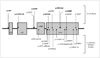
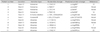
Ten different mutations of SPAST-comprising 4 missense, 4 nonsense, 1 in-frame deletion, and 1 frameshift mutations-were identified in 11 probands (Table 1, Fig. 1). Six of these (c.760A>T, c.131C>A, c.1351_1353delAGA, c.376_377dupTA, c.1114A>G, and c.1372A>C) were novel, and the others (c.734C>G, c.1496G>A, c.1741C>T, and c.1196C>T) have been described previously.7,16,19,20 Two novel missense mutations and one in-frame deletion mutation (c.1114A>G, c.1372A>C, and c.1351_1353delAGA) were located in the ATPases Associated with a wide variety of Activities (AAA) cassette domain. Two novel nonsense mutations and one frameshift mutation led to a premature termination codon, resulting in the production of a truncated protein. None of the mutation variants were found in 100 normal controls in PCR-RFLP analysis. No mutations in ATL1 and REEP1 were found, and no copy-number variants were detected among all three genes.
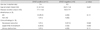
SPAST mutations were present in 66.7% (10/15) of the AD-HSP patients and 57.9% (11/19) of those with pure HSP. All of the SPAST mutations were associated with pure HSP. The clinical features of patients with pure HSP with SPAST mutations were compared to those without mutations (Table 2). In the SPAST-mutation-positive group, although most patients (90.9%, 10/11) had an AD inheritance pattern, the correlation was not statistically significant (p=0.111). Furthermore, various clinical parameters did not differ significantly between the SPAST-mutation-positive and -negative groups, and there was no significant correlation between disease duration and disability stage (Spearman's rho=0.239, p=0.324).
Discussion
This study identified SPAST mutations in ten AD-HSP patients and one sporadic HSP patient among 27 unrelated pure and complicated HSP probands. Six of these mutations have not been described previously. The frequency of SPAST mutations in our AD-HSP patients (66.7%) is higher than those reported previously (range, 18-42%).15,21,22,23 Park et al.7 also reported a similar frequency of SPAST mutations in Korean patients with uncomplicated AD-HSP. However, they did not perform MLPA to identify exon deletions or duplications. According to previous studies, exon deletions of SPAST are not rare and have been found in 18% and 20% of the point-mutation-negative patients.24,25 However, in our patients MLPA analysis of SPAST revealed no copy-number variants.
Mutations of ATL1 and REEP1 are relatively common in other ethnic groups and have been variously identified in up to 9% and 6.5% of HSP patients.13,26 Copy-number variations in ATL1 and REEP1 have also been reported.27,28,29,30 However, we did not identify any pathogenic mutations in ATL1 and REEP1. To date, only one Korean family with a missense mutation in ATL1 has been reported. These results suggest that SPAST mutations are responsible for most cases of genetically confirmed AD-HSP in Korean patients. The relatively high rate of SPAST mutations and the lower incidence of ATL1 and REEP1 mutations among Korean HSP patients imply the existence of ethnic differences in the subtype of HSP. However, our observation of the absence of ATL1 and REEP1 mutations needs to be confirmed in larger series.
Previous studies have demonstrated that most SPAST mutations are located in the AAA cassette domain, regardless of ethnicity.15,16,21,22,23,31,32 Six of the causative mutations identified in this study are located in the AAA cassette domain. It is remarkable that three of the nonsense mutations and the single frameshift mutation in our patients were outside of this domain. All of these mutations are predicted to affect the AAA cassette domain indirectly. Nonsense and frameshift mutations are expected to cause premature truncation before and within the AAA cassette domain, thereby producing dysfunctional spastin.
Mutations in SPAST, ATL1, and REEP1 are predominantly associated with pure forms of AD-HSP.1,2 However, some of them may have other neurologic deficits seen in complicated HSP.1,2 They can also appear sporadically due to low penetrance, de-novo mutations, premature death of transmitting parents, or underrecognition of family history.2 All of the SPAST mutations in our patients were associated with pure HSP. Most patients with SPAST mutations belonged to AD-HSP. The low incidence of SPAST mutations in the sporadic cases is consistent with the findings of previous studies.19,21 Among various clinical parameters of pure HSP, no significant correlation was found with the presence of a SPAST mutation in this study. Previous studies have found the onset age to be lower in SPAST-mutation-negative groups,32,33 and another study found that the disease progression was faster in patients with late-onset SPAST HSP than in those with early-onset HSP.15 In addition, wheelchair use and abnormal vibration sense in the lower limbs were more common in a SPAST-mutation-positive group.34 Further studies with larger numbers of patients are needed to elucidate the relationship between SPAST mutations and clinical phenotypes of Korean patients with HSP.
In conclusion, we report herein a mutational analysis of SPAST, ATL, and REEP1 in Korean patients with HSP. SPAST mutations were the found in most of the genetically confirmed AD-HSP patients. The findings of this study highlight the importance of SPAST mutation screening among Korean patients.
 XML Download