

 PDF
PDF ePub
ePub Citation
 Print
Print


Introduction
"When you have eliminated all which is impossible, then whatever remains, however improbable, must be the truth."
Arthur Conan Doyle, The Case-Book of Sherlock Holmes
Amyotrophic lateral sclerosis (ALS) is a universally fatal, neurodegenerative disease, characterized by cell death involving both upper motor neurons (UMNs) and lower motor neurons (LMNs) and subsequent catastrophic weakness of all voluntary muscles involving bulbar, limb, and respiratory regions.1 ALS seems to be a predominantly 'negative' disease, in that its clinical symptoms and signs develop from motor system destruction (death of UMNs and LMNs). However, ALS also exhibits unique 'positive' phenomena of hyperexcitability involving the motor system, specifically fasciculation, cramp, hyperreflexia, and spasticity. Despite some controversy as to whether hyperexcitability is a trigger of the disease or only a transient phenomenon that appears during the disease course, hyperexcitability is almost always encountered during the ALS disease course.2
At the time of the original description of ALS by Charcot, there were no in vivo tools for measuring the excitability of the human nervous system. Recently novel electrophysiological techniques for measuring motor networks were developed to enable evaluation of this relatively unappreciated issue in clinical neurology. Using these techniques, clinical neuroscientists may enhance understanding of the central and peripheral mechanisms associated with the development and manifestations of hyperexcitability in ALS. Additionally, in the clinical field, these techniques may yet be further developed as a diagnostic biomarker, that may predict prognosis in ALS. As such, the present Review will focus on the puzzling issue of the hyperexcitability in ALS and, similar to Conan Doyle's Sherlock Holmes, will attempt to dissect the clinical and pathophysiological determinants and thereby implications for ALS research.
What Really is Hyperexcitability in ALS?
The term 'hyperexcitability' may be considered more as an electrophysiological term rather than common clinical parlance. Put similarly, 'excitability' may be defined as the "capacity to be activated by or react to a stimulus".3 Thus, hyperexcitability means an increased or exaggerated response to a stimulus, which may usually have been expected to evoke a normal response. In ALS, a disease of both UMNs and LMNs, hyperexcitability denotes unique clinical and electrophysiological phenomena which may arise independently from the corticomotoneurons (CMs) and LMN.
Fasciculation and cramp are commonly observed clinical features in ALS and are considered to be representative features of LMN hyperexcitability.4 Of these, fasciculation, or a fasciculation potential in electromyography (EMG), has long been considered a clinical or electrophysiological hallmark of ALS.5 However fasciculation may be detected in various diseases of motor neurons or peripheral nerves, and sometimes even in healthy subjects.6,7 Perhaps more intriguingly, there has also been the suggestion that some fasciculations may originate from lesions involving the central nervous system (CNS).8-11 More recently, the identification of fasciculations in ALS have been in incorporated into diagnostic criteria aimed at improving the diagnostic certainty of ALS, the Awaji-Shima criteria.12
Identification of UMN hyperexcitability in ALS in the clinic always seems rather challenging. Irrespective of LMN pathology in ALS, which can give rise to various degrees of muscle wasting and weakness, hyperexcitability may seem closely related with the pathophysiological abnormalities that follow damage to descending motor pathways and to motor neurons and interneurons in the ventral horns of the spinal cord.5,13 For example, deep tendon reflexes (DTRs) in UMN lesions are typically increased (brisk), sometimes with clonus, representing unconstrained activity in the feedback loop to the hyperexcitable anterior horn cells from the Ia afferents imposed during sudden muscle stretching. In ALS, DTRs may be decreased when there is marked LMN weakness involving the muscles activated by the respective DTRs. However the H-reflex, an electrophysiological equivalent of DTRs, can sometimes be elicited from weak or wasted muscles such as intrinsic hand muscles, in contrast to difficulties in demonstrating this response in healthy 3 subjects.14 Presynaptic inhibition of the group Ia synapse on motor neurons has been found to be significantly reduced in ALS,15 a finding not explained by motor neuron dropout in the disease, but likely to be due to loss of interneurons at the spinal level.
How Can We Objectively Assess Hyperexcitability in ALS?
In some ALS patients, fasciculations and cramps precede the disease by many months, well prior to the development of muscular weakness.16 Cramps can occur in unusual regions, such as in the abdomen and hands. Fasciculations are sometimes persistent, diffuse, and readily observed. This may suggest that early identification of a hyperexcitable motor system might help in the predication of the development of ALS and as such, may provide a chance to make some early "neuroprotective" intervention early in the disease process.17 Such findings may also argue for need to develop sensitive and objective tools for the early detection of hyperexcitability in ALS.
Using EMG to evaluate the LMN system, Krarup18 conducted a longitudinal study in ALS and observed that denervation was more common in weak muscles; fasciculation potentials were more frequent in strong and proximal limb muscles; and muscles with normal strength usually had fasciculation potentials and neurogenic motor unit potentials (chronic partial denervation with signs of reinnervation). According to this study, fasciculation, a feature of LMN hyperexcitability, occurred mainly in less-involved or compensated regions, typically during the early stages of the disease. In turn, these observations may suggest that, clinically, hyperexcitability may be overlooked frequently during the early stages of ALS because of the lack of overtness of the developing phenotype.
Recently, two major advances have been introduced in the field of in vivo assessments of excitability in the human motor system: transcranial magnetic stimulation (TMS) and nerve excitability test (NET). TMS provides a method of stimulating the cerebral cortex and has revealed hyperexcitability of CMs.19,20 Automated NET,21 using similar stimulating and recording techniques to a conventional nerve conduction study, enables in vivo tracking of axonal excitability as either an independent measurement or various consequent changes to the conditioning manipulation. With these two major advances, we can now identify and measure both the central and peripheral excitability of the motor system in normals and in neurological disease states.
What is Origin of the Hyperexcitability in ALS?
Although there has been debate regarding the origin of ALS,22-24 there is now increasing support to the theory that hyperexcitability develops due to the cortical origins of ALS. Hyperexcitability in ALS seems to develop in a manner closely related to the disrupted integrity between two different parts of the human neocortex, the projection neurons (CMs, excitatory) and the interneurons (inhibitory). Although recent research has provided roles in ALS pathophysiology for other elements in the neocortex, such as glial cells,25 several studies support the concept that an imbalance between γ-aminobutyric acid (GABA)ergic interneurons and glutamatergic CMs in ALS causes the increased cortical excitability, particulary in the early stages of the disease.
Role of glutamate
Glutamate excitotoxicity and subsequent motor neuronal death has been increasingly recognized as an important pathophysiological factor in ALS, although the specific mechanisms underlying the selective degeneration of motor neurons in this disease remain unknown. Additionally, it remains unknown whether it is essential or an incidental bystander in the final disease.26,27 Glutamate is the major excitatory neurotransmitter in the CNS, and binds to ionotropic N-methyl-D-aspartate receptors and α-amino-3-hydroxy-5-methyl-4-isoxazoleproprionic acid receptors on postsynaptic membranes.28,29 Excessive activation of these postsynaptic receptors by glutamate, a process known as glutamate-induced excitotoxicity, can incite neurodegeneration through activation of calciumdependent enzymatic pathways.30,31 Glutamate-induced excitotoxicity can also result in the generation of free radicals, which, in turn, may cause neurodegeneration by damaging intracellular organelles and up-regulating proinflammatory mediators.32,33
Role of GABAergic interneurons
In the neocortex, GABAergic, inhibitory interneurons are morphologically distinct from excitatory pyramidal cells, and account for 20-25% of all neocortical neurons. GABAergic neocortical interneurons are interconnected by gap junctions. Most often, coupling is bidirectional and occurs between interneurons of the same type. In the mammalian brain, where interneuronal communication is based mainly on chemical synapses, electrical synapses between GABAergic interneurons have only been demonstrated in a limited number of regions of the adult brain (cerebral cortex, hippocampus, striatum, thalamus, cerebellum, inferior olive, dorsal cochlear nucleus).34 Electrical synapses between cells have been considered an important factor in synchronous activity.35,36 Physiologically, GABAergic neurotransmission in the cortex has been considered to provide a protective effect for the excitatory CMs. Release of inhibitory neurotransmitter GABA is largely a function of interneurons, at both the cortical and spinal levels, exerting a regulatory role over neuronal excitation of neurons and their networks. Thus, studies in cell culture models have shown the neuroprotective effects of GABAergic inhibition.37,38 The functional degradation of cortical neurons in old animals has largely been attributed to decreased intracortical GABAergic inhibition.29,30
The paradox of cortical hyperexcitability in motor neuron death: 'dying but overactive'
The threshold of the motor cortex may be reduced early in the course of ALS, despite the continuous loss of CMs.10,39 The motor cortex in ALS becomes hyperexcitable, even during the disease course marked by catastrophic destruction of neurons. This is a theoretically unique feature, which may suggest several possible mechanisms related to the complex interaction between glutamatergic CMs and GABAergic interneurons.
First, it may be explained by a theory associated with the widespread degeneration of the motor system of neighboring cells as well as CMs. Death of interneurons might disinhibit CMs and subsequently lead to the hyperexcitable state, despite the continuous loss. Importantly, in addition to CM hyperexcitability, weakness in GABAergic inhibitory transmission has also been documented in electrophysiological and pathological studies of ALS patients.40 If this explanation is correct, then progressive depletion of specific subpopulations of GABAergic neurons occurs even in the early course of the disease because hyperexcitability seems to be an early phenomenon in ALS. A wide range of evidence from clinical, histological, genetic, neurophysiological, neuroimaging, and neuropsychological studies, suggests that a loss of CNS inhibitory neuronal influence is a contributing factor in ALS pathogenesis.13
A second explanation stems from the debate as to whether widespread degeneration of motor system would be a feature of the early stages of ALS. While cortical hyperexcitability is a well-accepted finding in the early stages of ALS, it is hard to assume that there is widespread degeneration of cortical integrity in the early stage of ALS. In this sense, preferential loss of GABAergic interneurons may be an alternative mechanism for 'dying but overactive' CMs.
A pathological study demonstrated that GABAergic inhibitory neurons were reduced in the motor cortex of ALS, in comparison with control subjects.40 A previous in vivo study, using TMS and PET, proposed that reduced cortical inhibition and reduced binding of flumazenil, a benzodiazepine antagonist, in ALS patients might be due to a loss of GABAergic interneurons.10,41 Additionally, a recent study using magnetic resonance spectroscopy technique supports these electrophysiological findings by demonstrating differences in GABA content in the motor cortex of patients with ALS.42 This in vivo demonstration of selectively reduced GABAergic activity in ALS might suggest the selective involvement of inhibitory GABAergic interneurons and subsequent production of hyperexcitability of CMs, which are in a relatively intact state in the motor cortex.
The last theory concerns the susceptibility of the CMs themselves to GABA. The level of glutamic acid decarboxylase mRNA, as a marker of GABAergic interneurons, has been found to be similar in tissue from controls and ALS.43 However, a reduced alpha1-subunit mRNA level was developed due to decreased mRNA expression in large CMs. Together, the PET data,41 showing reduced flumazenil binding, and a reduced mRNA level of the alpha1-subunit of the GABAA receptor, suggests that alterations in the subunit assembly of inhibitory neurotransmitter receptors occur in CMs. This relatively less highlighted theory may provide a plausible explanation regarding the primary pathology in CMs, with disordered interneuronal inhibition even in the early stages of the disease. Recently, many genes have been found that are related to RNA metabolism in ALS.44 In the future, it may be demonstrated that certain mutations may induce various subunit changes in receptors involved in neurotransmission with subsequent excitability changes in the motor system.
Evolution of Hyperexcitability
Spatial evolution
In terms of the mechanism by which glutamate excitotoxicity may induce motor neuron degeneration, ALS may seem primarily a disorder of the CMs, which connect monosynaptically with anterior horn cells.24 Dysfunction in these CMs has been proposed to result in alterations in glutamate metabolism and, thereby, anterior horn cell degeneration, by means of an anterograde process. Extreme overactivity of CMs or reduced intracortical inhibition in the motor cortex by glutamate transmission consequently creates an excessive impulse burden on connected LMNs in the ventral horn of the spinal cord. According to this theory, the axonal properties of LMNs are adapted to the pattern and extent of impulse traffic from CMs. If the burden on UMNs exceeds the tolerance for LMN adaptation, then subsequent pathological changes may appear to develop in LMNs. Such a "dying forward" hypothesis has been supported by the clinical observations that the oculomotor, abducens, and Onuf's motor nuclei, all lacking direct CM connections, remain spared in ALS for a prolonged period.24
Studies utilizing novel threshold tracking TMS (TT-TMS) established that cortical hyperexcitability was inversely correlated with measures of LMN burden, which would suggest that cortical hyperexcitability is an early feature of ALS, providing further indirect support for a dying forward process.45 Furthermore, the fact that cortical hyperexcitability was evident in ALS patients with advanced disease [Compound muscle action potential (CAMP) <4 mV] suggested that cortical hyperexcitability was a persistent and ongoing process.
There has been some debate against this spatial progression of hyperexcitability. Clinically, the detection of hyperexcitability before clinical signs of motor neuron loss does not signify that the hyperexcitable CMs will cause preferential degeneration of the LMN, to which they have a direct synapse.46 For example, the muscles showing more cramps are not necessarily those that will first become weak and wasted. However, some authors have insisted that it is possible that the cortical hyperexcitability in ALS represents cortical plastic changes consequent to LMN degeneration or abnormal peripheral axonal membrane excitability.17
Temporal evolution
The onset of ALS remains a poorly defined issue, but it is widely believed that neurodegeneration, whether in the motor cortex or in the LMNs, begins long before the onset of symptomatic weakness.47,48 Some have proposed that the disease starts some years prior to clinical onset, possibly in adolescence, or even earlier, when there are marked alterations in anatomy, physiology, and biochemistry of the neocortex.11 During adolescence, there is synaptic pruning, increases in white matter, possibly related to continued myelination, and the formation of extensive neural circuits.
The present lack of an identifying biomarker makes it impossible to diagnose sporadic ALS at a pre-clinical stage when there might be the opportunity to normalize dysfunctional neuronal elements before irreversible cell death occurs.49,50 Certainly, when muscle weakness and wasting become apparent, there has already been loss of as many as 80% of the anterior horn cells in the involved motor neuron pool.
One possible method for overcoming this problem is a study design with familial ALS (fALS) subjects. If a certain difference was found in fALS carriers at a pre-symptomatic stage, it could be judged as an early change in ALS. In this respect, TT-TMS was utilized in fALS carriers who were presymptomatic.48 In copper-zinc superoxide dismutase type 1 (SOD-1) mutation carriers, who subsequently developed clinical features of the disease, the short-inhibitory cortical interval (SICI) was clearly abnormal. Thus, hyperexcitability in the motor cortex was identified as an early feature in some proportion of SOD-1 fALS subjects. To investigate the earliest structural change in this type of ALS, as a subsequent study, seven asymptomatic SOD-1 mutation carriers were taken from a cohort studied previously.51 Both DTI and TT-TMS studies were performed in these SOD-1 mutation carriers. The combined anatomical and functional investigations established that in asymptomatic SOD-1 mutation carrier subjects, the integrity of CMs was apparently normal. These findings suggest that hyperexcitability (as a physiological insult) preceded the structural changes in the motor system in this type of fALS. As such, hyperexcitability may precede the clinical features of ALS and may serve as a good therapeutic target for early intervention in the disease course.
Hyperexcitability and Its Clinical Implications in ALS
Cortical hyperexcitability
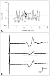
Paired-stimulation TMS has a potential role in the evaluation of functional integrity between CMs and interneuron. In the conventional paired-pulse technique, the conditioning and test stimuli are kept at constant intensity, and changes in the motor evoked potential (MEP) amplitude are then measured. A major limitation of this technique is the marked variability in the MEP amplitude with consecutive stimuli. In TT-TMS, the target MEP is of a predetermined fixed amplitude and changes in the test stimulus intensity required to generate this target response, when preceded by a subthreshold conditioning stimulus, are tracked automatically (Fig. 1). The threshold tracking strategy used in the previous study is described elsewhere.52
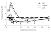
A common TMS finding in ALS and fALS is the development of cortical hyperexcitability, as established by TT-TMS with a decrease in SICI - the ability of a subthreshold conditioning stimulus to suppress the response to a later suprathreshold test stimulus by interstimulus intervals of up to 10 ms (Fig. 2).45,51 The mechanism of SICI appears to be mediated by GABA-secreting inhibitory cortical interneurons, through GABA-A receptors.53,54 Reduction in SICI may develop through loss of cortical inhibitory interneurons, combined with glutamine-mediated downregulation in CMs. Of relevance, SICI can be partially restored in patients with ALS who are treated with riluzole,55,56 possibly through inhibition of glutamate. Furthermore, decreased SICI can be reversed by agents that potentiate GABAergic transmission, such as diazepam and gabapentin.57
Cortical threshold, another index of hyperexcitability, was reported to be lowest early in the course of ALS, and to subsequently increase as the disease progressed.58 The duration of the cortical silent period seems to be uniformly decreased in all clinical ALS phenotypes.45 The duration of the cortical silent period is mediated in the early phase by inhibition of anterior horn cells59 and by cortical processes, through GABA-B receptors, in later phase.60 Thus, measurements of the cortical silent period could be refined to identify disinhibition of anterior horn cells15,61 or progressive dysfunction of cortical inhibitory interneurons that act through GABA-B receptors. Of relevance, the latter finding has been reported in patients with ALS during the course of the disease.62
In longitudinal studies in asymptomatic carriers of mutations in SOD1, as discussed earlier, cortical hyperexcitability develops before the clinical onset of ALS.51 This finding was also supported by flumazenil PET studies63; decreases in SICI in fALS could thus be a primary, presymptomatic event. Cortical hyperexcitability is absent in patients with Kennedy's disease, an X-linked recessive, LMN spinobulbar, neurodegenerative disorder that is slowly progressive.64 However, hyperexcitability is present in patients who have LMN-predominant flail arm ALS,65 suggesting that it is important to clarify the pathophysiology of this variant and it could be useful in distinguishing ALS from Kennedy's disease. In terms of clinical-prognostic correlation, one previous study showed that the amount of the SICI decrease correlated with disease duration and motor deficit score.66 However, studies with TT-TMS or NET showed that functional status or survival was instead correlated with peripheral NET or NCS parameters, such as CMAP or strength-duration time constant (SDTC).45,67
Peripheral axonal excitability in ALS
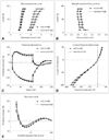
In terms of developing a novel peripheral biomarker in ALS, measurements of axonal excitability have been validated as a sensitive biomarker of the resting membrane potential. Changes in excitability produced by a combination of test and conditioning currents could be used to establish membrane potential and the biophysical properties of peripheral axons. This process is now possible using threshold-tracking protocols that provide insight into the ionic mechanisms underlying the pathophysiology of axonal dysfunction (Fig. 3).
Although studies of peripheral excitability in patients with ALS have, so far, been limited, an increase in persistent Na+ conductances was been identified across patients with a range of clinical phenotypes, as shown by a rise in SDTC.68-71 This increase in Na+ flow would tend to depolarize the axon and thereby contribute to LMN hyperexcitability (Fig. 3).69,72
ALS patients have been stratified according to disease severity and showed decreased slow K+ currents in the internode and increased SDTC.69 Increased SDTC and decreased slow K+ conductances would tend to indicate depolarization of the axolemma. Interestingly, these findings were most prominent in the mild to moderate stages of the disease, during which period most patients experience a degree if LMN hyperexcitability, such as fasciculation and cramp.
Two recent studies have shown further clinical utility of NET in ALS clinical studies, particularly as an excellent predictor of survival in ALS.67 Indeed, prolonged SDTC was strongly associated with shorter survival. This finding was consistent with a longitudinal EMG study, which showed that fasciculation potentials as well as fibrillation and sharp waves, were poor prognostic factors, as defined by survival rate.18
In another longitudinal study of axonal excitability, longitudinal changes in axonal excitability in ALS patients show increasing K+ channel dysfunction in motor axons.73 Thus, subsequent changes in axonal function with time in ALS may reflect the continually increasing demand for surviving motor units to compensate for progressive weakness.
Special Issues
UMN hyperexcitability and ionic biophysics
The precise mechanism of the persistent increased Na+ conductance in ALS remains unknown. However, it has been hypothesized that the increase in the Na+ influx into the cytoplasm would result in an increase in intracellular Ca2+ concentrations, via activation of the Na+/Ca2+ exchanger,74 possibly increasing motor neuronal death. Additionally, a continuous increase in Na+ influx would lead to overload of the Na+-K+ ATPase-dependent pump and excessive consumption of intracellular ATP and subsequent energy failure of the neuron.
Interestingly, hyperexcitability was demonstrated in CMs in a G93A mouse model, with a higher density of persistent Na+ currents in their CMs.75 In contrast to the peripheral Na+ current, cortical mechanisms underlying these changes in ionic biophysics remain to be clarified, although the finding of an increased channel density may favor transcriptional alterations over secondary functional modulation of channel behavior, as in LMN excitability.
Hyperexcitability and the ALS 'split hand'
The so-called ALS "split hand" is a peculiar pattern of selective muscle wasting in intrinsic hand muscles. Among the intrinsic hand muscles, wasting predominantly affects the 'lateral (thenar) hand', involving both median innervated muscles (APB and opponens pollicis) and ulnar innervated muscles [first dorsal interosseous (FDI), adductor pollicis and flexor pollicis brevis], with relative sparing of the ulnar innervated hypothenar muscles (abductor digiti minimi, ADM).76-78
In terms of mechanism, it has been hypothesized that, in humans, use of the APB muscle is greater than that of the ADM, which could render the former muscle (involved in split hand sign) subject to greater oxidative stress and metabolic demands at both UMN and LMN level.79,80 In this regard, a difference in axonal excitability between the thenar and hypothenar muscles has been established in normal subjects.81 Nodal persistent Na+ currents, estimated by SDTC and latent addition, were markedly greater for APB and FDI motor axons than for ADM axons. These findings may suggest that motor axons with physiologically greater persistent Na+ conductance, with subsequently depolarized axons, may be more vulnerable to degeneration in ALS, thereby contributing to the development of the split hand.
Recently, there was a report about the 'split-hand plus sign', which indicated the preferential saving of the flexor pollicis longus (FPL) muscle, even in advanced stages of ALS.82 This unique feature was investigated from a central aspect using TT-TMS. It was confirmed that there were significant differences in SICI between APB and FPL CMs. Such a finding may suggest that differences in intracortical integrity of excitatory and inhibitory neurons, and related excitability changes, can result in dissociated involvement of small hand muscles in ALS. These findings may in turn relate the development of the ALS 'split hand' to the relatively large cortical representation of hand muscles involved particularly in the human pincer grip.
Is hyperexcitability a shared feature of ALS and frontotemporal dementia (FTD) spectrum diseases?
The recent recognition of clinical, pathological, and genetic overlaps between FTD and ALS supports the view that FTD and MND form the extremes of a disease spectrum, with a predominance of cognitive symptoms at one end and motor dysfunction at the other83,84 Genetically, the ubiquitination of misfolded proteins that aggregate in affected cells are pathological hallmarks of ALS and FTD: misfolded TAR DNA-binding protein (TDP-43), which is involved in mRNA processing and the TARDBP gene, which encodes TDP-43, are frequently identified in both diseases.84-89 More recently, mutations in the fused sarcoma gene (also known as "translocated in liposarcoma"), an RNA-processing gene that is functionally related to TARDBP, were identified in large ALS families, linked to chromosome 16q12.90,91 Furthermore, many other RNA-binding proteins have been shown to be involved in ALS and TDP-43 aggregation, a core feature of both ALS and FTD, in pathological findings: C9ORF72, VCP, UBQLN2, and PFN1.44
Substantial motor system dysfunction has also been identified in a large cohort of patients with FTD.92 Although clinically detectable motor system involvement was relatively minor in patients with FTD, there was significant cortical and LMN dysfunction across most FTD subtypes using TT-TMS. Thus, the average SICI of FTD subtypes were decreased, at an intermediate level between ALS and normal controls. These findings in FTD, showing subclinical dysfunction in cortical excitatory-inhibitory integrity, may indicate that hyperexcitability of the motor cortex can be a shared component in the pathophysiology of ALS and FTD spectrum disease.
Conclusions
Hyperexcitability develops across both UMN and LMN compartments in ALS and appears as a unique phenomenon, absent or less prominent in other neurodegenerative disorders and other types of motor neuron disease. Although the exact mechanismsand substantial clinical implication remain to be determined, the identification and interpretation of UMN and LMN hyperexcitability are clearly developing as promising tools in the realm of ALS research.
Recent advances in the genetics of both ALS and FTD have opened new chapters in pathophysiology at the molecular level and have challenged the traditional concepts of disease causation and indeed, clinical phenotype. Abnormal RNA products and related erroneous transcriptional interventions into various cortical elements, and correlating such changes with the components related to the underlying excitability in CMs or interneurons, may yet clarify the exact mechanism of hyperexcitability at the molecular level in ALS.
Ideally, however, much research work needs to be completed in this complex area, to better facilitate new treatments for this most therapeutically resistant neurodegenerative disease. Returning to the beginning, perhaps the final word should be with Sherlock Holmes:
 XML Download