

 PDF
PDF ePub
ePub Citation
 Print
Print


Introduction
Congenital hypotonia is not uncommon, well-recognized disease entity for pediatric neurologists. Various etiologies underlie congenital hypotonia, many of which have genetic backgrounds. Recent advances in molecular genetics have made it possible to noninvasively and rapidly diagnose certain neuromuscular disorders, including congenital myotonic dystrophy, spinal muscular atrophy, and several forms of congenital myopathies.1
Myotubular myopathy (MTM), also known as centronuclear myopathy, is an inherited neuromuscular disorder characterized by clinical features of congenital myopathy and centrally placed nuclei in muscle fibers.2-4 This disease was first described by Spiro et al.2 in 1966 in an adolescent boy with progressive ocular, facial, and generalized limb muscle weakness with muscle pathology resembling fetal myotubes. Several case reports have been published since then, and the responsible gene mutations have been identified.5-7 MTM can be inherited as an autosomal dominant, autosomal recessive, or X-linked disorder.3 X-linked MTM (XLMTM) is the most severe form, with affected males presenting at birth with marked hypotonia and muscle weakness. XLMTM is caused by mutations in the myotubularin 1 gene (MTM1) on chromosome Xq28, whereas the autosomal dominant and autosomal recessive forms have been associated with mutations in the dynamin 2 gene (DNM2) on chromosome 19p13.2 and in the amphiphysin 2 gene (BINI) on chromosome 2q14, respectively.4
Here we report two patients with XLMTM, who were diagnosed by genetic analysis of MTM1 without muscle biopsies.
Case Report
Case 1
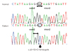
A male neonate was born by cesarean section after 35 weeks of gestation due to polyhydramnios that required repetitive amniocenteses during the fetal period. He was the second baby of nonconsanguineous parents. The first baby, a male, showed floppiness with poor respiration at birth and required ventilator support; he died at age 10 months. The second baby of the parents also showed generalized hypotonia and respiratory difficulty at birth. His Apgar scores were 3 and 5 at 1 and 5 minutes, respectively. His birth weight was 1800 g (10th percentile), length was 45.5 cm (25-50th percentiles), and head circumference was 32 cm (25-50th percentiles). He was resuscitated immediately and placed on ventilator support. He showed severe floppiness with no spontaneous active movement. His face was elongated with thin, narrow cheeks. He had a high-arched palate, and inverted V-shaped upper lips. The results of chest and abdomen examinations were normal, but both testes were undescended. A chest X-ray showed thin ribs and clavicles. On blood examination, the serum creatine kinase level was 31 IU/L. The results of tandem mass spectroscopy including amino acids, organic acids, fatty acids metabolic disorders, and a brain ultrasonogram were unremarkable. Due to failure to wean off the ventilator support, a tracheostomy was performed at 3 months of age. Although we strongly recommended a muscle biopsy for confirmative diagnosis, it was refused by his parents. With the suspicion that the patient might suffer from the X-linked congenital myopathy, especially XLMTM, a genetic study of MTM1 was performed, which revealed a splicing variant: c.63+1G>C (IVS2+1G>C) (Fig. 1). This variant has not been reported previously, and is expected to lead to aberrant splicing. At the latest evaluation at 29 months of age he was still on a mechanical ventilator.
Case 2
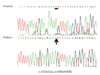
A 6-month-old male infant was transferred to our hospital due to failure to extubate. He was born at 38 weeks of gestation with a birth weight of 2590 g. His Apgar scores were 2 and 4 at 1 and 5 minutes, respectively. He was the third baby of non-consanguineous parents, and his two female siblings were reported to be healthy. No history of maternal disease or family history of neuromuscular diseases was reported by the parents. Reduced fetal movements or polyhydramnios were not noticed during pregnancy. At birth he was severely hypotonic with poor respiration, and required immediate ventilator support. He experienced 10 episodes of extubation failure up to 6 months of age. On admission to our hospital at the age of 6 months, the baby was extremely floppy with an absence of deep tendon reflexes. He had a characteristic myopathic face with paucity of facial expression. A high arched palate was also noted, but tongue fasciculation was not observed. His serum creatine kinase level was 31 IU/L. Brain magnetic resonance imaging revealed nonspecific findings, and so we suspected that the patient had peripheral hypotonia. Molecular genetic tests were performed for the diseases that can cause peripheral types of congenital hypotonia, including congenital type of muscular dystrophy type 1, spinal muscular atrophy, and Prader-Willi syndrome; all of the results were negative, whereas the study for MTM1 revealed a novel deletion mutation, c.473delA (p. Lys158SerfxX28) (Fig. 2), which is presumed to result in the premature truncated myotubularin protein. A muscle biopsy was refused by the parents. His mother is a carrier for this mutation. The patient is now 9 months old and is being maintained on a home ventilator.
Discussion
Congenital hypotonia can be caused by various diseases, including central nervous system disorders, neuromuscular diseases, and genetic disorders. A specific diagnosis is important because the clinical outcome and genetic counseling differ according to the causative disease, and specific treatments may be possible for some diseases.
MTM is one of the diseases causing congenital hypotonia, and manifests as a severe infantile form of congenital myopathy. MTM is characterized in a muscle biopsy by centrally placed nuclei surrounded by a perinuclear halo devoid of myofilaments.2,4 This feature resembles the myotubes of fetal muscles (and hence results in the name of "myotubular myopathy"). Some authors have attributed this alteration to an arrest in myofiber maturation, while others have suggested that either failure of myofiber maturation or neurogenic causes are implicated in the process. Despite similarities in morphology, three distinct modes of inheritance have been described: autosomal dominant, autosomal recessive, and X-linked recessive forms. Both autosomal dominant and autosomal recessive forms exhibit relatively benign courses, whereas the X-linked recessive form, known as XLMTM, is more severe, presenting at or soon after birth. Affected males exhibit profound global hypotonia and weakness, accompanied by respiratory difficulties that often require ventilation. The clinical features of the two cases presented herein did not differ from those of previously reported cases.3,5-8 Most of these patients die during infancy or early childhood, but some survive into later childhood or even adulthood. De Angelis et al.8 found a 74% mortality rate in the first month of life and 10% additional deaths within the first year in their 84 patients with XLMTM. In addition to classic clinical features, Joseph et al.9 reported several interesting clinical findings of their patients with XLMTM, including a birth length >90th percentile and a large head circumference with or without hydrocephalus (in 70% of cases), a narrow, elongated face (80%), and slender, long digits (60%). They suggested that these features can aid the early clinical diagnosis of XLMTM. In a study of long-term survivors, premature adrenarche, pyloric stenosis, spherocytosis, gallstones, nephrocalcinosis, and bleeding diatheses have been reported.10 The involvement of tissues other than skeletal muscle in XLMTM survivors suggests that myotubularin is also important in these tissues.
MTM1 is located on chromosome Xq28, and its mutations have been identified in most of the patients with XLMTM.3,11,12 This gene consists of 15 exons and encodes the myotubularin protein, which consists of 603 amino acids. The myotubularin protein seems to play an important role in controlling critical aspects of gene expression related to the growth control and differentiation of skeletal muscles. To date, about 200 mutations have been identified in MTM1, of which 30% are missense mutations, 25% are small insertions or deletions, 20% are nonsense mutations, 20% are splicing variants, and 5% are large deletions.3 The two mutations identified in our patients-c.63+1G>C, which is expected to cause aberrant splicing, and p. Lys158SerfxX28, which leads to a premature truncated protein are presumed to significantly alter the function of myotubularin. Moreover, both of these mutations are novel.
While a definitive diagnosis of XLMTM is normally performed based on characteristic findings in a muscle biopsy, our patients were diagnosed with XLMTM by genetic analysis without performing a muscle biopsy, suggesting that genetic testing can be performed as a confirmative method of diagnostic testing before performing an invasive procedure such as a muscle biopsy. With the positive results from genetic analysis, a muscle biopsy-which has the risk of complications as an invasive procedure-might be unnecessary for the diagnosis. Furthermore, genetic testing can provide useful guidance to parents considering prenatal genetic testing for their future pre-gnancy outcomes.
In conclusion, we have described two patients with XLMTM, which was confirmed by the genetic testing of MTM1. Although a muscle biopsy remains an essential diagnostic tool for neuromuscular diseases, advanced molecular genetic testing can provide a correct diagnosis while avoiding invasive procedures. Further studies on genotype-phenotype correlations and the function of myotubularin will provide new insights into this disorder.
 XML Download