

 PDF
PDF ePub
ePub Citation
 Print
Print


Introduction
Parkinson's disease (PD) is one of the most common neurodegenerative disorders, affecting about 1% of the general population older than 60-years.1,2 In spite of marked progress in the scientific understanding of this condition, its diagnosis continues to be based upon key clinical features.3,4 A definitive diagnosis can only be made by pathologic confirmation of dopaminergic neuronal loss and the presence of Lewy bodies or Lewy neurites in autopsied brain tissue, which has led to continued debated about the accuracy of clinical diagnoses.3,5-7
The Lewy body, which was first described by Friedrich Heinrich Lewy, is located in the neuronal cytoplasm and is a pathological hallmark of and dementia with Lewy bodies.8 This filamentous spherical inclusion is composed of ubiquitin, ubiquitin-C-terminal hydroxylase, microtubule-associated protein, DJ-1, tau, α-synuclein, and various proteins.9,10 α-synuclein has been regarded as the key pathological molecule for several reasons. First, genetic alterations (including point mutations and multiplications) of the α-synuclein gene cause familial PD.11-14 Second, α-synuclein can take the form of a soluble oligomer, fibril, or sheet structure by self aggregation, depending on the microenvironmental conditions.15-17 Recent studies have shown that the soluble oligomeric form of α-synuclein induces neuronal cell injury and that the Lewy body might be an end-product of the sequestration of this toxic molecule with other proteins, as a type of cell-protective mechanism.18,19 Third, α-synuclein can be a seed molecule, which may explain the propagation of Lewy body pathology from the lower brainstem to the cortex.20-22 Vesicular secretion of α-synuclein has been found without evidence of neuronal injury, and adjacent glial and neuronal cells could uptake the secreted α-synuclein.23-25
The finding of an extracellular location of α-synuclein and the search for a reliable biological maker for PD is now drawing attention to this protein. Using the enzyme-linked immunosorbent assay (ELISA), Lee et al.26 found that the levels of plasma α-synuclein were higher in patients with PD and multiple system atrophy than in age-matched healthy controls. In contrast, Western blot analysis revealed that the plasma levels of α-synuclein were lower in PD patients than in control subjects.27 Assay of α-synuclein in cerebrospinal fluid (CSF) produced similar conflicting results to those seen in plasma; Mollenhauer et al.28 reported that the CSF level of α-synuclein was lower in patients with PD and dementia with Lewy bodies than in those with Alzheimer's disease, while other researchers have found no such difference.29 These discrepancies might reflect conformational changes in α-synuclein and the presence of other cross-reactive molecules.
A novel method for detecting oligomeric α-synuclein has yielded notable results. El-Agnaf et al.30 reported that the plasma level of α-synuclein oligomer was significantly higher in PD patients than in age-matched controls. Their study used a specific double-ELISA method for α-synuclein oligomers, which demonstrated diagnostic specificity, sensitivity, and positive predictive values of 0.852, 0.529, and 0.818. The detection of oligomeric α-synuclein in brain extracts and in viable cells using various methods and the blocking of these oligomeric processes has yielded promising results.31-33 However, although such studies targeting oligomeric α-synuclein have produced significant results, α-synuclein is known to undergo conformational changes according to the surrounding conditions. Factors such as membranes, metals, and oxygen metabolites such as dopamine could therefore contribute to the pathologic potential of α-synuclein.18,34-36 Currently there is little known about the native structure of α-synuclein in living cells, and it is possible that dopaminergic treatment and other environmental exposures during the clinical course of the disease induce conformational changes in this protein.
Therefore CSF and plasma levels of α-synuclein were analyzed in the present study using different ELISA methods for both the total and oligomeric forms of the protein in drug-naïve patients with PD in order to determine the potential of α-synuclein as a biological marker for PD.
Methods
Subjects
Drug-naïve patients with PD (23 patients, age 62.4±12.7 years, mean±SD; 11 males) were consecutively recruited for this study. All patients showed mild-to-moderate symptom severity (Hoehn and Yahr stage 1.5-3.0, median 2.5), and the diagnosis of the patients was confirmed according to clinical criteria during follow-up visits for more than 6 months. Lumbar puncture and plasma sampling were conducted in the early morning after an overnight rest, prior to the administration of dopaminergic medications. The control group consisted of 11 symptomatic patients (age 72.7±6.9 years; 6 males) who complained of slow movement and/or tremor and/or gait disturbance, and were diagnosed as having drug-induced parkinsonism (4 patients), hydrocephalus (6 patients), or vascular parkinsonism (1 patient). The symptomatic patients were diagnosed based on drug withdrawal (for the patients with drug-induced parkinsonism), radioisotope cisternography, and therapeutic CSF drainage (for the patients with hydrocephalus). Movement specialists confirmed the diagnosis during the clinical follow-up visits for more than 6 months. An additional 18 subjects (age 52.4±15.4 years; 10 males), comprising 8 with diabetic ophthalmoplegia and 10 normal adults, formed the neurologic control group. CSF and plasma samples from 15 patients with multiple sclerosis (age 41.3±9.6 years; 6 males) were also analyzed as an additional control group. CSF samples were collected in sterile polypropylene tubes, centrifuged for 10 minutes at 3,200 rpm, and stored as aliquots at -80℃. Plasma samples were collected using plastic pipettes after being centrifuged at 3,000 rpm for 15 minutes and were stored in aliquots at -80℃. The local Institutional Review Board approved the processes and all patients or family members provided signed written informed consent to participate.
Materials
A mouse monoclonal antibody that recognized amino acid residues 121-125 of human α-synuclein, as well as a rabbit polyclonal antibody raised against the full-length α-synuclein of human origin were purchased from Santa Cruz Biotechnology (Santa Cruz, CA, USA). Recombinant human α-synuclein was obtained from Calbiochem (San Diego, CA, USA). Sulfo-NHS-LC-Biotin was purchased from Pierce (Rockford, IL, USA) and Bio-Spin-6 columns were obtained from Bio-Rad Laboratories (Hercules, CA, USA). Other chemicals including ExtrAvidin alkaline phosphatase and 4-hydroxyazobenzene-2-carboxylic acid/avidin reagent were purchased from Sigma (St. Louis, MO, USA).
Preparation of aggregated α-synuclein
Recombinant human α-synuclein was solubilized with sterilized phosphate-buffered saline [PBS; 137 mM phosphate buffer and 150 mM NaCl (pH 7.4)] to produce a final concentration of 25 µM. An Eppendorf tube containing 25 µM α-synuclein solution was tightly sealed with Parafilm and incubated at 37℃ for 4 days with continuous mixing (1,000 rpm). The resultant aggregated α-synuclein was aliquoted and stored at -80℃ until needed.
Preparation of the biotinylated antibody
Sulfo-NHS-LC-Biotin was reacted with the mouse monoclonal antibody in PBS, at a 50-fold molar excess of biotin over antibodies, for 2 hours. The mixture was desalted inn Bio-Spin-6 columns to remove any excess uncoupled biotin. The extent of biotinylation per mole of antibody was determined with the 4-hy-droxyazobenzene-2-carboxylic acid/avidin assay. The biotinylated antibodies were stored at 4℃ until use.
ELISA to measure α-synuclein oligomer
An ELISA plate was coated by overnight incubation at 4℃ with 1 µg/mL nonbiotinylated mouse monoclonal antibodies in 200 mM NaHCO3 (pH 9.6) containing 0.02% (w/v) sodium azide. The plate was then washed three times with PBS containing 0.05% Tween 20 (PBST), and blocked with PBST containing 2% bovine serum albumin for 2 hours at 37℃. After three further washes with PBST, 100 µL of α-synuclein monomer, aggregated α-synuclein, and samples to be tested were added to each well. CSF and plasma samples were used without dilution. The plate was incubated at 37℃ for 2 hours. After washing three times with PBST, 100 µL of biotinylated mouse monoclonal antibody diluted to 1 µg/mL in blocking buffer was added, and the mixture was incubated at 37℃ for 2 hours. The wells were washed three times with PBST and incubated for 1 hour at 37℃ with 100 µL per well of ExtrAvidin-Alkaline phosphatase diluted 1 : 2,000 in blocking buffer. The wells were then washed three times with PBST and the enzyme substrate p-nitrophenyl phosphate was added. The reaction was allowed to proceed for 30 minutes at room temperature, and then the absorbance was read at 405 nm on a microplate ELISA reader (EL312e, Bio-Tek).
ELISA to measure total α-synuclein
The same ELISA protocol was applied to measure the total α-synuclein in the samples, except that a rabbit polyclonal anti-α-synuclein antibody (1 µg/mL) and biotinylated antirabbit antibody were used as detecting antibodies.
Statistical analysis
Data analysis was performed with SPSS 14.0 software (SPSS, Chicago, IL, USA). The significance of intergroup differences was evaluated by one-way analyses of variance, while Student's t-test was used to evaluate single comparisons. Pearson's correlation was used to evaluate the association between the levels of α-synuclein and the clinical characteristics of the cohort. Differences were considered significant at p<0.05.
Results
Characteristics of the subjects
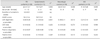
The control subjects were matched for age and sex with the PD patients. The initial CSF and plasma laboratory values did not differ significantly between the two groups (Table 1). When the control subjects were divided into symptomatic control and neurologic control subgroups, the only significant difference from the PD group was found for age in the symptomatic control group (Table 2).
Validation of ELISA methods for total and oligomeric form of α-synuclein
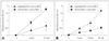
Aggregated α-synuclein was detected more sensitively by ELISA for oligomeric α-synuclein than by the conventional method that detects total α-synuclein (Fig. 1). The conventional ELISA method for total α-synuclein gave an optical density (OD) reading of 1 for monomeric α-synuclein at a concentration of 5 µM, but the same method produced an OD reading below 0.5 for aggregated α-synuclein at the same concentration. The OD using the ELISA method for oligomers was 1 for aggregated α-synuclein at a concentration of 10 µM, for monomeric α-synuclein at the same concentration it was only 0.5. These results show that the ELISA method for oligomeric α-synuclein is a sensitive one for the measurement of levels of oligomer in aggregated α-synuclein. Therefore, the subsequent results are presented as the OD value, of which α-synuclein oligomer was measured by the oligomeric ELISA method, and total α-synuclein was measured by the conventional ELISA method.
Total and oligomeric forms of α-synuclein in the CSF and plasma
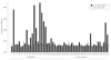
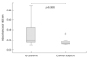
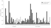
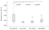
The absorbance of CSF oligomeric α-synuclein was significantly higher in patients with PD than in the control subjects (Table 3)(Figs. 2 and 3). However, there was no difference in total α-synuclein from the CSF and plasma oligomeric and total α-synuclein (Figs. 2 and 4). A subgroup comparison of the control subjects with the PD patients revealed no significant difference (Table 3)(Fig. 5); when the control group was subdivided into symptomatic and neurologic control subgroups, the level of α-synuclein did not differ between the control subgroups. Although subjects in the symptomatic control group were older and those in the neurologic control group were younger than the patient group, the CSF α-synuclein level was significantly higher in the patients group than in the control groups. The total level of α-synuclein in the CSF and plasma did not differ between the patient group and the subgroups of control subjects. Moreover, the CSF level of α-synuclein oligomer was significantly higher in patients with PD than in the multiple sclerosis patients.
The absorbance of neither oligomeric nor total α-synuclein in the sample of CSF or plasma was not correlated with any of the clinical parameters (i.e., age, duration of symptoms, or severity of motor symptoms; data not shown).
Discussion
α-synuclein is a classical cytoplasmic protein that exists unfolded in its native form.37 However, numerous transient structures can be found from monomers to oligomers and filamentous forms, depending on the environment.38 Although the functional role of α-synuclein has not yet been established, its membrane binding-affinity and presynaptic location of α-synuclein indicate a role in synaptic transmission.39,40 The classical concept is one of a cytoplasmic location for α-synuclein, and also the pathologic implications of α-synuclein have been widened with recent studies that have produced evidence for an extracellular location for α-synuclein in the body fluids of patients with PD. Along with the suggestion of Braak et al.,20 pathologic studies have shown a Lewy body-like pathology in grafted neurons of autopsied brains in patients with PD after embryonic nigral cell transplantation, which might indicate disease propagation from host to graft.41,42 These findings imply the importance of extracellular α-synuclein in the pathogenesis of PD, but no role for α-synuclein in the body fluids has been established yet.
Using different ELISA methods to detect both the total and oligomeric forms α-synuclein, we found that levels of the α-synuclein oligomer in the CSF were significantly higher in PD patients than in their non-PD counterparts; however, the plasma levels of α-synuclein did not differ between the PD patients and the controls. These findings are contrary to some previous studies that found increased plasma and decreased CSF levels of α-synuclein.26,28,43 However, those studies were based on a single method for detecting α-synuclein, which disregarded the conformational status of this protein. In addition, the study patients had been treated with dopaminergic drugs. The study found elevated levels of α-synuclein oligomer in the patients' plasma using the oligomer-detecting method also recruited patients who had already been exposed to dopaminergic drugs.30 The authors of that report found elevated levels of α-synuclein oligomer in postmortem CSF samples of patients. As mentioned above, the precise native structure of α-synuclein in living cells remains to be determined. These conflicting results might therefore be attributable to the unstable structure of α-synuclein. However, the design of our study employing a dual ELISA method for simultaneous measurement of total and oligomeric α-synuclein eliminated the possible confusion arising from such conformational changes in α-synuclein. We were also able to exclude the potential confounding effect of dopamine, which is known to be an inducer of α-synuclein oligomerization, since our patients had not been exposed to that drug.44
The meaning of elevated CSF levels of α-synuclein oligomer in PD patients is not easy to interpret. For example, we do not know whether α-synuclein oligomer levels are elevated in the CSF itself, or whether the oligomerization of α-synuclein is elevated in the milieu of the CSF. Although elevated levels of α-synuclein oligomer in the autopsied brain extracts of patients with dementia with Lewy bodies has been reported recently, the prevailing intracellular environment would differ from that of the CSF.31 However, the finding of an intravesicular localization of α-synuclein, and this being more prone to aggregation and secretion from cells by exocytosis, could provide an explanation for our results.23 There is also evidence of a role of extracellular α-synuclein oligomer in the pathology of in vitro neuronal cell death, and the aggregation process appears to be dependent upon the concentration of α-synuclein.25,45-47 These findings support the importance of elevated α-synuclein oligomer in the extracellular space and in the CSF, as indicated in our results. In fact, it has been suggested that neuronal cells are subject to the worst pathologic conditions during the early stage of PD, prior to medication with dopaminergic agents, because the increased turnover of dopamine in the brain could damage the surrounding tissues. This condition could also contribute to the increase in α-synuclein in the brain, which would lead to increases in CSF α-synuclein oligomer. Therefore, we suggest that the CSF level of α-synuclein oligomer is a useful biological marker for PD, especially in the dopaminergic drug-naïve condition.
Some reports have indicated that the level of α-synuclein is correlated with disease duration, cognitive function, and age, and suggested that synaptic loss or neuronal degeneration could be related to the level of α-synuclein.29,43 We did not find any correlations between these clinical factors and the level of α-synuclein in either the plasma or the CSF. The CSF level of α-synuclein oligomer was significantly higher in the patients with PD than in both the younger control group and the patients with multiple sclerosis who already had multiple brain and spinal cord lesions. These findings suggest that age and lesions of the brain or spinal cord were not associated with the level of α-synuclein, although pathological differences do exist between multiple sclerosis and PD.
Our study was subject to several limitations. First, even though movement specialists diagnosed the patients with parkinsonian conditions of different etiologies and confirmed the diagnosis over a clinical follow-up period of longer than 6 months, there is a possibility of diagnostic uncertainty. Objective diagnostic tests (e.g., dopamine-transporter imaging) and longer clinical observation periods of the patients groups could increase the reliability of any noted intergroup differences. Second, there was a wide range of absorbance reading for the CSF level of α-synuclein oligomer. Nearly half of the subjects exhibited an overlap, as shown in Figs. 4 and 5. Although these overlaps diminished with increments of age in symptomatic control subjects, more sensitive measures are needed for this to be considered a reliable marker. As suggested in the consensus regarding Alzheimer's disease, an ideal marker should detect fundamental features of the disease neuropathology and should have a diagnostic sensitivity and specificity of over 80%. Third, the exact target for different ELISA methods should be addressed. Since we do not know the precise form in which α-synuclein exists in cells, intercellular spaces, or body fluids, target specification in the controlled condition might shed light on α-synuclein dynamics.


 XML Download