

 PDF
PDF ePub
ePub Citation
 Print
Print


Introduction
Vascular dementia is a subtype of dementia, and its prevalence is second to that of Alzheimer's disease in westernized societies.1 Vascular dementia causes many neuropsychiatric and physical problems, and represents a significant economic burden. Brain imaging studies have revealed obvious changes in the cerebral white matter (WM) rather than in the cortex, and hence WM lesions are thought to be the core pathology for cognitive declines in patients with vascular dementia.2,3 It is known that WM damage is caused by widespread demyelination and axonal loss in the WM;4 however, the mechanisms underlying the pathologic changes are not fully understood.
Cerebral WM lesions are probably caused by chronic cerebral ischemia, and they can indeed be experimentally induced in the rat brain by permanent occlusion of both common carotid arteries,5 which can affect cognitive function,6 and this model is similar to that of vascular dementia.7 This technique can decrease the blood flow in the cerebral cortex and hippocampus by up to 40-82%8-10 for several months, which induces certain learning disorders. Thus, this technique can be used to study vascular dementia and cerebral WM lesions.
It has been reported that citicoline (exogenous cytidine-5-diphosphocholine), a substance synthesized by Eugene Kennedy in 1956, is effective in treating ischemia in the central nervous system, brain trauma, and Parkinson's disease.11 Citicoline is a precursor of phosphatidylcholine (PtdCho) biosynthesis, and an increase in PtdCho biosynthesis prevents apoptosis of neurons and protects against neuronal damage.12 In addition, citicoline maintains cardiolipin and sphingomyelin levels, stimulates glutathione biosynthesis, activates glutathione reductase, and reduces lipid peroxidation.12 Therefore, citicoline is expected to prevent apoptosis and delay disease progression in the brains of rats with chronic ischemia.
In the present study, we investigated the therapeutic effects of citicoline in chronic cerebral hypoperfusion induced in rats by conducting a histopathologic investigation and cognitive-behavior test.
Methods
Experimental animals
All procedures used in this study were approved by the Institutional Animal Care and Use Committee (IACUC) at the College of Medicine, The Catholic University of Korea. Ten-week-old male Wistar rats weighing 280-300 g (Joongang Lab, Seoul, Korea) were used in this study. The rats were housed at a temperature of 23-26℃ and a humidity of 50% with a 12-h light-dark cycle, and they were allowed free access to food and water except when performing the eight-arm radial maze.
Operation
All surgical procedures were carried out under inhalation anesthesia using 2% isofluorane with 30% O2 and 70% N2, and the body temperature was maintained during the operation at 37.0-37.8℃ using a temperature controller. After fixing a rat in the prone position on a board, a skin incision about 2 cm long was made and a cervical central line was placed. The bilateral common carotid arteries were exposed by isolating them from the vagus nerve and its sheath, and they were double ligated with 4-0 silk at 8-10 mm below the visible region of the external carotid artery.
Administration
Animals were divided into immediate- and delayed-treatment groups. Those in the immediate-treatment group received a sham operation (n=8), citicoline (Somazina, Bukwang, Korea) administered intraperitoneally (500 mg/kg/day) for 3 weeks immediately after the operation (n=9), or phosphate buffered saline (PBS)(n=7).
Rats in the delayed-treatment group were administered intraperitoneally with 500 mg/kg/day citicoline (n=8) or PBS (n=8) for 21 days beginning on the 8th day after the operation.
Rats in the immediate-treatment group were given 500 mg/kg/day citicoline for 3 weeks beginning immediately after the operation.13 PBS was administered at the same dosage in control animals. In addition, the chronic effects of citicoline on the progression of vascular change were examined in the delayed-treatment group.
Spatial memory test
The spatial memory test was performed in an eight-arm radial maze comprising a center with a radius of 34 cm and eight arms (60 cm long and 12 cm wide) with walls 20 cm high. Rats were trained twice daily for 10 days before model production, and rats with less than two total errors (TEs) were selected for further testing. The rats were then fed ad libitum for 3 hours and then fasted. Four of the eight arms in the maze contained 40-50 mg of feed that was placed in the same place every day. Considering the visual loss after model production, the floor paper in each arm had a different texture in order to allow the rats to identify the arms containing feed.14 The rats were placed in the center of the maze, and a test was carried out for 10 minutes to see if the rats ate all of the feed. A TE, reference memory error (RME), or walking memory error (WME) was considered to have occurred if the rat went to the arm without feed, went to the arm without feed for the first time, or returned to the arm without feed after eating, respectively. The time taken to eat all of the feed was recorded.
Tissue preparation
Rats were deeply anesthetized with 30% urethane, perfused transcardially with 0.01 M PBS, and then perfused with a fixative containing 4% paraformaldehyde in 0.1 M phosphate buffer (pH 7.4) containing 0.2% glutaraldehyde. The brain was extracted and fixed again with the fixative for 4 hours and soaked in a 30% sucrose solution until it sank. It was then quick-frozen with liquid nitrogen and stored at -70℃. Slices with thicknesses of 25 and 6 µm were then made by cutting at 2.8-3.14 mm posterior to the bregma using a cryomicrotome in accordance with the atlas of Paxinos and Watson.15
Klüver-barrera staining
Each 25-µm-thick section was soaked in 0.1% Solvent Blue 38 (Sigma, USA) solution at 60℃ overnight, and the dye was removed (except for that in the WM region) using lithium carbonate solution, distilled water, and 70% ethanol. The section was soaked in 0.1% cresyl violet (Sigma) solution for 15 minutes, and then in 95% ethanol followed by 100% ethanol and xylene for dehydration and transparency. After mounting, pathological findings in the optic tract, internal capsule, and corpus callosum were observed using an optical microscope at 200× magnification. The severity of the WM lesion was classified as normal (grade 0), disarrangement of nerve fibers (grade 1), formation of marked vacuoles (grade 2), and disappearance of myelinated fibers (grade 3).16
The terminal deoxynucleotidyl transferase biotin-dUTP nick end labelling staining
The terminal deoxynucleotidyl transferase biotin-dUTP nickend-labeling (TUNEL) assay was performed on 6-µm-thick brain sections using fluorescein with the In Situ Cell Death Detection Kit (Roche, Germany). TUNEL-positive cells were then examined using a fluorescent microscope.
Results
Spatial Memory Test
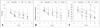
The eight-arm radial maze test was carried out for 5 days before sacrificing the animals. In the immediate-treatment group on the first day, the number of TEs did not differ significantly between sham (5.56±1.45) and citicoline (5.05±0.64) treatments but it did differ between PBS treatment (7.21±1.17) and sham and citicoline treatments. The total time spent and number of RMEs were somewhat higher for PBS treatment (338.57±35.24 s and 3.21±0.28, respectively) than for sham (278.88±56.50 s and 2.5±0.39) and citicoline (294.88±34.03 s and 2.66±0.25) treatments. In the last 5 days, there were decreases in TEs, time spent, and RMEs for all treatments: 1.25±0.42, 113.06±16.96 s, and 0.75±0.18, respectively, for sham treatment; 2.61±0.68, 131.06±26.76 s, and 1.27±0.33 for citicoline treatment; and 4.42±1.52, 246.64±49.11 s, and 2.00±0.34 for PBS treatment (Fig. 1). No significant difference was found in the frequency of WMEs, but the frequency was too low to determine the effect of citicoline on short-term memory disorder.
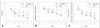
In the delayed-treatment group, the number of TEs did not differ significantly between citicoline (6.25±1.34) and PBS (6.31±0.87) treatments on the first day. However, from the second day, the difference in the number of TEs between citicoline and PBS treatments gradually increased over time, eventually becoming significant (at 3.12±0.65 and 4.00±0.64, respectively) on the 5th day. The time spent did not differ between citicoline and PBS treatments on the 1st day (423.81±58.76 and 426.25±47.93 s, respectively) but it did on the 5th day (178.37±37.05 and 223.43±40.80 s). In addition, the frequency of memory errors eventually differed significantly between citicoline treatment (2.87±0.30 and 1.81±0.33 on the 1st and 5th days, respectively) and PBS treatment (3.12±0.24 and 2.68±0.32)(Fig. 2). The frequency of WMEs did not differ significantly in the delayed-treatment group.
Pathologic examination
Examination of white matter changes
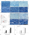
Rats in the immediate-treatment group showed marked differences in the optic tract, some differences in the corpus callosum (Fig. 3A and B), and few changes in the internal capsule and other WM regions. The extent of WM changes in the optic tract and corpus callosum of rats was graded according to the standard of Wakita et al.13 as follows: 0.23±0.04, 1.52±0.13, and 1.85±0.15 for sham, citicoline, and PBS treatments, respectively, in the optic tract; and 0.20±0.04, 0.50±0.08, and 0.88±0.17 in the corpus callosum (p<0.05).
In the delayed-treatment group, WM changes were observed in the optic tract and corpus callosum, but there was no significant difference between citicoline and PBS treatments: 2.09±0.09 and 1.98±0.12, respectively, in the optic tract; and 0.83±0.09 and 0.96±0.13 in the corpus callosum (Fig. 3D and E).
Observation of TUNEL-positive cells
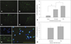
There were TUNEL-positive cells around cortex and lateral ventricles (Fig. 4C and D) as well as in the WM (Fig. 4A and B), but not in the hippocampus.
The number of TUNEL-positive cells that were also stained with DAPI (excluding positive cells in vessels of sections)17 (Fig. 4E) was significantly higher for citicoline (12.66±4.29 cells) and PBS (17.42±6.94 cells) treatments than for the sham treatment (2.62±0.88 cells) in the immediate-treatment group (Fig. 4F). Similar numbers of TUNEL-positive cells were observed in the delayed-treatment group, but there was no significant difference between citicoline and PBS treatments (12.75±1.71 and 14.37±2.42, respectively)(Fig. 4G).
Discussion
In the immediate-treatment group, WM damage and apoptosis were lower for citicoline treatment than for PBS treatment. Phosphatidylcholine is a cell membrane component that is degraded during cerebral ischemia into free fatty acids and free radicals.18,20 Because citicoline is an intermediate in the synthesis of phosphatidylcholine, it might counter the progression of ischemic damage by reducing the release of free fatty acids18,20 and by recovering activities of mitochondrial ATPase and membrane Na+/K+ ATPase.18 Also, it prevents apoptosis by improving membrane recovery.19 These findings are consistent with our results. Therefore, citicoline improves memory and learning ability by reducing the number of ischemic lesions and exerting neuroprotective effects on ischemia via the above mechanisms.
In the delayed-treatment group, WM lesions and apoptosis did not differ between citicoline and PBS treatments, but there was an improvement in cognitive ability. These results indicate that citicoline acts via a different mechanism in addition to neuroprotection in cognitive improvement. Although the exact mechanism of cognitive improvement is not known, citicoline might prevent cognitive impairment by increasing phospholipids, which have an important role in neurotransmission,21 and by increasing the levels of norepinephrine and dopamine,22 which are known to be neurotransmitters involved in cognition. Further study is needed to elucidate the actual mechanism.
Citicoline is used as an immediate treatment for neuroprotection in patients with acute stroke, but the effects of delayed treatment of citicoline on cognition have been unclear. This study confirms that delayed treatment improves the cognitive decline that is a typical symptom in dementia. Therefore, it is likely that citicoline can be applied more widely in patients with vascular dementia.
 XML Download