

 PDF
PDF ePub
ePub Citation
 Print
Print


Foot drop can be defined as a weakness in ankle and toe dorsiflexors.1 It usually results from lesions affecting the peripheral nervous system, from the lumbosacral radicles to the deep peroneal nerve.1 Isolated foot drop caused by an upper motor neuron lesion is rarely reported,2-7 and we are not aware of any report on isolated foot drop caused by cerebral infarction. We present such a patient here.
CASE REPORT
A 68-year-old hypertensive man visited our department complaining of weakness in dorsiflexion of the left toe and ankle that had been present for 2 days. He described having a steppage gait on walking, especially when climbing stairs. He claimed to have no history of diabetes, heart disease, stroke, alcohol abuse, or recent trauma to the left lower limb.
On neurologic examination he was alert and oriented. He showed no dysarthria or dysphagia, and all cranial nerves were intact. The results from a funduscopic examination were normal. His motor strength was normal throughout the upper and lower limbs except for the weakness of the left ankle and toe dorsiflexors. Individual muscle strength testing revealed that the power of the left tibialis anterior, extensor digitorum longus, and extensor digitorum brevis power was grade II, while the left quadriceps femoris, gastrocnemius, iliopsoas, and gluteus maximus muscle strengths were within normal limits. Eversion and inversion movements of both ankles were also unremarkable. The deep tendon reflex (DTR) was normoactive in all extremities. His sensory perception of light touch, pinprick, joint position, and vibration were normal including on the left foot dorsal surface. Rombergand cerebellar function tests were negative, and there were no signs of ataxia. The patient had a slight slapping gait that involved dragging his left foot on ambulation, which was aggravated when walking uphill. His straight leg raising was normal and there was no tender area in the lumbosacral area.
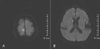
The nerve conduction velocity (NCV) and electromyogram (EMG) were normal: no denervation potentials were detected in any of the examined muscles, including the left tibialis anterior, peroneus longus, short head of biceps femoris, and lumbosacral paraspinal muscles. The only abnormal findings were decreased recruitment patterns in the left tibialis anterior and extensor digitorum brevis muscles. Brain magnetic resonance imaging (MRI) was performed 5 days after the onset to identify any central lesion responsible for the patient's symptoms. Diffusion-weighted imaging (DWI) revealed a focal, round high-intensity signal in the right precentral gyrus at the high convexity and right periventricular white matter (PVWM). Magnetic resonance angiography (MRA) showed mild stenosis in the A1 segment of right anterior cerebral artery (ACA) and steno-occlusive change along the right distal vertebral artery (Fig. 1). The electrocardiogram and echocardiogram were unremarkable except for mild left ventricular hypertrophy. Two weeks later, his ambulation difficulty completely resolved without any residual deficit.
DISCUSSION
Foot drop can result from lesions affecting any point along the neural pathways that supply the dorsiflexor muscles.1 Compression of the common peroneal nerve around the fibular head is the most common cause of foot drop.1 Other causes include damage to the peripheral nerve, leg compartment syndromes, peripheral polyneuropathies, and systemic diseases such as connective tissue disorders, vasculitis, and diabetes mellitus.1
In our patient, the above causes were excluded by clinical examination, laboratory findings, and normal NCV and EMG results. Findings inconsistent with a peripheral cause of foot drop include the lack of sensory deficit or paresthesia in the dorsal surface of the left foot and preserved DTR. This prompted us to perform an MRI examination, which revealed a cortical infarction.
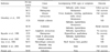
There have been anecdotal case reports of lesions located in the parasagittal area producing acute foot drop.2-7 To our knowledge, 13 cases with foot drop due to central nervous system lesions have been reported, including our patient (Table 1). The etiologies include brain tumor, multiple sclerosis, hemorrhagic contusion from gunshot wound, head trauma, and brain abscess.2-7 Most previous reported cases had accompanying upper motor neuron signs or symptoms such as hemiparesis, hyperreflexia, ankle clonus, and Babinski signs.2,5,6 However, there are rare cases in which the clinical presentation resembles a peripheral type of foot drop.3,7 As far as we are aware, this is the first report of sudden isolated foot drop caused by a cortical infarction. DWI in our patient revealed a cortical infarction in the high convexity of the right precentral gyrus and a small infarction in the periventricular white matter. The somatotopic ankle and toe topography has previously been established in these parasagittal regions by electro stimulation localization.8 The lesion in our patient was considered consistent with the alleged motor homunculus. The periventricular white-matter lesion identified in our patient was considered to be insignificant.
The infarction was in the ACA territory, with MRA revealing mild stenosis in the Al segment of the right ACA. Although the exact pathogenesis of the stroke remained unclear, an artery-to-artery embolism was suspected.
In conclusion, the patient reported here illustrates that a small infarction may present with isolated foot drop mimicking a peripheral lesion. Thus, central nervous system lesions should be considered in such cases, especially in the presence of findings that are atypical for peripheral lesions.
 XML Download