

 PDF
PDF ePub
ePub Citation
 Print
Print


Increasing antimicrobial resistance can threaten public health, which leads to make a global action plan by world health organization (WHO), in order to strengthen the knowledge and evidence to reduce the incidence of drug resistant bacterial infection through research, and to optimize the use of antimicrobial agents [1]. The emergence of carbapenem-resistant bacteria in the clinical setting has become a major public health concern also in Korea. In particular, spread of carbapenem-resistant Gram-negative bacteria in healthcare environment has been focused due to the higher mortality and lack of optimal treatment [234]. To overcome this, researchers have developed several approaches to combat carbapenem-resistant bacteria, and animal models have been established to evaluate the efficacies of new antibiotics, antimicrobial agent combinations, proper route of antimicrobial administration, or in vivo pharmacokinetics/pharmacodynamics study.
Several animal infection models for carbapenem-resistant Gram-negative bacteria of clinical isolates have been reported [567891011], each of these models involved different methods and diverse mouse strains. BALB/c, C57BL/6, Swiss-Webster, and C3 mice have been used, and the site of bacterial administration has varied (e.g., intranasal, intratracheal, intraperitoneal, and intravenous). Diabetic and neutropenic models using BALB/c, ICR or A/J mice have been also developed [12]. Accordingly, the aim of this study was to design a murine peritonitis model using carbapenem-resistant Gram-negative bacteria that were clinical isolates from Korea.
The four bacterial strains used in this study were clinical isolates of carbapenem-resistant Gram-negative bacteria. Escherichia coli (EC-1), Klebsiella pneumoniae (KP-4), and Pseudomonas aeruginosa (PA-3) were obtained from the Korea Centers for Disease Control and Prevention. Acinetobacter baumannii (Aci_100087) was selected from the clinical isolate collections of hospitals affiliated with the Catholic University of Korea. Each bacterial strain was grown at 37°C on blood agar (Hanil Komed, Co., Seongnam, Korea) and Mueller-Hinton broth (Becton Dickinson, Sparks, MD, USA) was used for antimicrobial susceptibility testing and the animal infection model. The minimum inhibitory concentrations (MICs) of ciprofloxacin (CIP), ceftazidime (CAZ), imipenem (IPM), colistin (CST), ampicillin (AMP), amoxicillin-clavulanic acid (AMC), gentamicin (GEN), and trimethoprim-sulfamethoxazole (TMP/SMX) for the four isolates were determined by the broth microdilution method, in accordance with the Clinical and Laboratory Standards Institute (2015) recommendations [13]. All antimicrobials were obtained from Sigma-Aldrich Korea (Seoul, Korea). All carbapenem resistant Gram-negative bacteria were screened for the presence of blaNDM-1, blaVIM, blaAmpC, and blaKPC by polymerase chain reaction (PCR) and blaOXA by multiplex PCR using previously described primers. The primer sequences of the genes used in this study were described previously by Kim et al. [14].
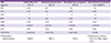
The MIC ranges of four carbapenem-resistant Gram-negative strains are presented in Table 1 with the genetic characteristics of antimicrobial resistance of the bacterial strains. E. coli, K. pneumoniae, and P. aeruginosa carried blaNDM-1, blaKPC, and blaVIM, respectively. Two distinct blaOXA-like genes, blaOXA-23-like and blaOXA-51-like, as well as blaAmpC were identified for A. baumannii by multiplex PCR [14].
Six-week-old specific-pathogen-free, female ICR/Swiss and BALB/c mice weighing 23–27 g (Orient Bio, Sungnam, Korea) were used in this study. All animal experiments and animal care were carried out in accordance with the criteria of the Laboratory Animals Welfare Act, the Guide for the Care and Use of Laboratory Animals, and the Guidelines and Policies for Rodent Survival Surgery provided by the Institutional Animal Care and Use Committee (IACUC) of the College of Medicine, The Catholic University of Korea (approval no. CUMC-2015-0002-04). This study was also exempted from judges of Institutional Review Board, Seoul St. Mary's Hospital as we used stock strains (KC15SISI0111). Mice were housed in rooms with controlled temperature (18–25°C) and humidity (40–70%). Broth cultures of freshly plated bacteria were grown to logarithmic phase overnight to an absorbance at 600 nm of 0.04–0.05 (SpectraMAX190; Molecular Devices, Sunnyvale, CA, USA), corresponding to 1 × 10⁸ CFU/mL. Hog gastric mucin (Sigma-Aldrich, St. Louis, MO, USA) was used as a virulence-enhancing factor for bacteria. Bacterial cells were adjusted to 108 CFU/mL with phosphate-buffered saline. The challenge dose, 107 CFU, was prepared by mixing an equal volume of 6% hog mucin (dissolved in phosphate-buffered saline) with an equal volume of the bacterial suspension immediately before experiments. Infections by each of the isolates and inocula were produced by intraperitoneal injection of 0.2 mL of each inoculum into isoflurane-anesthetized (JW Pharmaceutical, Hwasung, Korea) mice. Mice were observed until they were fully recovered from anesthesia and showed no signs of sudden death from mistakenly placed injections into a site other than the peritoneal cavity for 10 min. Each experimental group consisted of three mice, and the tests were repeated twice for each condition to confirm reproducibility. The survival rates of mice were monitored for up to 24 h at 0, 12, and 24 h after the intraperitoneal bacterial injection. Mice that survived the experiments were sacrificed by CO2 asphyxiation after completion of experiments.
We compared the lethality of each murine model at 24 h; 1) peritonitis model without hog gastric mucin vs. with hog gastric mucin, regardless of mice species 2) peritonitis model using ICR mice vs. BALB/c mice, 3) peritonitis model with mucin using ICR mice vs. BALB/c. The categorical variables were compared using the Fisher's exact test or Chi-square analysis. All statistical analyses were done using SPSS software version 24.0 (SPSS Korea, Seoul, Korea). A P value of ≤0.05 was considered statistically significant.
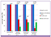
Comparison of survival rates of several murine peritonitis model by each carbapenem-resistant Gram-negative strains were shown in Figure 1. E. coli (EC-1) with 3% hog gastric mucin failed to infect ICR mice but completely killed BALB/c mice at 24 h after injection. For K. pneumoniae (KP-4) and P. aeruginosa (PA-3) infections, both ICR (45% vs 100% in K. pneumoniae [P = 0.036], 45% vs 100% in P. aeruginosa [P = 0.036]) and BALB/c (17% vs 100% in K. pneumoniae [P = 0.001], 34% vs 100% in P. aeruginosa [P = 0.009]) mice treated with mucin showed significantly higher mortality. For A. baumannii (Aci_100087), both ICR (11% vs 100%, P = 0.015) and BALB/c (34% vs 100%, P = 0.009) mice treated with mucin exhibited higher lethality than those without mucin treatment. Although ICR mice exhibited higher mortality than BALB/c mice, there were no significant differences in lethality in both mouse strains.
Taken together, these findings demonstrated that the use of 3% hog gastric mucin could increase infection rates in mice. However, there were no significant differences in fatalities between ICR and BALB/c mice infected with any of the bacterial strains, with the exception of E. coli (EC-1, P <0.001).
In this study, we infected ICR mice since this strain is known to have good reproductive performance and has been used extensively in animal experiments. BALB/c mice, which are frequently used for a variety of immunological studies, including vaccine development, and in studies of infectious diseases [15], were also evaluated. Based on the result of our previous research, in which injection of 105 CFU virulent pneumococcus without mucin completely killed both ICR and BALB/c mice, we established our first experimental conditions that did not use hog gastric mucin. However, we failed to induce infection with clinical isolates of carbapenem-resistant Gram-negative E. coli (EC-1), K. pneumoniae (KP-4), P. aeruginosa (PA-3), and A. baumannii (Aci_100087) in both mouse strains. Several reports have described the use of hog gastric mucin as a virulence-enhancing factor. Mucin is the macromolecular component of mammalian mucus, and hog gastric mucin is purified from the hog gastric mucosa of healthy pigs. In 1932, researchers found that the pathogenicity of bacteria was increased by admixture with hog gastric mucin, whereas mucin alone did not cause death in mice if injected into the peritoneal cavity [16]. Although the exact pathophysiological mechanism through which mucin increases bacterial virulence is still uncertain, after numerous hypotheses and observations, researchers have concluded that intraperitoneal infection with mucin can be attributed to its coating effect on bacteria. Keefer et al. showed that administration of mucin profoundly lowered the levels of the normal serum bactericidin, properdin, resulting in enhanced virulence of the bacteria [17]. In another study mucin was employed to replenish nutrition for Enterococci just before infecting mice [18]. To determine the bacterial inoculum in this study, we referred to a journal of Fattom et al. [19], whose group also used mucin to induce peritonitis. According to their method, we infected ICR and BALB/c with 105-107 CFU of carbapenem-resistant Gram-negative bacteria using mucin.As a result, mice showed lethal effect with inoculum 10
7 CFU. Given this information, we designed our intraperitoneal infection model with inoculum 107 CFU plus 3% hog gastric mucin for the second trial. Finally, our research results demonstrated that carbapenem-resistant K. pneumoniae (KP-4), P. aeruginosa (PA-3), and A. baumannii (Aci_100087) could successfully infect both ICR and BALB/c mice when mucin was added. The differences in lethality between ICR and BALB/c for the three bacterial strains were statistically insignificant. However, the carbapenem-resistant E. coli (EC-1) could infect only BALB/c mice even with mucin treatment, showing statistically significant difference between ICR and BABL/c mice. It is unclear whether there is a significant relation between murine species and bacterial pathogens, which needs to be further studied in the future. Hence, when testing carbapenem-resistant strains including E. coli, intraperitoneal injection of the bacterial strains with 3% hog gastric mucin to BALB/c mice would be suitable. Furthermore, the animal model developed in this study could be widely and easily applied to investigate the pathophysiology of carbapenem-resistant Gram-negative bacteria detected in Korea and to determine the usefulness of testing when aiming to cure infectious diseases caused by these bacterial strains.
 XML Download