

 PDF
PDF ePub
ePub Citation
 Print
Print


Introduction
Severe fever with thrombocytopenia syndrome (SFTS) is an emerging infectious disease caused by the newly discovered SFTS Bunyavirus (SFTSV) [1]. SFTS can be life threatening and is characterized by sudden onset of fever, thrombocytopenia, gastrointestinal symptoms, and leukopenia. First identified in China in 2010, cases of SFTS have also been reported in South Korea and Japan since 2013 [123]. While the mortality rate of SFTS reported in China ranges from 6 to 12%, the mortality rate in South Korea and Japan is much higher at 45 to 55%, although the patient population of these two countries is relatively small [1245].
Hemophagocytic lymphohistiocytosis (HLH) is another potentially fatal syndrome involving pathological immune activation, which is characterized by clinical signs and symptoms of extreme inflammation [6]. It is associated with a variety of infections, among which Epstein–Barr virus (EBV) is the most frequent cause.
Upon examining the relevant clinical literature published, we found a few publications reporting patients with SFTS presenting with HLH [78]. So far, there has been no case report on SFTS associated with HLH in the English literature. Herein, we report the first case of SFTS associated with HLH in Korea.
Case report
On July 2, 2013, a 73-year-old male farmer, residing in the countryside near Ulsan, South Korea, developed fever. His fever did not subside over the next 3 days, so he was admitted to a local hospital on July 5. One day after admission, the patient experienced a sudden drop in blood pressure. Thus, the patient was transferred to our facility (Ulsan University Hospital, an 800-bed teaching hospital), while receiving dopamine infusions.
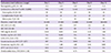
When the patient was admitted to our facility, he was awake and alert. The patient had myalgia, headache, and diarrhea with a frequency of 3–4 times per day. He had no medical history other than a 10-year history of hypertension, for which he took medication. The patient did not recall being bitten by any insect. Physical examination revealed no lymph node enlargement or rash. His temperature was 38.5°C, blood pressure 122/70 mmHg with dopamine infusions at the rate of a 10 µg/kg/min, and heart rate 87 beats per minute. Laboratory test results showed bicytopenia and elevated serum aspartate aminotransferase, alanine aminotransferase, creatinine kinase, lactate dehydrogenase, and ferritin (Table 1). Prothrombin time, activated partial prothrombin time, and C-reactive protein levels revealed normal results. A neck, chest, and abdominal computed tomography (CT) scan did not reveal any abnormal findings. Ceftriaxone and doxycycline therapy was started on the first day of admission.
Table 1
Laboratory test results of the patient
On the second day of admission, the patient’s blood pressure stabilized; thus, we stopped the dopamine infusion. However, the patient became drowsy and disoriented regarding time and location, although he was able to recognize the persons around him. Cerebrospinal fluid analysis revealed no erythrocytes or leukocytes, and chemistry profile was normal. Brain CT showed no signs of hemorrhage, infarction, or other abnormalities. Bone marrow was examined because of the presence of pancytopenia. Bone marrow aspirate revealed proliferation of histiocytes with prominent hemophagocytosis. (Fig. 1). Natural killer (NK) cell activity had decreased to 7.6% (normal range: 18–40%) and triglyceride levels had increased to 320 mg/dl (normal range: 0–250 mg/dl). However, fibrinogen was normal at 262 mg/dl (normal range: 200–400 mg/dl).
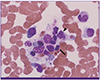
Figure 1
The smear of the bone marrow aspiration showed hemophagocytosis of erythroid (arrow) and myeloid precursors (arrowhead) by histiocytes (Wright-Giemsa stain, 1,000×).
Regarding the cause of fever, repeated blood culture and serology test results did not indicate the presence of leptospira, Orientia tsutsugamushi, Hantaan virus, hepatitis A/B/C virus, human immunodeficiency virus, EBV, cytomegalovirus, or parvovirus. On the fifth day of admission, reverse transcriptase polymerase chain reaction (RT-PCR) test results from the Korean Centers for Disease Control and Prevention, which we had requested on the third day of admission, indicated the presence of the SFTSV. At this point, the patient no longer had fever. A phylogenetic tree was constructed by the neighbor-joining method based on the partial M (Fig. 2A) and S segment (Fig. 2B) sequences of the strain from our patient and 15 SFTSVs from China and Japan registered in GenBank. The tree indicated that the strain was closely related to the SFTSV isolates from China and Japan. With supportive care, the patient’s mental state had fully recovered by the seventh day of admission. As the white blood cell count, platelet count, and aminotransferase levels had normalized on day 11 of admission, the patient was discharged.
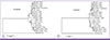
Figure 2
Phylogenetic analysis of the SFTSV KAGBH3 strain (the strain from our patient) based on the partial viral genome sequences (560 bp), which were included in glycoprotein Gn of M segment sequences (A) and nucleocapsid protein of S segment sequences (B). The phylogenetic trees that are shown were generated by MEGA version 5.2 software from aligned nucleotide sequences of 16 isolates of phleboviruses, including the identified SFTSV. Heartland virus was used as the outgroup. The phylogenetic analysis is comparing M and S partial nucleotide sequences of SFTSV KAGBH3 strain with homologous sequences of previously characterized SFTSVs. Sequences were analyzed by the neighbor-joining method based on the maximum composite likelihood model. The minimal length trees shown were supported as the majority rule consensus tree in 5,000 replicates. The bootstrap replicates supporting each node are indicated. KAGBH3 strain is marked with black circles.
Discussion
Our patient underwent bone marrow biopsy because he presented with a fever of unknown cause with severe neutropenia (absolute neutrophil count of 332/mm3) and thrombocytopenia. He presented with fever, pancytopenia, hyperferritinemia, hypertriglyceridemia, low NK-cell activity, and hemophagocytosis in the bone marrow, fulfilling the diagnostic criteria for HLH [6]. We could find a clinical report that describes patients having SFTS presenting with HLH [8]. Weng et al. reported that among 12 patients diagnosed with SFTS, five cases were confirmed as having HLH [8]. In addition, reports regarding the bone marrow biopsy findings of SFTS patients are available. According to QuanTai et al., among 10 patients who underwent bone marrow biopsy, none exhibited hemophagocytosis in the bone marrow aspirate [7]. In contrast, Takahashi et al. reported that among five SFTS patients who underwent bone marrow biopsy, all five exhibited hemophagocytosis in the bone marrow aspirate [2]. Compared with the rarity of HLH presenting with hemorrhagic fever with renal syndrome due to Hantavirus of the same Bunyaviridae family, SFTS may be commonly accompanied by HLH, as per the reports of Weng et al. and Takahashi et al. [28].
Both SFTS and HLH are potentially life-threatening syndromes. According to Weng et al., two of the five patients that presented with HLH did not survive, and while the number of SFTS patients with HLH is low, their mortality rate of 40% is quite high. The known factors indicating poor prognosis of SFTS include old age, central nervous system (CNS) symptoms, and disseminated intravascular coagulation [1234]. Poor prognostic factors for HLH also include old age, CNS symptoms, and high ferritin levels [69]. Although the present patient was old and exhibited CNS symptoms, he recovered fully with supportive care such as inotropic agents and intravenous fluid therapy.
Currently, there is no specific treatment for SFTS other than supportive care, although treatments for patients with HLH whose clinical condition is deteriorating rapidly include chemotherapy and immunotherapy to reduce inflammation [6]. As such, treatments targeting HLH could be considered for SFTS patients with associated HLH whose clinical condition is deteriorating. Thus, if an SFTS patient presents with severe neutropenia and high ferritin levels, a bone marrow biopsy could be conducted to test for the presence of HLH. In this case, the clinical condition of the patient gradually improved, and no treatment, such as chemotherapy or steroids, was administered.
We report a case of a patient with SFTS presenting with HLH, who recovered fully with only supportive care, despite having poor prognostic factors, including old age and altered mental status. We recommend that clinicians consider the possibility of HLH in patients with SFTS presenting with severe neutropenia and high ferritin levels.
 XML Download