

 PDF
PDF ePub
ePub Citation
 Print
Print


Introduction
Cytomegalovirus (CMV) disease is an important cause of morbidity and mortality among patients with impaired cellular immunity, including patients who have received solid organ transplants (SOT) or hematopoietic stem cell transplants (HCT), are undergoing chemotherapy for malignant disease, or are HIV-infected [1]. Of the various end organ diseases of CMV infection, CMV retinitis is the most common, and is the leading cause of disability in HIV-infected patients [2]. In addition, since the numbers of patients with SOT, HCT, and malignancy undergoing chemotherapy are increasing, the incidence of CMV retinitis is expected to increase [3]. Hence, accurate diagnosis and treatment of CMV retinitis is important in HIV-uninfected immunocompromised patients as well as in HIV-infected patients.
CMV retinitis is diagnosed by experienced ophthalmologists based on the patient’s medical history, an ophthalmoscopic appearance typical of CMV retinopathy, and laboratory assessment of immune status that eliminates HIV retinopathy, toxoplasma retinitis, acute retinal necrosis, progressive outer retinal necrosis, and syphilitic retinochoroiditis [4]. The characteristic appearance of the infection is sufficiently distinctive that other diagnostic procedures are rarely needed. However, atypical cases, or infections with other organisms in patients at high risk of CMV retinitis, may require invasive diagnostic tests such as vitreous sampling. Furthermore, even invasive diagnostic tests may fail to establish the diagnosis. Since 1980, non-invasive diagnostic methods, such as the CMV antigenemia assay, have been widely used as adjunct tests for the diagnosis of tissue-invasive CMV diseases. However, data on the diagnostic value of the CMV antigenemia assay for CMV retinitis are limited [5]. Therefore, to assess its value, we compared the sensitivity of the CMV antigenemia assay to diagnose CMV retinitis in HIV-infected and HIV-uninfected patients.
Materials and Methods
1. Patient selection and method of CMV antigenemia assay
We reviewed the medical records of patients diagnosed with CMV retinitis between January 2005 and December 2013 in a 2,700-bed tertiary-care hospital in Seoul, Korea, where the prevalence of HIV infection is low. The CMV antigenemia assays used were the Light Diagnostics CMV pp65 antigenemia assay (Millipore Corp., Temecula, CA, USA), used from January 2005 to February 2012, and the CINAkit Rapid Antigenemia assay (Argene, North Massapequa, NY, USA), used from March 2012 to December 2013. These were performed as previously described [6]. Briefly, about 5 mL of blood was mixed with 1.5 mL of 6% dextran in saline and allowed to sediment. The polymorphonuclear leukocyte-enriched supernatant was extracted. Contaminating erythrocytes were lysed with a 0.8 NH4Cl solution. After two washing steps, leukocyte suspensions were counted and adjusted to 1 × 10⁶ cells/mL. A total of approximately 2 × 10⁵ cells were spotted on a slide by cytocentrifugation, fixed in a 5% formaldehyde solution, and stained by immunofluorescent assay using a monoclonal antibody directed against the pp65 CMV antigen [7]. Counts were expressed as positive cells per 2 × 10⁵ leukocytes [89].
In kidney transplantation recipients, repeated CMV antigenemia assays were performed to monitor CMV infection at 1, 2, 3, 4, 6, 8, 12, 16, 20, and 24 weeks after transplantation [10]. In patients who underwent HCT, CMV antigenemia assays were performed weekly from day 21 to day 100 post-HCT, then monthly until one year after HCT [11]. Surveillance testing using the CMV antigenemia assay in patients with other underlying diseases was performed at the discretion of the attending physician.
2. Definition of CMV retinitis and outcomes
CMV retinitis was defined as presence of the characteristic ophthalmoscopic picture of necrotizing retinitis with or without hemorrhage, as determined by an ophthalmologist [4]. The diagnosis of CMV retinitis was confirmed by review of fundoscopic findings by an experienced retinal specialist JY Lee.
Best corrected visual acuity (BCVA) was measured using Snellen charts. BCVA was assessed whenever patients visited the ophthalmologist. Stable vision was defined as either the maintenance or improvement of BCVA subsequent to receiving a diagnosis of CMV retinitis. Worsening vision was defined as BCVA declining by ≥1 line. Visual loss was defined as a complete lack of light perception. “Default” was defined as being lost to follow-up because of death or transfer to another hospital. “Other” was defined when the result could not be analyzed because of the presence of other ophthalmologic diseases.
3. Literature Review
We searched PubMed (which contains citations over the 30 years from 1985 to September 2014) for English-language literature on HIV-uninfected patients aged older than19 years with CMV retinitis. We used the search terms "cytomegalovirus", "retinitis", "NOT acquired immunodeficiency syndrome", and "NOT HIV". A total of 196 articles were found; 12 included the results of the CMV antigenemia assays. In addition, we searched the Korean-language literature in KoreaMed in a similar manner.
4. Statistical analysis
All statistical analyses were performed using SPSS version 21.0 (SPSS, Chicago, IL, USA). Categorical variables were compared using Fisher’s exact test or Pearson chi-square test, as appropriate. Continuous variables were compared using the Mann-Whitney U-test or Student’s t-test. All tests were two-tailed and differences were considered significant at P <0.05.
Results
Fifty-two patients with CMV retinitis were identified. Eight were excluded because they had no CMV antigenemia assay performed. Of the remaining 44 patients, 31 (70%) were HIV-uninfected. The cutoff value for a positive CMV antigenemia assay was set at one positive cell per 2 × 10⁵ leukocytes. The results were positive in 29 (66%) of 44 patients and negative in the remaining 15 (34%) patients. The baseline clinical characteristics and outcomes of the patients with positive and negative CMV antigenemia results are presented in Table 1. The overall sensitivity of the CMV antigenemia assay was 66% (95% confidence interval [CI], 50–80%) (Table 2), showing a tendency to be higher in the HIV-infected patients (85% [11/13]) than in the HIV-uninfected patients (58% [18/31]), but this effect did not reach statistical significance (P = 0.16). Of the 31 HIV-uninfected patients, 10 had undergone SOT and 13 had undergone HCT. CMV retinitis was diagnosed at a median interval of 190 days (interquartile range [IQR] 140–430 days) and 84 days (IQR 44–233 days) post-transplantation in those who received SOT and HCT, respectively. Of these 23 patients, 5 (22%) had neutropenia (absolute neutrophil count ≤1.5 cells × 109/L) at the time they were diagnosed with CMV retinitis and 2 (9%) had severe neutropenia (absolute neutrophil count ≤0.5 cells × 109/L). The sensitivity of the CMV antigenemia assay in neutropenic patients was 40% (2/5) and in non-neutropenic patients was 61% (11/18). If patients with concurrent CMV diseases (n = 9) were excluded from the analysis, the sensitivity of the CMV antigenemia assay was 57% (20/35, 95% CI 40–74%) (Table 2). Twenty-seven of the 35 patients without concurrent CMV diseases were HIV-uninfected; the sensitivity of the assay in HIV-uninfected and HIV-infected patients was 52% (14/27) and 75% (6/8), respectively.
Table 1
Clinical characteristics and outcomes in patients with CMV retinitis who showed positive and negative CMV antigenemia result
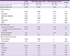
Data are presented as number (%), unless otherwise indicated.
aTwo patients had thymomas and one had a pancreatic cancer.
bOne patient without underlying disease was treated in the intensive care unit for one month because of complicated methicillin-sensitive Staphylococcus aureus bacteremia with multiple metastatic infections; another had dyskeratosiscongenita, and the third had interstitial lung disease; the latter two patients were treated with cyclosporine.
CMV, cytomegalovirus; SD, standard deviation; HCT, hematopoietic stem cell transplants; HIV, human immunodeficiency virus; GI, gastrointestinal.
Table 2
Comparison of the sensitivity of the CMV antigenemia assay in HIV-uninfected and HIV-infected patients with CMV retinitis

Overall, 23 (52%) patients had stable visual outcomes. Two of these underwent vitrectomy due to vitreous hemorrhage; their BCVAs improved. During the treatment of CMV retinitis, BCVA declined in 8 patients (17%) and 4 (9%) had no light perception. Three patients (7%) with CMV-related retinal detachment underwent retinal re-attachment surgery. The BCVA had deteriorated in 2 of these patients, and the remaining patient had no light perception. There were no statistically significant differences in outcomes between the HIV-infected and HIV-uninfected patients. The median follow-up duration of the 35 patients who did not default was 16.7 months (IQR 157–1,086 months).
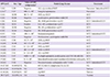
We performed a literature review to determine the performance of the CMV antigenemia assay in CMV retinitis in HIV-uninfected patients (Table 3). CMV antigenemia assay results were available for a total of 18 patients among all the patients included in the 12 articles identified. The overall sensitivity of the CMV antigenemia assay in the literature review was 61% (11/18).
Table 3
Literature review of the results of the CMV antigenemia test in HIV-uninfected patients with CMV retinitis
| UPN [ref] | Sex / Age | Value of CMV antigenemiaa | Underlying disease | Treatment |
|---|---|---|---|---|
| 1 [12] | F/20 | negative | ALL s/p unrelated BMT | Foscarnet / intravitreal GCV |
| 2 [13] b | F/27 | 600 / 2 × 10⁵ | Lung transplantation | Foscarnet |
| 3 [14] | M/58 | 66 / 5 × 10⁴ | NHL s/p ASCT | GCV |
| 4 [15] | M/26 | Negative | azathioprine, prednisolone with CVI | GCV |
| 5 [16] | M/42 | Negative | FK, MMF, prednisolone s/p KT | GCV / intravitreal GCV |
| 6 [16] | M/31 | Positive | CsA, MZ, prednisolone s/p KT | GCV |
| 7 [16] | M/50 | Negative | FK, MZ, prednisolone s/p KT | GCV |
| 8 [16] | M/42 | Negative | FK, MMF, prednisolone s/p KT | GCV |
| 9 [17] | F/51 | >50 / 4 × 10⁵ | Immunocompetent | GCV / intravitreal GCV |
| 10 [18] | F/51 | Negative | Prednisolone, azathioprine with DM | GCV |
| 11 [19] | F/36 | 12 / 2 × 10⁵ | AML s/p BMT | GCV |
| 12 [20] | M/38 | 10 / 1.5 × 10⁵ | ALL s/p BMT | GCV |
| 13 [20] | F/43 | 6 / 1.5 × 10⁵ | NHL s/p BMT | GCV |
| 14 [20] | F/57 | 28 / 1.5 × 10⁵ | AML s/p BMT | GCV / intravitreal GCV |
| 15 [20] | F/41 | 4 / 1.5 × 10⁵ | ALL s/p BMT | GCV |
| 16 [21] | F/57 | 2400 / 5 × 10⁵ | Cyclophosphamide, azathioprine, anti-TNF Ab with RA | GCV |
| 17 [22] | F/61 | 9 / 2 × 10⁴ | Intravitreal bevacizumab injection with diabetes retinopathy | GCV |
| 18 [23] | F/52 | Negative | Dexamethasone, cyclosporine with T-LGLL | GCV / intravitreal GCV |
| 19-27 [24] | M (88%)/NA | NA | KT (1), SLE (1), lymphoma (3), AA (4) | NA |
| 28-42 [25] | M (93%)/36 | NA | AIDS | various |
| 43-58 [26] | M (81%)/33 | NAc | s/p BMT | various |
| 59-77 [27] | M (63%)/14 | NA | various | various |
aData are number of positive cells per number of leukocytes.
bOnly UPN 2 had another concurrent CMV disease: CMV pneumonia.
cSix among 16 CMV retinitis patients (38%) showed positive results in either CMV antigenemia assay or CMV PCR.
UPN, unique patient number; CMV, cytomegalovirus; ALL, acute lymphoblastic leukemia; BMT, bone marrow transplantation; GCV, ganciclovir; NHL, non-Hodgkin's lymphoma; ASCT, autologous; stem cell transplantation; CVI, common variable immunodeficiency; FK, tacrolimus; MMF, mycophenolate mofetil; KT, kidney transplant; CsA, cyclosporine A; MZ, mizoribine; DM, dermatomyositis; AML, acute myeloid leukemia; TNF, tumor necrosis factors; RA, rheumatoid arthritis, T-LGLL, T cell large granular lymphocytic leukemia; s/p, status post; SLE, systemic lupus erythematosus; AA, aplastic anemia; AIDS, acquired immunodeficiency syndrome; NA, not applicable
Discussion
The CMV antigenemia assay has been used to detect CMV reactivation in a variety of clinical settings [528]. It has the advantages of providing results within 24 hours and being quantifiable. Its introduction has considerably improved the clinical management of CMV disease in allogeneic HCT and SOT recipients, using a preemptive approach. However, studies of the diagnostic value of the CMV antigenemia assay in CMV retinitis are limited. We previously showed that the CMV antigenemia test was positive in 54% (26/57; 95% CI 41–68%) of patients with CMV gastrointestinal disease and 69% (25/36; 95% CI 52–84%) of patients with CMV pneumonia, suggesting that it has low sensitivity for ruling out these 2 conditions [89]. Xharrd et al. [3] reported that the CMV antigenemia test or CMV PCR were negative in all of 6 hematopoietic stem cell transplant patients with CMV retinitis. Eid et al. [29] also found that only 4 of 9 (44%; 95% CI 18–74%) solid organ transplant recipients with CMV retinitis tested positive for CMV PCR or CMV culture. In the present study, the test’s sensitivity in HIV-uninfected patients was 58% (18/31; 95% CI 42–74%). If patients with other concurrent CMV infections were excluded from the analysis, the sensitivity was 52% (14/27; 95% CI 33–70%), suggesting that the assay is not useful for ruling out CMV retinitis. Interestingly, there was a statistically non-significant trend for the sensitivity in HIV-uninfected patients to be lower (58%) than in HIV infected patients (85%, P = 0.16). Such variable sensitivity might possibly reflect differences in the causes of CMV retinitis in HIV-uninfected patients compared with HIV-infected patients. Further studies are needed in this area.
Our study had several limitations. First, by virtue of its retrospective design, we could not gather clinical data at the time of presentation in all of the patients. Furthermore, because the patient population was heterogeneous, follow-up was not standardized. Second, due to the specific circumstances of the Korean national medical insurance system, CMV PCR was not routinely performed for surveillance. The CMV antigenemia assay does not require expensive equipment and the associated costs are low. But it is time-consuming, labor-intensive, and requires a high level of technical expertise. In addition, there is no assay standardization and poor inter-institutional correlation exists. False negative results for CMV antigenemia can occur in patients with leukopenia [28303132]. Furthermore, CMV antigenemia has a lower sensitivity than CMV PCR. However, in the light of previous results, it can be assumed that the use of PCR would not have yielded very different results [11]. Third, because patients who did not have CMV antigenemia assay results were excluded from the study population, there may have been some selection bias. Lastly, CMV retinitis is a rare disease. Therefore, the study patients were collected over a 9-year period. The medical management of patients after transplantation or on immunosuppressive therapy clearly changed over this period.
In conclusion, the CMV antigenemia assay has limited value as a noninvasive diagnostic adjunct test for CMV retinitis. Because of the high cost of a false-negative diagnosis of CMV retinitis the results of the assay need to be interpreted in the context of the underlying disease, clinical presentation, and ophthalmoscopic findings.
 XML Download