

 PDF
PDF ePub
ePub Citation
 Print
Print


Abstract
Background
There have been some investigations concerning the role of p53, p21 and bcl-2 protein expressions for the tumorigenesis of thyroid cancer. It had been debated that these protein expressions were associated with aggressive features of papillary thyroid carcinoma. We studied to evaluate the prevalence of these protein expressions and their clinical significances in papillary thyroid carcinoma.
Methods
We selected 49 patients with papillary thyroid carcinoma who had been operated on at Samsung Medical Center during the last 10 years. Immunohistochemical staining for p53, p21 and bcl-2 was done by the use of paraffin embedded tissues. We analyzed the results of immunohistochemical staining for p53, p21 and bcl-2 and the correlation with the patients' age, gender, tumor size, multifocality, tumor invasion to both lobes, extrathyroidal invasion, cervical lymph node invasion, distant metastasis and the clinical outcomes.
Results
Immunohistochemical staining for p53 was positive in 10 patients (20%), p21 was positive in 36 patients (73%) and bcl-2 was positive in 18 patients (37%). The p53 and bcl-2 expressions were not associated with the clinical parameters. Tumor multifocality and extrathyroidal invasion were significantly higher in the p21 positive group (both P < 0.05).
Figures and Tables
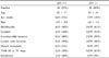
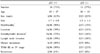
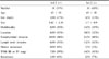
Table 1
Analysis of clinicopathologic data according to the p53 immunohistochemical stains in papillary thyroid carcinoma
References
1. Farid NR, Shi Y, Zou M. Molecular basis of thyroid cancer. Endocr Rev. 1994. 15:202–232.
2. Ito T, Seyama T, Mizuno T, Tsuyama N, Hayashi T, Hayashi Y, Dohi K, Nakamura N, Akiyama M. Unique association of p53 mutations with undifferentiated but not with differentiated carcinomas of the thyroid gland. Cancer Res. 1992. 52:1369–1371.
3. Patel KN, Singh B. Genetic considerations in thyroid cancer. Cancer Control. 2006. 13:111–118.
4. Basolo F, Pollina L, Fontanini G, Fiore L, Pacini F, Baldanzi A. Apoptosis and proliferation in thyroid carcinoma: correlation with bcl-2 and p53 protein expression. Br J Cancer. 1997. 75:537–541.
5. Moore D, Ohene-Fianko D, Garcia B, Chakrabarti S. Apoptosis in thyroid neoplasms: relationship with p53 and bcl-2 expression. Histopathology. 1998. 32:35–42.
6. Godballe C, Asschenfeldt P, Jorgensen KE, Bastholt L, Clausen PP, Hansen TP, Hansen O, Bentzen SM. Prognostic factors in papillary and follicular thyroid carcinomas: p53 expression is a significant indicator of prognosis. Laryngoscope. 1998. 108:243–249.
7. Omar E, Madhavan M, Othman NH. Immunohistochemical localisation of RET and p53 mutant protein of thyroid lesions in a North-Eastern Malaysian population and its prognostic implications. Pathology. 2004. 36:152–159.
8. Harper JW, Adami GR, Wei N, Keyomarsi K, Elledge SJ. The p21 Cdk-interacting protein Cip1 is a potent inhibitor of G1 cyclin-dependent kinases. Cell. 1993. 75:805–816.
9. el-Deiry WS, Tokino T, Velculescu VE, Levy DB, Parsons R, Trent JM, Lin D, Mercer WE, Kinzler KW, Vogelstein B. WAF1, a potential mediator of p53 tumor suppression. Cell. 1993. 75:817–825.
10. Michieli P, Chedid M, Lin D, Pierce JH, Mercer WE, Givol D. Induction of WAF1/CIP1 by a p53-independent pathway. Cancer Res. 1994. 54:3391–3395.
11. Parker SB, Eichele G, Zhang P, Rawls A, Sands AT, Bradley A, Olson EN, Harper JW, Elledge SJ. p53-independent expression of p21Cip1 in muscle and other terminally differentiating cells. Science. 1995. 267:1024–1027.
12. Johnson TL, Lloyd RV, Thor A. Expression of ras oncogene p21 antigen in normal and proliferative thyroid tissues. Am J Pathol. 1987. 127:60–65.
13. Mizukami Y, Nonomura A, Michigishi T, Noguchi M, Nakamura S, Hashimoto T. Differential (Ha-, K- and N-) ras p21 expression in benign and malignant human thyroid tumors: an immunohistochemical study. Anticancer Res. 1995. 15:755–759.
14. Okayasu I, Osakabe T, Onozawa M, Mikami T, Fujiwara M. p53 and p21 (WAF1) expression in lymphocytic thyroiditis and thyroid tumors. Clin Immunol Immunopathol. 1998. 88:183–191.
15. Akslen LA, Varhaug JE. Oncoproteins and tumor progression in papillary thyroid carcinoma: presence of epidermal growth factor receptor, c-erbB-2 protein, estrogen receptor related protein, p21-ras protein, and proliferation indicators in relation to tumor recurrences and patient survival. Cancer. 1995. 76:1643–1654.
16. Basolo F, Pinchera A, Fugazzola L, Fontanini G, Elisei R, Romei C, Pacini F. Expression of p21 ras protein as a prognostic factor in papillary thyroid cancer. Eur J Cancer. 1994. 30:171–174.
17. Pilotti S, Collini P, Rilke F, Cattoretti G, Del Bo R, Pierotti MA. bcl-2 protein expression in carcinomas originating from the follicular epithelium of the thyroid gland. J Pathol. 1994. 172:337–342.
18. Siironen P, Nordling S, Louhimo J, Haapiainen R, Haglund C. Immunohistochemical expression of bcl-2, Ki-67, and p21 in patients with papillary thyroid cancer. Tumour Biol. 2005. 26:50–56.
19. AJCC cancer staging manual. 2002. 6th ed. New York: Springer-Verlag Press;77–79.
20. Goto A, Sakamoto A, Machinami R. An immunohistochemical analysis of cyclin D1, p53, and p21waf1/cip1 proteins in tumors originating from the follicular epithelium of the thyroid gland. Pathol Res Pract. 2001. 197:217–222.
21. Hay ID. Papillary thyroid carcinoma. Endocrinol Metab Clin North Am. 1990. 19:545–576.
22. Furth ME, Aldrich TH, Cordon-Cardo C. Expression of ras proto-oncogene proteins in normal human tissues. Oncogene. 1987. 1:47–58.
23. Suarez HG, Du Villard JA, Caillou B, Schlumberger M, Tubiana M, Parmentier C, Monier R. Detection of activated ras oncogenes in human thyroid carcinomas. Oncogene. 1988. 2:403–406.
24. Wright PA, Lemoine NR, Mayall ES, Wyllie FS, Hughes D, Williams ED, Wynford-Thomas D. Papillary and follicular thyroid carcinomas show a different pattern of ras oncogene mutation. Br J Cancer. 1989. 60:576–577.
25. Namba H, Rubin SA, Fagin JA. Point mutations of ras oncogenes are an early event in thyroid tumorigenesis. Mol Endocrinol. 1990. 4:1474–1479.
26. Konishi N, Enomoto T, Buzard G, Ohshima M, Ward JM, Rice JM. K-ras activation and ras p21 expression in latent prostatic carcinoma in Japanese men. Cancer. 1992. 69:2293–2299.
27. Ito Y, Kobayashi T, Takeda T, Komoike Y, Wakasugi E, Tamaki Y, Tsujimoto M, Matsuura N, Monden M. Expression of p21 (WAF1/CIP1) protein in clinical thyroid tissues. Br J Cancer. 1996. 74:1269–1274.
 XML Download