

 PDF
PDF Citation
 Print
Print


INTRODUCTION
To use the immune system as a weapon against cancer had always been a dream of oncologic research in the last decades. The major traditional approach was to stimulate the immune system. A vast arsenal of strategies was developed, including tumor vaccines, dendritic cell activation and immunostimulants. Despite extensive efforts, no groundbreaking progress has been made. Even though activation of the immune system can be achieved with these traditional techniques, response and tumor cell elimination remain limited. This can be explained by the existence of tumor related immune-escape mechanisms. The immune system provides a strong defense against cancer cells. But once cancer becomes clinically apparent, those defence mechanisms are already breached. In this setting, stimulating the immune system can be compared with stepping on the gas without putting the engine into gear. The cancer has already found ways to surpass the immune system, and therefore a higher level of immune activity is unlikely to reverse that process. The introduction of nivolumab and other checkpoint-inhibitors was the first step to change this situation. This novel approach targets the immune-escape mechanisms. It re-activates the immune defences and enables T-cells to re-gain their anti-tumor-toxicity. This new therapeutic approach has revolutionized the management of tumor entities like melanoma, in which no other standard therapy modality was able to demonstrate survival benefit. The ongoing development has raised high hopes, that this new group of agents could also revolutionize the field of gynecologic oncology. In 2015 the first experience with nivolumab in ovarian cancer (OC) was reported [1]. But to understand the complex background and the new challenges that are associated with the use of these novel agents, several aspects have to be considered.
THE ROLE OF THE IMMUNE SYSTEM IN CANCER CONTROL
The mutation of physiological cells into cancer cells is a phenomenon that occurs on a daily basis in every human. Without the elimination of these cells by the immune system, life would not be possible. A finely orchestrated interaction is neccessary to secure the day-to-day elimination of all tumor cells without the occurence of autoimmune toxicity. The immune system can be divided into two major systems: The much older hereditary “innate system” and the newer “adaptive system.” Both factions generate a humoral as well as a cellular immune response against tumor cells. The major weapon against cancer cells in both systems are the cellular factions. The hereditary system is not specific. It mainly uses natural killer (NK) cells as its cellular agents. These NK-cells check the self major histocompatibility complex class I molecules (MHC-I) on the surface of all cells, which act as an ID-card to identify physiological cells to the immune-system. Cancer cells can loose this MHC-I expression, a phenomenon that was described by Klas Kärre as “missing self” as early as 1980 [2]. NK-cells use a general and basic approach to eliminate all tumor cells with missing self. A more specific strategy is used by the cellular agents of the adaptive system: the T-cells. The name originates from the localisation of their differentation: the thymus. These cells direct their activity against cancer cells, that still express an MHC-I complex. They bind cells with their T-cell-receptor at the MHC-I site, identify tumor specific antigens and eliminate tumor cells by lysis or apoptosis-induction. Taking into account the daily occurence of cancer cell mutation and the horrific growth rate of tumor cells that results from exponential duplication, the immune system is a marvelous tool of unmatched efficiency and balance. It very rarely underperforms allowing cancer to become clinically apparent or overperforms which results in autoimmune toxicity. In exeptional cases of underperformance, the cause can be immune-suppression or hereditary malfunction, but this is generally compounded by cancer-related cleverly devised methods of immune escape.
CANCER-RELATED METHODS OF IMMUNE-ESCAPE
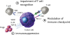
Despite extensive autonomous defense mechanisms, cancer cells can find a way to breach that defense, become clinically apparent and multiply uncontrollably until eventually the host dies. The whole spectrum of immune-escape pathways used by cancer remains unkown, but several methods have been discovered. The current perception of cancer-related methods of immune-escape can be divided into three main strategies (Fig. 1) [34]. The first being the impairment of tumor cell recognition by T-cells. This invisible-cloak-strategy is based on the down regulation of antigen-presentation and loss of MHC-I-complex expression by tumor cells. The second method involves the creation of an immunosuppressive micro-environment. This is facilitated by cancer cells with the secretion of immuno-active agents like interleukin (IL)-10, vascular endothelial growth factor (VEGF), C-C motif chemokine ligand 21 (CCL-21), indoleamine 2,3-dioxygenase (IDO), and transforming growth factor (TGF)-β. Immunosuppresion can be achieved by these agents themselves, as well as through the recruitment, acceleration of differentiation and expansion of immunosuppressive cells, namely regulatory T-cells. This cancer-initiated immunosuppression is essential for the early survival of cancer cells, since it initially enables enough cancer cells to withstand the attack of the immune-system to multiply. But this early cancer survival strategy itself can pose a vital threat to the host, because newly created cells will start secretion of immunosuppressive agents. This synergy can culminate into a snowball-effect and results in total immunosuppression. At this point, the host can be very vulnerable to standard infection, a state that can regularly be witnessed in the clinic in cases of advanced cancer with a high tumor burden. The last strategy uses a safety feature of the host's immune-system called “the immune checkpoint”. This checkpoint mechanism is designed to prevent cytotoxic T-cells from inflicting autoimmune damage to healthy tissue. Cancer evolution has somehow highjacked that mechanism and used it to block T-cell aggression against tumor cells.
Fig. 1
Tumor-related methods of immune-escape. (A) Downregulation of MHC-I complex or antigen presentation (B) Deactivation of cytotoxic t cell via the PD1-PD-L1 pathway (C) Creation of an immunosuppressive environment by excretion of immunosuppressive agents or recruitment of immunosuppressive cells like Tregs. Modified after Töpfer et al. [3] and Nurieva et al. [4].
IL, interleukin; MHC-I, major histocompatibility complex class I molecules; PD1, programmed cell death protein 1; PD-L1, programmed death-ligand 1; TCR, T-cell receptor; TGF, transforming growth factor; Tregs, regulatory T cells; VEGF, vascular endothelial growth factor.
CHECKPOINT INHIBITION (CI): THE MODE OF ACTION
Activated T-cells are able to destroy or severly damage other cells. The T-cell is activated when it recognizes a specific antigen. The main target of T-cell related cytotoxicity are virus-infected cells or tumor cells. To prevent T-cells from destroying healthy cells, T-cells carry mechanisms to prevent activation despite specific antigen recognition. These mechanisms are complex arrangements of receptors and ligands on the surface of the T-cell and other cells referred to as “immune-checkpoints”. This checkpoint-controlled interaction between T-cells and healthy cells is essential. Misfunction of this vital mechanism results in autoimmune reactions against healthy tissue. The immune checkpoint consists of a complex array of receptors and ligands and remains not fully understood. One pivotal element of that checkpoint mechanism is represented by programmed cell death protein 1 (PD-1)/ programmed cell death-ligand 1 (PD-L1) interaction. PD-1 (or cluster of differentation 279) is a cell surface receptor, found mostly — if not exclusively — on T-cells. PD-L1 (or cluster of differentation 274) is a transmembranous protein found frequently on the surface of human cells. PD-1/PD-L1 interaction results in deactivation of T-cells preventing healthy cells from being a target of T-cell-toxicity. It also plays a major role in pregnancy. Fetal cells carry a completely foreign genetic signature, but because of high PD-L1 expression on fetal cells of the syncythiothrophoblast and the placenta usually no destruction takes place. Cancer cells also take advantage of this mechanism to evade the hosts immune defence by overexpressing PD-L1 and deactivating cytotoxic T-cells. Checkpoint-Inhibitors target this mechanism to restore the T-cells of the host to full anti-tumor activity. While several receptors and ligands have been chosen as targets for CI, the PD-1/PD-L1 pathway remains in the spotlight of pharmaceutical research.
SUBSTANCES IN USE AND IN THE PIPELINE
A variety of antibodies against both PD-1 and PD-L1 have been developed. Most clinical experience up to this point has been gathered with an anti-PD1 blockade, namely with nivolumab and pembrolizumab. Nivolumab is a fully human immunoglobulin G4 (IgG4) anti-PD-1 monoclonal antibody that binds to the PD-1 receptor on the hosts T-cells and blocks ligand activation on the tumor cells PD-L1 and PD-L2 ligand. Pembrolizumab is a humanised anti-PD-1 IgG4 antibody. The hope to create checkpoint-inhibitors with less auto-immune toxicity inspired the development of anti-PD-L1 antibodies, such as atezolizumab, avelumab and durvalumab. While nivolumab and pembrolizumab have reached Food and Drug Administration (FDA)-approval in some entities, the availiability of anti-PD-L1 antibodies is currently limited to clinical studies.
DIAGNOSTIC TOOLS FOR THE DETERMINATION OF PD-1/PD-L1 EXPRESSION
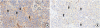
No established biomarkers for the prediction of a response (or a resistance) to checkpoint inhibitors are available to date. Due to function of PD-L1 as a ligand and activator of PD-1, PD-L1 expression, particularly in cancer cells, is the favorite biomarker in this regard and is used as a selection or stratification marker in some clinical trials. However, a clear predictive effect of PD-L1 expression in cancer has not been determined yet. This might be due to the fact that antibodies, evaluation methods and cut-off points for determining positivity vary between studies. A large number of antibodies directed against human PD-L1 are commercially available to date, and their number is increasing continuously. Typically, PD-L1 expression is membranous or membrane-accentuated and frequently a cytoplasmic stain is seen as well. (Fig. 2) Intensity and rate of stained tumor cells varies significantly across tumor entities but also within a given entity (median positivity rate in solid tumors 52.5%) [5]. In a systematic evaluation of PD-1 and PD-L1 expression in 200 primary high-grade serous ovarian carcinomas, a PD-L1 staining of any rate and intensity was seen in 90% of tumors. Applying a more stringent cut-off of 50% of stained cells, the positivity rate was 30% [6]. This is in line with other reports, particularly in lung cancer, where this cut-off has been repeatedly used [5]. The present reports on PD-L1 expression in cancer however are difficult to compare as staining protocols and cut-offs vary significantly. PD-L1 is also detected in tumor-infiltrating lymphocytes (TILs) and typically constitutes a small proportion of the total TILs infiltrate (median numbers in high-grade serous ovarian carcinoma: 8 per 1 mm2 tumor), but also in other immune cells such as macrophages and dendritic cells. PD-1 expression is most often restricted to TILs, however an expression in cancer cells (membranous) has been reported in ovarian and lung carcinoma and seems to occur by the use of particular antibodies [7].
Fig. 2
PD-L1 and PD-1 expression in high-grade serous ovarian carcinoma. (A) A moderate expression is seen in cancer cells. Staining of cell membranes is typically incomplete. Additionally, TILs are stained for PD-L1 (black arrows indicate examples of positive intra-epithelial TILs, white arrows show positive stromal TILs). (B) Numerous PD-1-positive TILs are seen. Arrows indicate examples of positive intra-epithelial TILs.
PD1, programmed cell death protein 1; PD-L1, programmed death-ligand 1; TIL, tumor-infiltrating lymphocyte.
An attempt to standardize PD-L1 and PD-1 expression in cancer cells and TILs is currently being made by the German Societies of Pathologists in their preparation of a round robin trial. Further translational research in current and future clinical trial cohorts on checkpoint inhibitors are necessary to determine whether PD-L1 expression in cancer cells or alternative biomarkers (e.g. PD-L1/PD-1 expression in TILs, mutational burden/microsatellite instability, infiltration of CD8+ T cells) may be optimized as predictive markers for patient selection [89101112].
CI IN OC: CURRENT DATA
To our knowledge, only one study on checkpoint-inhibition in OC has reached full publication up to this point. This single-center, phase II clinical trial by Hamanishi et al. [1] has assessed the efficacy of nivolumab in patients with platinum-resistant, recurrent or advanced OC. The trial was conducted in 20 patients, who were sequentially allocated to a low-dose cohort (cohort 1: 1 mg/kg, n=10) followed by a high-dose cohort (cohort 2: 3 mg/kg, n=10). Both cohorts received nivolumab intravenously biweekly for up to 1 year or until disease progression. The median follow-up time was 11 months and the median treatment time with nivolumab was 3.5 months. Cohort 2 showed a better efficacy with an overall response rate (RR) of 20% compared to cohort 1 (RR=10%). Interestingly, 2 patients in cohort 2 had a complete response (CR). One of the patients had clear-cell carcinoma and continues to experience CR following the 1-year trial. Expected treatment-related adverse events where not different between cohorts. These findings are similar to previous trials of nivolumab in other solid tumors [513]. Therefore, the authors suggest the high-dose cohort may be more favorable in OC as it showed better efficacy and similar toxicity in comparison to the low-dose cohort. Moreover, the durability of the anti-tumor response exhibited across the cohorts (RR=15%; 3/20 patients) was significant, with a median progression-free survival (PFS) of 3.5 months and a median overall survival (OS) of 20 months. Hamanishi et al. [1] noticed that one of the long-term complete responders had clear-cell carcinoma, which has a worse prognosis than serous OC with fewer available effective chemotherapies for recurrent disease. The evidence shows that clear-cell ovarian and renal carcinomas share similar gene expression profiles [14]. Therefore, the authors suggest that nivolumab, which is approved for renal cell carcinoma may also be beneficial in treating clear-cell carcinoma of the ovary.
Another research group surrounding Andrea Varga [15] reported experience with pembrolizumab in OC from the Keynote-028 multi-cohort trial. In this non-randomised basket study a great diversity of PD-1 positive solid tumor entities was enrolled, along with 26 patients with OC. The majority of the patients was in recurrent disease (84.6%) and more than one third (38.5%) were heavily pre-treated (>5 prior lines of chemotherapy). Six patients showed stable disease. Three patients showed response (2 partial response [PR], 1 CR) and all responding patients had a long duration of response of >24 weeks. The best overall response was 11.5% and 23.1% had evidence of tumor reduction.
Infante et al. [16] conducted a study on atezolizumab, an anti-PD-L1 antibody. In contrary to nivolumab and pembrolizumab, it does not bind to the immune cell, but to the T-cell. The number of participating patients is even smaller, but the results are very promising. All of the patients included were heavily pretreated and platinum-resistant. The PD-L1 expression of the immune cells infiltrating the tumor tissue was measured and the patients were allocated into groups accordingly (IC0: <1%, IC1: ≥1 to <5%, IC2: ≥5 to <10%, and IC3: ≥10%). A vast majority of the patients was PD-L1 positive (83%). The study showed an association between the success of atezolizumab therapy and PD-L1 expression: the patient group with the highest PD-L1 expression benefited most from the atezolizumab treatment. While the median progression free survival for the patients in the IC-2 group was 11,3 months, the IC-3 group had a better result with 17,4 months. The overall response rate in this study lies at 25%.
Durvalumab is another anti-PD-L1 antibody [17]. A small study conducted on only 15 heavily pretreated and platinum-resistant patients combined the antibody (administered either every 2 or 4 weeks) with either olaparib or cediranib. A partial response or a stable disease (>4 months) could be achieved in both groups (durvalumab/olaparib: 83%, durvalumab/cediranib: 86%). It is important to mention that Lee [17] did not publish the overall response rate in this study.
The results from the JAVELIN Basket study are especially significant as they included a large number of patients (124, heavily pretreated and platinum-resistant). Disis et al. [18] administered the anti-PD-L1 antibody avelumab biweekly IV as a monotherapy. The overall response rate was 9.7% and there was no statistical significance between the PD-L1 positive and negative group like in atezolizumab. The disease control rate (either partial response or stable disease) was 54% [18].
In 2018 new data were presented at the American Society of Clinical Oncology (ASCO) annual meeting: Konstantinopoulos and colleagues [19] presented data from the TOPACIO trial, in which 62 patients with mostly platinum resistant OC were treated with a combination of pembrolizumab and niraparib. A overall response rate of 25% and a disease control rate of 67% were reported. A group from Norway reported even more promising results for treatment of 18 platinum resistant patients with a PD-1 inhibitor with an overall response rate of 39% and a disease control rate of 44% [20].
In conclusion, these trials demonstrate an activity of CI in recurrent OC. The patients who respond to CI have a longterm benefit from the treatment. Although these first results show overall response rates in OC that no other mono-immunotherapy was able to achieve so far, they cannot be compared to the sensational results in other solid tumors.
The safety profiles in patients with OC seem to be similar to other solid tumors. The key to extraordinary treatment success in OC could lie in patient selection. Identifying patients with a more immunogenic disease and patients with a still functioning immune-system that is not impaired by tumor-related immunosuppresion might be a possible way of selection. Another way to make immune checkpoint inhibitor therapy in OC more successfull may be a combination of CI with other targeted therapies such as cediranib. It has to be stated, that none of the trials reported to date, have combined CI with chemotherapy. Nevertheless, we suggest that this combination could create significant benefitial synergism. Chemotherapy has been proven to induce PD-L1 overexpression in colorectal cancer. It could also deplete immunosuppressive leucocytes such as regulatory T-cells and myeloid-derived supressor cells. But most importanly, chemotherapy will significantly improve response rates and result in output of tumor antigen. All these synergistic effects suggest a strong rationale to combine CI and chemotherapy in OC.
PSEUDOPROGRESSION: A NEW PHENOMENON IN RESPONSE-EVALUATION
The efficacy of oncologic therapy is historically evaluated with imaging techniques. Response Evaluation Criteria In Solid Tumors (RECIST) criteria have provided a platform for therapeutic management decisions based on measurement of tumor size or appearance of new lesions [21]. The development of novel immunotherapeutics such as nivolumab changes the common practice in response evaluation. While some patients under immunotherapeutics show a stable disease or response that can be evaluated with classic RECIST criteria, others present a phenomenon called pseudoprogression [22]. This phenomenon consists of a transient increase in tumor size and even development of new lesions, with a consecutive and delayed shrinkage and response of tumor manifestation below the pre-treatment level. This delayed response can be explained by the underlying mechanism of anti-tumor activity by modern immunotherapy [22]. The efficacy is based on tumor cell destruction by immune cells. This mode of action is often linked to an infiltration of immuno-competent cells into solid tumors leading to edema and necrosis. But even if the therapy is efficient and tumor cells are destructed, immune cell infiltration and edema can lead to growth in tumor size and therefore translate into a mixed response or pseudoprogression in RECIST-based imaging. Even though the expected incidence of pseudoprogression is reported to be low with approximately less than 10% of all patients under immunotherapy, it poses a problem to the clinician [22]. While historically tumor growth was consistent with treatment failure, now the phenomenon of pseudoprogression has to be considered. Standard imaging techniques and RECIST criteria can not evaluate this phenomen sufficiently. This leads to the implementation of immune-related response criteria (irRC), which are used in most current studies on immunotherapeutics as a supplement to the standard RECIST criteria in order to evaluate treatment efficacy [23]. One of the major changes in this novel response evaluation system is that it takes into account a follow up imaging study no less than 4 weeks from the date first documented to evaluate delayed response. Even though the irRC-system remains an attempt to fully understand and objectify response to immunotherapy, it has to be considered when using these novel agents.
BRCAness AND CI
DNA single-strand-breaks occur in every human cell on a daily basis. These breaks are repaired by poly-ADP-ribose-polymerase (PARP). A second repair mechanism, called homologous recombination (HR), acts as a safety feature. In patients with BRCA mutation, this pathway of HR is dysfunctional, resulting in frequent DNA breaks and mutation. Even though BRCA mutation is the best known condition, other patients can also display a HR-deficiency despite being BRCA negative. This phenomenon is referred to as BRCAness and translates into a highly multiplied risk for ovarian and breast cancer just as BRCA positive women.
The concept of CI is based on the recognition of cancer cells by the immune system. Higher mutational load and excessive presentation of neoantigens makes a tumor more immuogenetic and an ideal target for CI [10,11]. Tumor entities such as melanoma are defined by a high mutational burden and neoantigen presentation. Spontaneous regression of the primary site due to immune anti-tumor activity — quite often observed in malignant melanoma — is an expression of the immunogenicity of melanoma. This optimal background for CI in melanoma might explain the revolutionary results of nivolumab in this entity. Even though there is currently no proof of this hypothesis for BRCA mutation, patients with OC and underlying BRCAness could possibly be more sensitive to CI due to a higher mutational load and more neoantigen presentation as a result of HR deficiency. Le and colleagues [11] investigated patients with mismatch repair deficiency (Lynch-Syndrome) in colorectal cancer, a form of DNA repair disorder, similar to BRCA mutation. They found, that mismatch repair deficiency was strongly correlated to treatment success with checkpoint inhibitors. Since germline and somatic BRCA mutation is frequent in OC and is prevalent in approximately 50% of all high-grade serous ovarian carcinoma, this could be clinically revelant [2425]. In the only published experience of nivolumab in OC, BRCA mutation was not assessed [1]. Future trials on CI in OC should consider this background and further investigate this hypothesis.
TOXICITY AND SIDE EFFECT MANAGEMENT
Toxicity of anti-PD1 checkpoint-inhibitors can be explained by the mode of action and therefore result in autoimmune toxicity, that is the reason for most side effects of these agents. The main toxicities can be classified into pulmonary, gastrointestinal, hepatic, renal, endocrinologic and cutaneous side effects. In one of the first phase-I-studies on nivolumab in melanoma reported in 2014, the most common side effects were fatigue (33%), rash (23%), and diarrhea (18%) [13]. Toxicity does not seem to be cumulative and usually occurs within 6 months after start of therapy [13]. Despite the very diverse effector organs of auto-immune toxicity, therapy remains consistent due to the same immunologic pathomechanism that causes the side effects. Most toxicities are treated symptomatic when Common Terminology Criteria for Adverse Events (CTCAE) grade I. Steroids are used in case of toxicities of CTCAE grade II or higher independent of the effector organ. In case of pulmonary, hepatic, and renal toxicities grade II, 0.5–1.0 mg/kg/body weight methylprednisolone or equivalent and pause of CI is recommended. Since endocrinologic toxicities can not be rated in a CTCAE grading system, it is recommended to treat with 1.0–2.0 mg/kg/body weight methylprednisolone in cases of symptomatic endocrinopathies (e.g. hypothyroidism, diabetes) and to postpone application of the ceckpoint-inhibitor. In cases of endocrinologic crisis, discontinuation is mandatory. Steroid doses can be escalated up to 1.0–2.0 mg/kg/body weight in cases of grade IV toxicities independent from the effector organ, but the checkpoint-inhibitor should be permanently stopped at this point. As a general statement, it has to be recommended, that steroid application should be tapered down slowly over no less than 4 weeks. Abrupt discontinuation can lead to escalation of side effects. Before steroid commencement, infection or other possible causes of toxicities have to be excluded. Antibiotic prophylaxis has to be considered in cases of immunosuppressive steroid application.
CONCLUSION
The introduction of nivolumab and other checkpoint-inhibitors has revolutionized the treatment in different tumor entities, such as melanoma and renal cell cancer. Growing experience with checkpoint-inhibitors show difficulties in management of this type of agent. A novel side effect pattern, the missing standard for pathologic PD1/PD-L1 measurement and pseudoprogression pose a challenge to the clinician using checkpoint inhibitors.
The first published experience in OC shows very promising results and a much higher efficacy than expected from previously known mono immuno-therapeutics like vaccines or immunostimulants. However, the positive results in OC do not compare to the extraordinary results in other entities like melanoma, where CI changed the game. The key to extraordinary treamtent success in OC could be patient selection through molecular biomarkers like BRCA mutation. Another promising way to improve the efficacy might lie in a combination therapy with another targeted therapy or chemotherapy. Additionally, the results might improve if the CI could be administered at an earlier stage of disease as the majority of the studies conducted so far included only heavily pretreated and platinum-resistant patients. In the future, a potential way to predict response to CI in OC patients might be the evaluation of BRCA mutation. High neoantigen expression associated with deficient DNA repair pathways — quite similar to BRCA mutation — was reported to be a predictor of excellent response in colorectal cancer. Hence, patients with BRCA mutation might benefit much more from CI. Future studies in OC should investigate this hypothesis.
 XML Download