

 PDF
PDF ePub
ePub Citation
 Print
Print


INTRODUCTION
Uterine leiomyosarcoma (ULMS) is an uncommon mesenchymal neoplasm accounting for 1.3% of all uterine malignancies and 30% to 40% of all uterine sarcomas [1]. In contrast to endometrial carcinomas which most commonly metastasize to the lymph nodes, ULMS have a high propensity for hematogeneous spread most commonly to the lungs. The standard management of localized ULMS is surgical and includes total abdominal hysterectomy with or without salpingo-oophorotectomy. To date there are no data which demonstrate an overall survival benefit from either adjuvant radiotherapy or adjuvant chemotherapy. ULMS are aggressive tumors with poor 5-year survival rates which vary between 15% and 25% for all stages of disease [2]. The major factors determining the 5-year disease-specific survival (DSS) include demographic factors such as age and race, histopathological factors such as stage, mitotic index, and lymphovascular invasion [3,4].
Advances in imaging techniques in the last few decades have enabled timely detection of recurrent and metastatic disease in oncology practice. Data pertaining to the metastatic pattern of ULMS has not been adequately captured in any of the prior studies including the largest study of 1,396 patients with ULMS obtained from the Surveillance, Epidemiology, and End Results (SEER) data base [4]. Most of the existing studies have focused mainly on the management and prognostic factors of ULMS [3,5,6,7]. The predictors of metastases, however have not been analyzed in any of these studies. With respect to the metastatic pattern, the literature is replete with several individual case reports of atypical metastatic sites [8,9,10,11,12,13,14,15,16,17]. In the large series, the emphasis has always been on lymph nodal involvement and the need for lymphadenectomy [6]. The purpose of our study was therefore to review a large database of patients with ULMS at our tertiary cancer institute to describe the pattern of metastasis in ULMS and provide a correlation with various clinical and histopathologic parameters.
MATERIAL AND METHODS
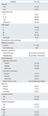
In this Health Insurance Portability and Accountability Act-compliant institutional review board-approved retrospective study, the electronic medical records and imaging studies of 113 women (mean age, 53 years; range, 29 to 72 years) with histopathology-confirmed ULMS seen at our institution between January, 2000 and December, 2012 were reviewed. Patients with leiomyosarcoma who were treated with morcellation were excluded from the study. The demographic data, the date of diagnosis of ULMS and the International Federation of Gynecology and Obstetrics (FIGO) stage 2009 either surgical in patients undergoing surgery or clinical in patients without surgery was recorded in all the patients. The presence of metastases at presentation, the time from diagnosis to metastases and local recurrence, the date of last follow-up and the outcome were also extracted from the electronic medical records. The histopathological features of the primary tumor including the size of the tumor, serosal and cervical involvement, mitotic count, nuclear atypia, lymphovascular invasion, and necrosis were recorded.
All the available computed tomography (CT) scans in the 113 patients were systematically reviewed in consensus by two cancer imaging fellowship-trained radiologists with 8 and 15 years of experience. The restaging CT scans were performed at 6 to 8 weeks interval according to the institutional protocol. In total 892 restaging CT scans were reviewed. The presence or absence of local recurrence, site, number, and size of the distant metastasis and the date of detection of the recurrent/metastatic disease were recorded for each patient. The recurrent or metastatic disease was confirmed either by biopsy or by the presence of unequivocal progression on follow-up imaging. Nodal metastases was confirmed either at histopathology or was presumed based on the typical anatomic location on imaging.
The predictors of development of metastases and impact of various clinical and histopathologic parameters on survival were statistically analyzed. The predictive value of age, tumor size, FIGO stage, presence or absence of necrosis, mitotic count, serosal involvement, cervical involvement, and local recurrence for development of metastatic disease was assessed using univariate and multivariate analysis; multivariate analysis was performed using logistic regression. For the purpose of this analysis, we divided the tumor stage into three groups, stage 1, stage 2 or 3, and stage 4. This was done partly because there were fewer patients in stages 2 and 3, but also because there is significant prognostic overlap between stage 2 and 3 by both FIGO and American Joint Committee on Cancer (AJCC) classifications [18,19].
Survival between the three FIGO stage-groups was compared using Log-rank test. The three FIGO stage-groups were also individually compared with each other for differences in survival. The effect of age, tumor size, stage, and presence of local recurrence on survival was assessed in a multivariate analysis using the Cox proportional hazard regression model. Time to local recurrence was correlated with size of the primary tumor using Spearman correlation.
RESULTS
1. Patient and tumor characteristics
Of the 113 patients, 76 patients (67%) were ≥50 years of age at the time of first diagnosis of ULMS. The mean tumor size was 10.9 cm (range, 0.5 to 25 cm) with 49 tumors >10 cm in size. Of the 113 patients, 54 (48%) had FIGO stage I tumors, 17 (15%) had stage II tumors, six (5%) had stage III, and 32 (28%) had stage IV tumors (Table 1). The FIGO stage was unknown in four patients. Surgery for primary disease was performed in 111 patients while two patients received only chemotherapy. Of the 111 patients who underwent surgery, 99 patients underwent total abdominal hysterectomy with or without salpingo-oopphrectomy, 10 patients had supracervial/subtotal hysterectomy and two patients underwent radical hysterectomy. In 12 patients, a myomectomy was performed initially which was followed by total hysterectomy due to detection of ULMS at histopathology (one patient diagnosed intraoperatively on frozen section specimen). Pelvic node assessment was performed in 33 patients with positive nodes seen at histopathology in one patient. Neoadjuvant chemotherapy was administered in three patients. Postoperative chemotherapy was administered in 92 patients 23 after development of recurrent/metastatic disease. Postoperative radiotherapy was administered in 56 patients, 33 after development of recurrent/metastatic disease. At histopathology, serosal involvement was present in 22, absent in 37 and not available in 54 patients. Cervical involvement was present in 13, absent in 76 and not available in 24 patients. The mean mitotic count was 24 per 10 high power fields (range, 0 to 97). Necrosis was present in 76 cases, absent in 13 cases and was not commented in 24 cases.
2. Distribution of metastases
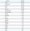
Of the 113 patients, 92 (81%) had radiographic metastasis at any point in the follow-up period of 45 months (range, 4 to 271 months), 12 (10.6%) of whom presented with metastatic disease. The time from diagnosis to metastases was 7 months (interquartile range [IQR], 1 to 21). Lung was the most common site of metastases (84 patients, 74%) followed by the peritoneum (46 patients, 41%), bones (37patients, 33%), and liver (30 patients, 27%). Table 2 shows the distribution and frequency of metastases in ULMS. At least one of the three most common sites of metastases, i.e., lung, peritoneum, and bone were involved in all the 92 patients with metastases. Thirty eight of the 92 patients had metastases in all the three common sites while 47/92 patients had metastases in the lung and peritoneum. In 64/92 patients with metastases, lung was the first site of metastases, nine of whom had concurrent metastases at other sites.
Lung metastases were seen as pulmonary nodules and masses varying in size between 5 mm to 14 cm with cavitation noted in larger nodules. Pleural metastases were noted in 27/84 patients with lung metastases. Peritoneal involvement in all 46 patients was in the form of peritoneal nodules and masses varying in size from 1 to 18 cm, located in the peritoneal cavity including the mesentery, omentum, paracolic gutters, bowel serosa, lesser sac, and pelvis. Two patients developed tumor-bowel fistula. Bone metastases, the third most common site of metastases seen in 37 patients, were lytic with soft tissue component in 30 patients and sclerotic in seven patients. There was associated epidural soft tissue in four cases. All 30 patients with hepatic metastases had multiple lesions which were low in attenuation.
The other sites of metastases were skeletal muscles (29 patients, 26%), lymph nodes (25 patients, 22%), and subcutaneous soft tissues (17 patients, 15%). Of the 25 patients with lymph nodal metastases, 12 had involvement of the supradiaphragmatic nodes mainly the mediastinal and hilar nodes; retroperitoneal node involvement was seen in five cases and pelvic nodal metastases in six cases. Renal and pancreatic metastases were seen in six cases each. Brain and cardiac metastases were found in five patients each. Adrenal glands, spleen, thyroid, breast, orbit, and vagina were uncommon metastatic sites.
3. Predictors of metastases
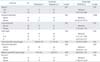
Analysis of the various predictors of metastases showed that age, serosal involvement, local recurrence, and FIGO stage had statistically significant predictive value (Table 3). Ad hoc analysis of age as predictor for metastases, showed that 67/76 patients with ≥50 years of age developed metastases while 25/37 patients <50 years of age developed metastases. The difference between these two groups was statistically significant (multivariate analysis: p=0.03). Distant metastases developed in 16/22 patients with serosal involvement (p=0.006) and 51/57 patients with local recurrence (p=0.01). With respect to FIGO stage, 40/54 patient with stage I, 20/23 patients with stage II/III and 28/32 patients with stage IV disease developed metastatic disease (p=0.03). The size of the tumor (p=0.81), cervical involvement (p=0.09), mitotic count (p=0.09), and necrosis (p=0.05) were not predictive of metastases.
4. Correlation of local recurrence with metastatic pattern
During the median follow-up of 45 months (range, 4 to 271 months), local tumor recurrence developed in 57 patients (50%). Distant metastases developed in 51 of these 57 patients with local recurrence. Distant metastases developed before local recurrence in 21/51 patients, after the local recurrence in 14/51 patients and at the same time as local recurrence in 16/51 patients. Analyses of the pattern of distant metastases in the 21 patients who developed distant metastases developed before local recurrence showed that lung was the first site of metastases in 19 patients, one of them having concurrent peritoneal metastases; bone and subcutaneous metastases were the first site in the one each remaining patient. In the 14 patients who developed local recurrence before distant metastases, lung was the first site of metastases in six patients, peritoneal cavity was the first site in four patients, liver in two patients, bone in one patient, and multiple sites (lung, peritoneum, bone) in one another patient. These differences were; however, not statistically significant (p>0.05). Over all, of the 57 patients with local recurrence, 42 developed lung metastases, and 34 developed peritoneal metastases. We correlated local recurrence with the five most common sites of metastases- lung, peritoneum, bones, liver, and muscles which showed statistically significant correlation between local recurrence and peritoneal metastases (Fisher exact test, p<0.001) and no significant correlation with the other sites of metastases. There was statistically significant correlation between lung metastases and other common sites of hematogeneous metastases including bone (Fisher exact test, p=0.0026), liver (Fisher exact test, p<0.001), and muscle (Fisher exact test, p=0.0065).
5. Predictors of survival
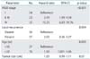
The median metastases-free survival in this cohort was 7 months (IQR, 1 to 21 months). At the time of last follow-up, 74/113 patients (65%) have died. Stage-wise analysis showed that 25/54 patients with stage I disease at presentation, 16/23 patients with stage II/III, and 29/32 patients with stage IV disease have died. The median overall survival for all patients was 45 months (IQR, 25 to 74 months). The median survival of patients with FIGO stage I, II/III, and IV tumors was 63.5 months (IQR, 35 to 121 months), 45 months (IQR, 31 to 61 months), and 23 months (IQR, 11 to 38 months), respectively (p<0.001). There was statistically significant survival difference between stage I and stage II/III (p=0.004), stage II/III and stage IV disease (p=0.02), and stage I and IV (p<0.001). Analysis of predictors of survival using the Cox proportional regression model showed that stage, local recurrence, and age adversely affected survival (Table 4). Patients with higher FIGO stage and patients with local recurrence had higher risk of death with hazard ratios of 2.19 (range, 1.56 to 3.08) and 2.03 (range, 0.26 to 3.27) respectively. Elderly patients also had statistically significant minimal increase in risk of death with hazard ratio of 1.03 (range, 1.001 to 1.06), although the clinical relevance of this minimal increase in risk is uncertain. The size of the primary tumor did not significantly affect the survival. There was no significant correlation between the size of primary tumor and local recurrence-free interval (Spearman rank correlation, 0.008).
DISCUSSION
The diagnosis of ULMS often occurs in retrospect after surgical resection of a presumed benign uterine neoplasm and therefore, patients often do not undergo preoperative staging work up. Bernstein-Molho et al. [5] found in their study of 33 patients that this lack of preoperative staging as one of the causes for the short disease-free interval in their study. The National Comprehensive Cancer Network (NCCN) clinical practice guidelines for uterine sarcomas recommend hysterectomy as the initial treatment of choice for all medically operable patients with disease limited to the uterus [20]. However, in patients with suspected extrauterine disease spread, additional surgical resection of metastatic disease, or systemic chemotherapy is recommended after radiological staging. This underscores the significance of the knowledge of the metastatic pattern which helps in planning treatment strategies.
In the autopsy series of 73 patients with uterine sarcomas by Rose et al. [21] which included 19 patients with ULMS, the most common site of metastases was peritoneal cavity followed by lung, lymph nodes, and liver. The most common sites of metastases in our study in descending order of involvement were lung, peritoneal cavity, bone, and liver. Though this pattern is slightly different from the autopsy series of Rose et al. [21] which can be due to inclusion of all types of sarcomas in their study and different management strategies, it is in agreement with other studies. In the study of 126 patients with ULMS by Gadducci et al. [6], of the 30 patients with distant metastases, lung, peritoneal cavity, liver, and bone were the most common sites of metastases. Similarly, in a study of 71 patients with ULMS, Mayerhofer et al. [7] found lung to be the most common site of metastases. The high incidence of lung metastases (64/92 patients in our study) justifies the inclusion of thoracic imaging in the follow-up of patients with ULMS.
The management of pelvic and para-aortic lymph nodes in ULMS has been controversial with no clear recommendations by the NCCN guidelines [20]. Though the overall incidence of nodal metastases in our study was 22%, the incidence of pelvic and para-aortic nodal metastases was 8.8% which is in agreement with earlier literature. In the largest study of 1396 ULMS patients till date by Kapp et al. [4], the incidence of nodal metastasis was 6.6% in the 348 patients who underwent lymphadenectomy. The presence of nodal metastases decreased the 5-year survival from 64% in node negative cases to 26% in node positive cases in their study. The incidence of intrathoracic nodal metastases (10.6%) reported in our study is lower than that reported by Rose et al. (23.6%) [21].
The metastasis-free interval was 7 months in our study. This was shorter than that observed by Gadducci et al. [6] (12 to 16 months) but similar to Mayerhofer et al. [7] (8 months), and may well be influenced by a referral bias to our tertiary center [8,13]. We analyzed the factors which can predict the risk of metastases using univariate and multivariate analysis. Increasing age was an important risk factor for distant metastases with patients ≥50 years having higher risk of metastases. Higher FIGO stage and presence of serosal involvement also translates into increased risk for metastases. Patients with higher FIGO stages, i.e., stage III and stage IV are by definition known to have extrauterine disease which usually occurs with aggressive histology [22]. We did not find a statistically significant correlation between the risk of metastases and histopathological parameters like cervical involvement, necrosis, and mitotic count. However, studies have shown that cervical invasion, in addition to high mitotic count portend poor prognosis in ULMS [18,19,23]. The discrepancy with respect to cervical involvement in our study can be in part related to the low number of patients with cervical involvement. Though size is an integral part of FIGO staging and has been shown in several studies to influence the patient outcome, it did not have significant predictive value for metastases in our study [7,22]. Presence of local recurrence increased the risk of distant metastases in our study.
We analyzed the correlation between local recurrence and the pattern of distant metastases. Although not statistically significant, we found that patients who developed distant metastases before local recurrence (21 patients) had a more predictable pattern of distant metastases in that lung was often (19/21) the first site of metastases in them. In contrast, in patients who developed local recurrence before distant metastases, the pattern of metastases was unpredictable. It can be hypothesized that local recurrence indicates presence of viable tumor cells in the surgical bed which makes alternate routes of spread including peritoneal spread, possible. This is further supported by the statistically significant correlation between local recurrence and peritoneal metastases (Fisher exact test, p<0.001) in our study. Rose et al. [21] hypothesized difference routes of spread in uterine sarcomas to explain the different patterns of metastases. While peritoneal disease was presumed to be due to direct implantation and nodal disease due to lymphatic spread, the lung and other distant sites of metastases were thought to result from hematogeneous spread. A similar observation was made in our study in that a statistically significant correlation was noted between presence of lung metastases and presence of bone (Fisher exact test, p=0.0026), liver (Fisher exact test, p<0.001), and muscle (Fisher exact test, p=0.0065), all of which share the common hematogeneous route of dissemination.
Giuntoli et al. [3] developed a risk assessment index for predicting the outcome in patients with ULMS based on the age of the patient, tumor size, FIGO stage, and grade of the tumor. In their study of 208 patients with ULMS, stage and grade were found to be the most important predictors of outcome. Kapp et al. [4] in the their study of 1,396 patients with ULMS concluded that African-American race, older age at diagnosis, higher stage, and grade at the time of diagnosis were associated with poor DSS. The results in our study were in agreement with some of these earlier observations. Stage specific survival showed that higher stage doubled the risk of death for stage IV disease compared to stage I disease. Similarly, presence of local recurrence and advanced age also shortened the survival, although the shortening of survival with elderly age was minimal. Larger tumor size was associated with decreased DSS and increased risk of local recurrence in the study by Giuntoli et al. [3]. However, in our study tumor size neither affected the survival nor the local recurrence-free interval.
The limitations of our study were the retrospective study design and the selection bias of our cohort treated at our tertiary cancer center which may differ from patients in the general population. Because of this selection bias, we did not calculate the incidence of metastasis in our study which may be over-estimated due to the frequent referral of patients with advanced disease. We did not assess the impact of certain parameters like cervical involvement which was not consistently evaluated in our study.
In conclusion, we found that ULMS metastasizes most frequently to the lung, peritoneum, bone and liver. Local recurrence was often associated with peritoneal spread and lung metastases with other sites of hematogeneous metastases in our study. The factors which predicted the risk of metastases in our study were advanced age and FIGO stage, presence of serosal involvement and local recurrence.
 XML Download