

 PDF
PDF ePub
ePub Citation
 Print
Print


INTRODUCTION
Gallbladder cancer (GBC), though generally considered rare, is the most common malignancy of the biliary tract, accounting for 80–95% of biliary tract cancers. GBC is considered the most aggressive of biliary cancers with the shortest survival time.1 Complete surgical resection offers the only chance for a cure; however, only 10% of patients with GBC present with early-stage disease and are considered surgical candidates. Advanced GBC is characterized by early local invasion, extensive regional lymph node metastasis, vascular encasement, and distant metastasis, leading to a poor prognosis from unresectable or metastatic disease.12
Accurate stratification for outcome prediction based only on anatomic stage is difficult. A more accurate and reliable prognostic system incorporating additional features of tumors, such as biological and molecular information, may be necessary to obtain a better prediction of prognosis and to choose the most appropriate treatment modality and follow-up plan for locally advanced and metastatic GBC. 18F-fluorodeoxyglucose positron emission tomography/computed tomography (18F-FDG PET/CT) is an increasingly available noninvasive test for malignancy based on glucose metabolism. It was recently demonstrated to be valuable for initial staging and for the detection of recurrent diseases in many kinds of tumors, including GBC.3
18F-FDG PET is useful not only for diagnosing and staging, but also for evaluating the proliferative activity and malignancy grades of tumors reflecting prognosis. The standardized uptake value (SUV) of the primary tumor, a semi-quantitative parameter derived from 18F-FDG PET, is a significant prognostic factor for various types of cancer.45 Despite being a popular landmark clinically, this parameter only has a single voxel value and cannot be used to indicate the metabolism of the whole tumor and metastatic lesions. In fact, many studies that have indicated SUV as a significant prognostic factor did not analyze parameters reflective of tumor volume. Recent studies have reported that volumetric PET parameters, such as metabolic tumor volume (MTV) and total lesion glycolysis (TLG), using a threshold-based automatic volume of interest (VOI) were better prognostic predictors for survival in patients with malignant pleural mesothelioma, esophageal cancer, and advanced head and neck squamous cell carcinomas.678 There is still little evidence that volumetric PET parameters are significant prognostic predictor in patients with GBC. This study sought to investigate the prognostic value of SUV and volume-based metabolic parameters on used 18F-FDG PET/CT in comparison to other clinical parameters in patients with advanced and metastatic GBC.
MATERIALS AND METHODS
Patients
A total of 83 patients at Gangnam Severance Hospital with biopsy-proven gallbladder adenocarcinoma who received 18F-FDG PET/CT pretreatment between January 2007 and December 2015 were included in this retrospective study. Exclusion criteria were patients with resectable early disease, double-primary malignancy, previous cholecystectomy, and other histology types, such as cystic neoplasms, neuroendocrine tumors, or lymphomas. The Institutional Review Board of Severance Hospital, Yonsei University Health System approved this retrospective study (IRB Number: 3-2015-0318) and waived the requirement to obtain informed consent.
The medical records of each patient were investigated for sex, age, histologic typing, performance status (PS), extrahepatic metastases, carcinomatosis, and treatment modality. Histologic typing was classified as well, moderately, or poorly differentiated, and PS was classified according to the Eastern Cooperative Oncology Group (ECOG) performance status. Computed tomography (CT) of the chest and abdominopelvic cavity, a radionuclide bone scan, and 18F-FDG PET/CT were performed to evaluate locally advanced disease or distant metastasis. Survival status was retrieved from our medical records or from attempts to contact the patients or their referring physicians. All follow-up evaluations ended on December 30, 2015.
18F-FDG PET/CT imaging protocols
Imaging and data acquisition for metabolic parameters was conducted using the PET/CT system (Biograph TruePoint 40, Siemens Healthcare, Munich, Germany). PET/CT was performed before treatment. The fasting time before the administration of 18F-FDG (Nambuk Medical, Seoul, Korea) was at least 6 hours, and serum glucose levels were not to have exceeded 150 mg/dL. After the injection of 5.18 MBq/kg (0.14 mCi/kg) of 18F-FDG, each patient waited in a warm, quiet, dim room for 60 minutes. An initial low-dose CT scan was followed by a PET scan from the skull base to the upper thigh level in the three-dimensional mode (1.5-min acquisition time per bed), and the scanned data were reconstructed using the iterative method, ordered subset expectation maximization, using two iterations and 21 subsets. Trans-axial spatial resolution of the PET system was 5 mm full-width at half maximum at the center of the field of view. The matrix size and thickness of the reconstructed PET image were 128×128 and 5 mm, respectively.
Analysis of PET/CT data
The metabolic parameters from 18F-FDG PET/CT data were evaluated by two experienced nuclear medicine physicians using dedicated software for the PET/CT workstation (Syngo VE32B, Siemens AG, Berlin, Germany). To define the contouring margins around the tumor, SUV >2.5 was used as previously reported.9 The contour around the target lesions within the boundaries was automatically generated and within the contour margin were combined to define the tumor volumes. MTV was defined as the sum of metabolic volumes of tumor tissues with increased FDG uptake. The SUV threshold value used in this study was 50% of the local maximum SUV intensity, identified as a reasonable value in phantom studies.910 We selected lesions with SUV >2.5, and selected regions within lesions with a SUV intensity greater than 50% for quantitative MTV measurement. TLG was representative of the metabolic activity throughout the entire tumor and was calculated by multiplying MTV and the mean SUV (SUVmean) of the MTV. Appropriately sized spherical VOIs, including each targeted locally advanced and metastatic lesion, were created by considering the tumor location in the trans-axial, sagittal, and coronal planes. Physiologic activities in the adjacent liver, stomach, and bowel loops were avoided. These parameters, SUV, and MTV were automatically calculated by the Syngo software (Siemens, Erlangen, Germany).
Statistical analysis
The primary end point of this study was overall survival (OS), which was measured from the date of diagnosis of GBC to the date of death from any cause. The Kaplan-Meier method was used for survival analysis, and the difference in the rate was compared using a log-rank test. A prognostic model was established by finding all of the variables that significantly influenced OS (p<0.05) in univariate analysis. The clinical variables included in the univariate analysis were age, sex, pathologic differentiation, ECOG PS, extrahepatic metastases, carcinomatosis, C-reactive protein (CRP), and tumor markers [serum carcinoembryonic antigen (CEA) and carbohydrate antigen 19-9 (CA19-9) levels]. Metabolic PET variables included the highest SUV of the locally advanced lesion (SUVLA max), the highest SUV among the distant metastatic lesions (SUVmt max), the MTV of the locally advanced lesion (MTVLA), the sum of the MTVs of all metastatic lesions (MTVmt total), the highest MTV among the metastatic lesions (MTVmt max), the sum of the MTVs of both the locally advanced and metastatic lesions (MTVtotal), the TLG of the locally advanced lesion (TLGLA), the sum of the TLGs of all metastatic lesions (TLGmt total), the highest TLG among the TLGmt (TLGmt max), and the sum of the TLGs of both locally advanced and metastatic lesions (TLGtotal). For metabolic parameters, the median value was used as the cut-off; for tumor markers, the normal range was used. A Cox proportional hazards regression analysis was performed to determine independent prognostic factors. Statistical analyses were performed using IBM SPSS Statistics for Windows, Version 23 (IBM Corp., Armonk, NY, USA). P values <0.05 were considered significant.
RESULTS
Patient characteristics
This study included 83 patients. The median clinical follow-up period was 9.9 months (range, 0.2–77.2 months). Baseline patient and tumor characteristics, including age, sex, pathologic differentiation, ECOG PS, extrahepatic metastases, carcinomatosis, CRP, CEA, CA19-9, SUVLA max, SUVmt max, MTVLA, MTVmt total, MTVmt max, MTVtotal, TLGLA, TLGmt total, TLGmt max, TLGtotal levels, and treatment modality are presented in Table 1.
Prognostic factors evaluated in univariate analysis
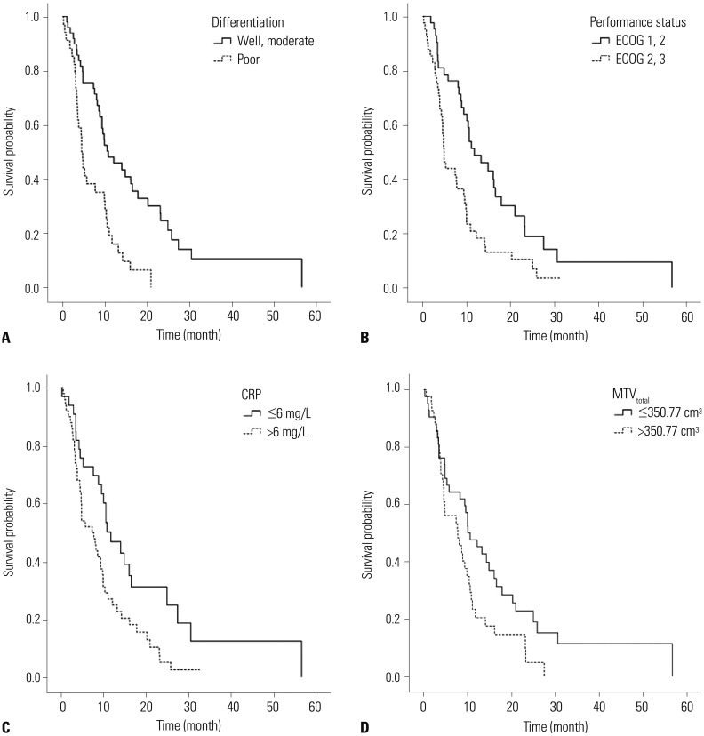
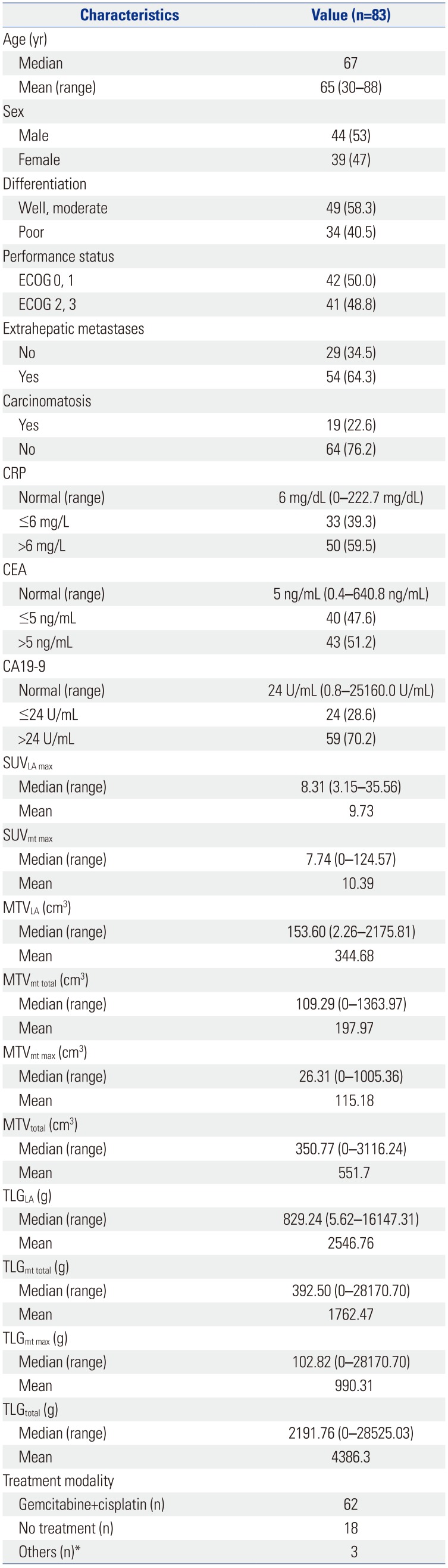
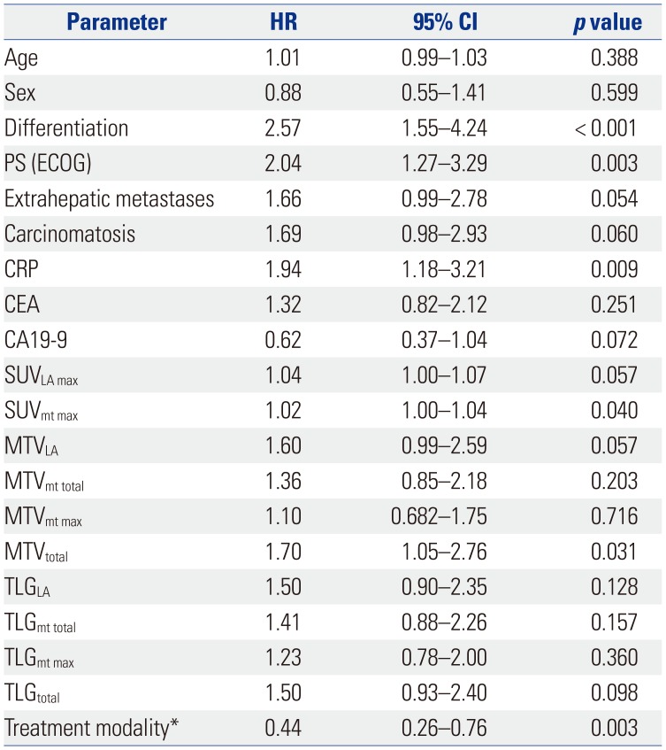
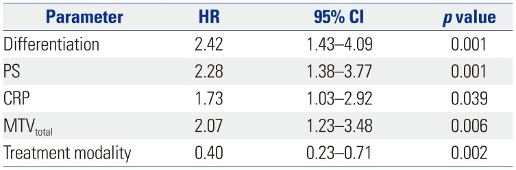
The cut-off levels of serum CRP, CEA, and CA19-9 levels were set to 6 mg/L, 5 ng/mL, and 24 U/mL, respectively, based on the normal values at our institution. The median age of 67 years was used as a cut-off. The median values of the PET and metabolic parameters were as follows: SUVLA max, 8.31; SUVmt max, 7.74; MTVLA, 153.60 cm3; MTVmt total, 109.29 cm3; MTVmt max, 26.31 cm3; MTVtotal, 350.77 cm3; TLGLA, 829.24 g; TLGmt total, 392.50 g; TLGmt max, 102.82 g; and TLGtotal, 2191.76 g. The patients were divided into two groups according to the median value of the parameters. Overall, 46 patients were younger than the median age of 67 years; 37 patients were older. Univariate analysis demonstrated that pathologic differentiation (p<0.001), PS (p=0.003), CRP level (p=0.009), SUVmt max (p=0.040), and MTVtotal (p=0.031) were significantly prognostic. In addition, as expected, chemotherapy with gemcitabine and cisplatin had a significant impact on prognosis (Table 2).
Prognostic factors evaluated in multivariate analysis
In multivariate analysis of adjusted treatment modalities, pathologic differentiation [HR=2.42 (well differentiated and moderately differentiated vs. poorly differentiated); p=0.001], PS [HR=2.28 (ECOG 0, 1 vs. 2, 3); p=0.001], CRP (HR=1.73; p=0.039), and MTVtotal [HR=2.07 (≤350.77 cm3 vs. >350.77 cm3); p=0.006] were independent prognostic factors for the prediction of OS (Table 3, Fig. 1). In patients with locally advanced and metastatic disease, MTVtotal, a volume-based metabolic PET parameter, was an important independent prognostic factor for OS, along with other PET parameters.
DISCUSSION
There have been various reports on the role of PET/CT in GBC diagnosis. In particular, 18F-FDG PET/CT seems to have a potential role in staging, as these cancers are intensely FDG-avid. PET/CT has an overall diagnostic accuracy of 95.9% for the primary disease and 85.7% and 95.9% for the detection of lymph nodes and metastatic lesions, respectively.11 Enhanced utilization of glucose than normal tissues, more aggressive malignancies, and higher rates of glycolysis than less malignant or benign tumors have been observed in cancer cells.121314 These glucose metabolism differences can be measured quantitatively in vivo by PET after FDG administration.
We investigated the prognostic values of volume-based metabolic parameters using 18F-FDG PET/CT in metastatic GBC, compared with conventional clinical parameters. Many prognostic factors for advanced and metastatic GBC have been proposed: most are clinico-pathological parameters. Clinical or pathologic staging, including tumor extension and nodal involvement, and blood CEA levels have remained good prognostic values for GBC.151617 However, these parameters cannot be categorized in detail or presented as objective numbers in unresectable GBC. In other words, a significant prognostic marker that can quantify molecular and metabolic parameters is required for the treatment decision of GBC patients.
Several recent studies have investigated the prognostic value of 18F-FDG PET/CT parameters in GBC patients. Despite an absence of standardized cut-off values, poorer survival has consistently been correlated with a high SUVmax of the primary lesion as measured on pretreatment 18F-FDG PET/CT scans of biliary tract carcinoma.18 Only one previous study investigated the utility of 18F-FDG PET/CT volumetric parameters to predict clinical outcomes in GBC. Yoo, et al.19 analyzed the various metabolic volume-based PET parameters of primary tumors, including maximum and average SUV, MTV, and TLG, measured on 18F-FDG PET/CT scans of 44 patients with GBC. They showed that those with an MTV cut-off of 135 cm3 (p=0.001) and a TLG cut-off of 7090 g (p<0.050) had significantly longer OS than those with lower MTV and TLG values. In the present study, pathologic differentiation, PS, and serum CRP levels were significant factors according to univariate analysis, while SUV and MTV were significant independent prognostic factors. MTVtotal was determined to be a meaningful independent prognostic factor in multivariate analysis with adjustment for treatment modality.
Histologically, the gallbladder does not have submucosa, and the cancer infiltrates directly into the muscularis propria. The gallbladder wall is thin, and the cancer is able to easily infiltrate to adjacent organs, such as the liver, duodenum, and pancreas. As GBC is associated with a high rate of local invasion and distant metastases, resulting in poor survival, we hypothesized that the metabolic activity of all primary and metastatic lesions on 18F-FDG PET/CT scans might be more helpful to guide treatment decisions than the primary lesion only. Thus, our study included locally advanced and metastatic GBC patients and measured the MTV and TLG of both metastatic and locally advanced primary lesions. In doing so, we deduced through multivariate analysis that the total MTV, including metastatic lesions, was the most significant prognostic factor.20
Quantified metabolic activity can provide valuable information to help prognosticate and assess treatment response in clinical oncology.21 While CT scan and MRI readily reveal the anatomic distribution of tumors, they do not permit the quantification of the metabolic activity of a tumor. Anatomically large tumors in CT scan can have low metabolic activity, and small lesions of metastases can be highly active. Therefore, it is thought that MTV measurement with PET/CT is an important factor for prediction of survival prognosis.2223 While SUVmt max (the highest SUV among the metastatic lesions) was significant in univariate analysis, it was excluded from multivariate analysis because SUVmt max, interpreted as a single voxel value, may not reflect the tumor's general metabolism due to tumor heterogeneity. In addition, TLG was not significant for survival prediction: it is calculated by multiplying the tumor volume by the SUVmean. Metastatic GBC can show diverse SUVs in both a locally advanced primary lesion and in multiple metastatic lesions. Therefore, as the TLG is calculated as the SUVmean, its importance in survival prediction may be weakened. MTV appeared to be more important for prediction of survival prognosis than SUV due to the variety of SUVs obtained.
Our present study had several limitations. First, it was designed as a retrospective study and included a relatively small number of patients. Therefore, our results may not be applicable to all patients with locally advanced and metastatic GBC. Second, it was difficult to clarify the boundary between the primary lesion and liver infiltrative lesion, since GBC readily invade the liver. Hence, we defined locally advanced GBC including liver invasion and obtained MTV according to locally advanced primary lesion. Third, the cut-off value of each PET parameter was set to a median value due to a small number of samples and a wide range of parameter values. It is necessary to find an accurate cut-off value with a larger number of patients.
Despite these limitations, our study is the first to demonstrate the clinical value of volume-based PET parameters of GBC in a metastatic clinical setting, and our results support a more detailed follow-up or stratification of aggressive therapy in high MTVtotal patients due to their poor prognosis. Additional larger-scale prospective studies using 18F-FDG PET/CT are required to validate our results.
 XML Download