

 PDF
PDF ePub
ePub Citation
 Print
Print


INTRODUCTION
Annually, more than 50000 individuals receive hematopoietic stem cell transplantations (SCTs) worldwide. SCT recipients are at higher risk of severe infections, since they often remain in a state of immunosuppression after transplantation, depending on such comorbidities as graft-versus-host disease (GVHD) and the need for immunosuppressive drug treatment.12 In particular, SCT recipients are susceptible to common respiratory viruses (CRVs) owing to their impaired T-cell immunity.3 CRVs are common causes of both upper respiratory tract disease (URD) and lower respiratory tract disease (LRD) in this population, and have been associated with significant morbidity and mortality.456 To date, there has been only a preliminary study of infections caused by CRVs among SCT recipients in Korea.78 However, the epidemiology of individual CRVs varies according to the region and the times and many advances in the diagnosis and treatment of infections caused by CRVs have been achieved. The present study aimed to describe the incidence, clinical courses, and risk factors for mortality associated with LRDs caused by CRVs in SCT recipients at a single SCT center in Korea.
MATERIALS AND METHODS
Patients
We retrospectively reviewed the medical records of 1038 patients aged ≥18 years who had undergone a SCT between January 1, 2007 and August 31, 2011 at the Catholic Blood and Marrow Transplantation Centre of Seoul St. Mary's Hospital, The Catholic University of Korea (Seoul, Republic of Korea). The aim was to identify LRDs caused by CRVs. CRVs associated with LRDs in patients included influenza A and B viruses, respiratory syncytial virus (RSV), human parainfluenza virus (HPIV) 1–3, human rhinovirus (HRhV), human adenovirus (HAdV), and human metapneumovirus (HMPV). Clinical information, including demographics, comorbidities, coinfections, and mortality were investigated. The endpoint of the study was set as January 31, 2012, or the time of death or loss to follow up. This study was approved by the Institutional Review Board of the Yeouido and Seoul St. Mary's Hospital (approval nos. SC10RISI0023 and KC13RISI0364).
Respiratory virus identification
SCT recipients with respiratory symptoms were screened for CRVs at the discretion of physicians. Specimens for diagnostic testing included nasal or throat swabs, sputum, tracheal aspirates, bronchial wash fluid, and bronchoalveolar lavage (BAL) fluid. Laboratory tests to identify CRVs included the respiratory virus polymerase chain reaction (PCR) multiplex panel (AdvanSure RV Real-Time PCR Kit; LG Life Sciences, Seoul, Korea) and rapid influenza antigen testing (BD Veritor System for Rapid Detection of Flu A+B; BD Diagnostics, Sparks, MD, USA). The respiratory virus PCR multiplex panel was used to test for influenza A and B viruses, RSV, HMPV, HPIV, HRhV, and HAdV. Influenza-specific reverse transcription PCR was used to detect the influenza A (H1N1) during the 2009 H1N1 pandemic.
Definitions
URD was defined as the detection of a CRV in upper respiratory secretions, such as nasal or throat swabs, in association with respiratory symptoms, including cough, rhinorrhea, and sore throat, and in the absence of new infiltrates on chest imaging.910 A LRD was defined as an acute respiratory illness with dyspnea, hypoxia, or pulmonary infiltrates occurring in association with the detection of a CRV in any respiratory secretions.10 Assessment of the radiographic findings, including chest radiography and computed tomography scans, was based on the formal reading by a radiology specialist. Hospital-acquired infection was defined as symptom onset 3 or more days after hospital admission.9 The presence of co-pathogens was defined as the isolation of pathogenic bacterial species, fungal species, or other opportunistic viruses, such as cytomegalovirus (CMV), from the respiratory specimen obtained within a month of detection of a CRV, in conjunction with consistent symptoms, and as confirmed by infectious disease specialists.5 Proven or probable invasive fungal disease was defined according to the criteria of the European Organization for Research and Treatment of Cancer/Invasive Fungal Infections Cooperative Group and the National Institute of Allergy and Infectious Diseases Mycoses Study Group Consensus Group in 2008.11 A mixed CRV infection was defined by the isolation of more than two CRVs during the same episode. Mortality due to LRDs caused by CRVs was defined as death resulting from respiratory failure, with no period of complete recovery between the onset of illness and death within 30 days, and without a documented co-pathogen.12 Overall mortality was defined as death from any cause within 30 days following diagnosis of CRV-LRD. An absolute lymphocyte count (ALC) of <200 cells/mm3 blood within the 2 weeks preceding a CRV infection diagnosis was defined as lymphopenia, and an absolute neutrophil count (ANC) of <500 cells/mm3 blood within the 2 weeks prior to a CRV infection was defined as neutropenia.6 The dosage of corticosteroid use was classified into three groups according to the highest daily dose taken by the patient during the 2 weeks preceding a CRV infection: high-dose corticosteroid use was defined as ≥1 mg/kg/day of prednisone and its equivalent dose of corticosteroids: low-dose use was defined as ≤1 mg/kg/day of prednisone; and the third group received topical corticosteroids.13 The grade of immunodeficiency was classified into three groups, including severe, moderate, and mild immunodeficiency.14 Severe immunodeficiency (SID) was defined as the presence of two or more of the following: allogeneic SCT (alloSCT) within 6 months, or autologous SCT (autoSCT) within 3 months, preceding the diagnosis of a CRV infection; acute GVHD (grade ≥2); ALC <200 cells/mm3 or ANC <500 cells/mm3 within 2 weeks, before the diagnosis; and a pre-engraftment period or administration of immunosuppressive therapy ≤2 weeks prior to the diagnosis. Moderate immunodeficiency (MID) was defined as the presence of only one criteria of SID or two or more of the following criteria: an alloSCT within 1 year (≥6 months) or an autoSCT within 6 months (≥3 months), prior to the diagnosis of a CRV infection; ALC between 200 and 500 cells/mm3, or ANC between 500 and 1000 cells/mm3, within 2 weeks before the diagnosis; and administration of immunosuppressive drugs within 1 month prior to the diagnosis. Patients who met only one criterion of MID were regarded as having a mild immunodeficiency state.
Statistics
All analyses were performed for the first episode of CRV infection after SCT. Categorical variables are described as number (percentage). Continuous variables are expressed as medians (interquartile range) or mean±standard deviation. Categorical variables were compared using chi-square or Fisher's exact tests, and continuous variables were compared using t-tests or Wilcoxon rank-sum tests. Using Cox proportional hazards regression models, all variables with a p<0.05 upon univariate analysis were included in a multivariate logistic regression analysis to determine the significance of the risk factors for mortality associated with CRV-LRDs. All p values were two-tailed, and statistical significance was set at p<0.05. Statistical analyses were performed using SAS version 9.2 (SAS Institute Inc., Cary, NC, USA).
RESULTS
Characteristics of patients with CRV-LRDs
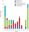
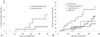
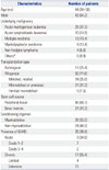
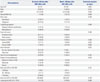
A total of 1038 patients received SCTs (273 autologous and 765 allogeneic) between January 1, 2007 and August 31, 2011. Over a 5-year period between 2007 and 2012, 71 CRV-LRDs were identified in 67 (6.5%) patients, and coinfection with more than two CRVs was observed in 4 (5.9%) patients. Influenza A virus and RSV coinfection was observed in two patients. HPIV and HRhV coinfection was observed in one patient, and HPIV and RSV coinfection was also observed in one patient. The baseline characteristics of the 67 SCT recipients with CRV-LRDs are shown in Table 1. During the winter, RSV and influenza A virus were the predominant CRVs, while the proportion of HPIV infection increased through spring and summer (Fig. 1). Analysis for cumulative incidences of first CRV-LRD episodes demonstrated that the most common causative pathogen of CRV-LRDs at both 100 days [cumulative incidence estimate, 23.5%; 95% confidence interval (CI), 3.3–43.7] and at 1 year after SCT (cumulative incidence estimate, 69.2%; 95% CI, 45.9–92.5) was HPIV. The cumulative incidence estimate of RSV was 6.9% (95% CI, 0.0–16.1), and only two patients presented with LRDs cau-sed by influenza A virus and HRhV (one patient each) before 100 days following SCT (Fig. 2A). The cumulative incidence estimates of LRDs caused by influenza virus, RSV, HPIV, and HRhV at 1 year after SCT were 42.9% (95% CI, 17.0–68.8), 21.6% (95% CI, 6.3–36.9), 69.2% (95% CI, 45.9–92.5), and 28.6% (95% CI, 0.0–62.1), respectively (Fig. 2B).
Clinical presentations and outcomes of CRV-LRDs
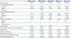
The characteristics of the LRDs caused by the various CRVs are described in Table 2. The most common type and extent of radiographic infiltration of influenza-caused LRDs were alveolar pattern (64.3%) and bilateral lung field (85.7%), respectively. Seven (50%) of 14 patients with influenza-LRDs received antiviral therapy, and all received it within 48 hours following influenza-LRD diagnosis. However, early antiviral therapy within 48 hours after symptom onset was performed for only 3 (21.4%) patients. One patient received combination therapy with peramivir (600 mg/day for 8 days) and oseltamivir (300 mg/day for 15 days). Five (35.7%) patients experienced mechanical ventilation, and the 30-day overall mortality rate of these patients was 35.7% (5/14). Two patients were diagnosed with the influenza A (H1N1) pdm09 virus, of which one died due to respiratory failure on day 28 following the diagnosis. Of the 31 patients with RSV-LRDs, 41.9% (13/31) were hospital-acquired and 9.7% (3/31) received antiviral therapy. The 30-day overall mortality rate was 25.8% (8/31) for these patients. Two patients with RSV-LRDs were treated with oral ribavirin, one with aerosolized ribavirin, and all received intravenous immunoglobulin (IVIG; 1–1.5 g/kg for 1–2 days). The patient who received aerosolized ribavirin and IVIG survived, whereas the two patients treated with oral ribavirin died. Seven (36.8%) of the 19 patients with HPIV-LRDs received oral ribavirin (800 mg/day for 11–26 days). The 30-day overall mortality rate of patients with HPIV-LRDs was 31.6% (6/19). Four (57.1%) of seven patients with HRhV-LRDs required mechanical ventilation, and the 30-day mortality rate for these patients was 14.3% (1/7). Co-pathogens isolated from respiratory specimens were detected in 33.8% (23/67) of patients with CRV-LRDs. The most common bacterial co-pathogen was Acinetobacter baumannii (n=7; 10.4%), followed by Pseudomonas aeruginosa (n=5; 7.4%) and Streptococcus pneumoniae (n=4; 5.9%). Aspergillus species were detected in 6 patients and CMV-PCR of the BAL fluid was positive for two patients. Only 2 patients died without pneumonia being the cause of death, one due to septic shock as a result of infectious colitis and one due to diffuse alveolar hemorrhage.
Risk factors for 30 day-mortality in SCT recipients with CRV-LRDs
The overall mortality at day 30 after the diagnosis of a CRV-LRD was 32.8% (22/67). In the univariate analysis, high-dose steroid usage (p=0.025), SID (p=0.033), and lymphopenia (p=0.006) were significantly associated with mortality within 30 days following a CRV-LRD diagnosis (Table 3). Multivariate logistic regression analysis revealed that the use of high-dose steroids [odds ratio (OR), 4.05; 95% CI, 1.12–14.61; p=0.033] and lymphopenia (OR, 6.57; 95% CI, 1.80–24.03; p=0.004) were independent risk factors for mortality within 30 days of CRV-LRD detection (Table 4).
DISCUSSION
There has been almost no data on respiratory virus infection among SCT recipients in Korea. This study aimed to retrospectively collect and analyze data regarding the epidemiology and risk factors of LRDs caused by CRVs in SCT recipients from the largest SCT center in Korea.
The incidences of influenza-associated LRD among SCT recipients reported in other studies ranged from 0.4–4.1%, and the mortality rates were 14.7–28%.561015 In previous studies, the proportions of progression to pneumonia among influenza virus-infected SCT recipients were reported as 29–43%.1516 The 30-day mortality rate of influenza-LRD at our center was 14.3%, which is consistent with the previous reports.
RSV infections have been reported to occur in 1.2–9.6% of adult SCT recipients.51718 The incidences and mortality rates of RSV-LRDs among SCT recipients were reported as 2.9–5.1% and 16.7–50%, respectively.561417 These findings are consistent with the incidence (2.7%) and mortality rate (22.5%) of RSV-LRD observed at our center, in spite of the low rate of ribavirin treatment (9.7%). Known risk factors of RSV-associated mortality in SCT patients included pre-engraftment, lymphopenia, alloSCT <1 month previously, SID, and an older age (>65 years).1419 The highest incidence of RSV occurred between January and April, which is consistent with our result that the majority of RSV-LRDs were detected between December and March.20
Symptomatic HPIV infections have been reported to occur in 1.4–7.1% of adult SCT recipients in previous studies.52122 The reported frequency and mortality rates of HPIV-LRDs among SCT recipients were 0.05–8.1% and 11.8–46%, respectively.56910222324 The 30-day mortality rate of HPIV-LRD at our center, 26.3% (5/19), was similar to that of previous studies, despite the low rate of antiviral treatment (36.8%). Since HPIV showed the highest cumulative incidence of LRDs at 100 days (23.5%; 95% CI, 3.3–43.7) and 1 year (69.2%; 95% CI, 45.9–92.5) following SCT, the performance of a laboratory examination is important due to non-specific respiratory symptoms. Independent predictors of HPIV-associated death that have been reported in the literature include steroid usage, cancer status, Acute Physiology and Chronic Health Evaluation II score, LRD, infection immediately following SCT (<30 days), mismatched donor, the need for mechanical ventilation, and the presence of co-pathogens.92223 Similarly, high-dose steroid usage at the time of diagnosis was one of the independent predictors of death among SCT recipients with CRV-LRDs in the present study. Furthermore, the present study demonstrated that HPIV infections were most prevalent in the summer months, followed by spring, which had been reported previously.922
Previous studies reported that the incidence of HRhV-LRD among SCT recipients is rare (0–1.4%), and that HRhV-LRDs are associated with low mortality rates.525 In the present study, the incidence of HRhV-LRD was 0.7% (7/1038 patients) and the associated 30-day mortality rate was 14.3% (1/7 patients). These results are consistent with the preceding studies that HRhV may cause severe infections in SCT recipients, with high rates of progression to LRD and mortality.182627 Ison, et al.28 reported that all 6 SCT recipients with pneumonia in whom HRhV was detected in BAL had significant coinfections and suggested that HRhV might be a cause of frequent superinfections, possibly through lytic infection or an indirect immunosuppressive effect. Our study also indicated that 42.9% (3/7) of patients with HRhV-LRDs were positive for the presence of co-pathogens.
HAdV has been shown to infect up to 3% of SCT recipients, and the subsequent mortality rate can be high (15–28%).29 HMPV-LRDs have been reported to occur in 0.7–1.2% of SCT recipients, of which 0–40% progressed to death.530 However, no cases of HAdV-LRDs or HMPV-LRDs were observed in our patient cohort.
CRV infection may contribute to sustained inflammation or activation of an inflammatory process that leads to irreversible airway damage, and can cause late airflow obstruction in SCT recipients.31 Furthermore, CRV infections frequently coexist with bacterial or fungal pathogens, contributing to the development of pneumonia. It may be the result of epithelial damage, impaired ciliary function, an altered immune response, or up-regulation of bacterial receptors on the CRV-infected respiratory tract.1632 In addition, there have been several reports that SCT recipients have shown a lower immune response to the influenza vaccine compared with that observed for the general population.3334 Severely immunocompromised patients, such as SCT recipients, have been reported to show prolonged influenza shedding, resulting in the development of antiviral resistance during prolonged antiviral therapy.
In the present study, high-dose steroid usage and lymphopenia were shown to be risk factors for mortality in SCT recipients with CRV-LRDs. Lymphopenia was previously reported as a risk factor for mortality among SCT patients with LRDs caused by influenza or RSV.61319 Additionally, high-dose steroid usage during the 2 weeks preceding a CRV-LRD diagnosis was reported as an independent predictor of mortality among SCT recipients with HPIV-LRDs.923 It is likely that these two risk factors are associated with a decreased T-cell-mediated immune response.22 Furthermore, high-dose corticosteroid use is known to be associated with a trend toward delayed viral clearance.35 Conversely, a few previous studies reported that the use of corticosteroids exerted protective effects against the progression to pneumonia and requirement for mechanical ventilation among SCT recipients with influenza-LRDs, which was hypothesized to occur due to a salutary immunomodulatory effect.1315
There are several limitations of the present study. As this was a retrospective study, there were no standardized guidelines for the screening and treatment of CRV infections. However, to overcome this limitation, only CRV-LRD cases were included. Due to the small number of cases, it was not possible to analyze the risk factors for mortality for individual CRV-LRDs. Another limitation was that it was difficult to determine whether the fatal outcome was solely attributable to the CRV or due to a combination of the CRV and co-pathogens. However, precise discrimination of the pathogen would likely have been meaningless, since damage to the respiratory epithelium as a result of a CRV infection may have been the key factor that facilitated the occurrence of superinfections.9 Recent studies showed that viral nucleic acid was detected in the bloodstream more frequently among SCT recipients with LRDs caused by influenza, RSV, HAdV, and HMPV, and that this was associated with increased mortality rates.363738 Therefore, viral nucleic acid detection in the bloodstream of SCT recipients with CRV-LRDs could be used to predict disease severity, poor outcome, and the need for intensified antiviral treatment in the future.
Our study demonstrated that CRV-LRDs have a significant morbidity and mortality among SCT recipients; thus, the implement of an active diagnostic approach for CRV infection is required for SCT recipients with respiratory symptoms. For SCT recipients with LRDs caused by influenza, RSV, or HPIV infections, those receiving high-dose steroid treatment or those with lymphopenia, early antiviral treatment should be considered; especially considering the fact that no vaccine is yet available for RSV and HPIV. Furthermore, infection control measures, including hand hygiene and respiratory droplet isolation, which reduce the occurrence of new infections and transmission, are important for CRVs without a specific antiviral treatment option, such as HRhV and HMPV. Finally, in order to prevent LRDs caused by influenza, which is the only CRV with an available vaccine, the annual seasonal influenza vaccination is recommended for all SCT recipients.

 XML Download