Journal List > Yonsei Med J > v.57(1) > 1031995

Kim, Yang, Kim, Park, Park, Uhm, Joung, Lee, and Pak: Clinical Significance of Additional Ablation of Atrial Premature Beats after Catheter Ablation for Atrial Fibrillation

Abstract

Purpose

The clinical significance of post-procedural atrial premature beats immediately after catheter ablation for atrial fibrillation (AF) has not been clearly determined. We hypothesized that the provocation of immediate recurrence of atrial premature beats (IRAPB) and additional ablation improves the clinical outcome of AF ablation.

Materials and Methods

We enrolled 200 patients with AF (76.5% males; 57.4±11.1 years old; 64.3% paroxysmal AF) who underwent catheter ablation. Post-procedure IRAPB was defined as frequent atrial premature beats (≥6/min) under isoproterenol infusion (5 µg/min), monitored for 10 min after internal cardioversion, and we ablated mappable IRAPBs. Post-procedural IRAPB provocations were conducted in 100 patients. We compared the patients who showed IRAPB with those who did not. We also compared the IRAPB provocation group with 100 age-, sex-, and AF-type-matched patients who completed ablation without provocation (No-Test group).

Results

1) Among the post-procedural IRAPB provocation group, 33% showed IRAPB and required additional ablation with a longer procedure time (p=0.001) than those without IRAPB, without increasing the complication rate. 2) During 18.0±6.6 months of follow-up, the patients who showed IRAPB had a worse clinical recurrence rate than those who did not (27.3% vs. 9.0%; p=0.016), in spite of additional IRAPB ablation. 3) However, the clinical recurrence rate was significantly lower in the IRAPB provocation group (15.0%) than in the No-Test group (28.0%; p=0.025) without lengthening of the procedure time or raising complication rate.

Conclusion

The presence of post-procedural IRAPB was associated with a higher recurrence rate after AF ablation. However, IRAPB provocation and additional ablation might facilitate a better clinical outcome. A further prospective randomized study is warranted.

INTRODUCTION

Although radiofrequency catheter ablation (RFCA) for atrial fibrillation (AF) is an effective strategy for rhythm control, there remains a substantial recurrence rate.123 The majority of AF cases are triggered in patients with paroxysmal AF (PAF) derived from pulmonary veins (PVs), although 10-30% of PAF patients and the majority of patients with persistent AF (PeAF) have non-PV triggers.1456 Therefore, post-procedural detection and elimination of non-PV triggers might result in a better clinical outcome. Testing for immediate recurrence of AF after cardioversion is the common method for the detection of AF triggers.78 However, a post-procedural test for immediate recurrence of AF requires time, isoproterenol infusion,6 sedation, and electrical cardioversion. Moreover, repetitive electrical cardioversion may increase the risk of peri-procedural stroke in high-risk patients.9 Therefore, the clinical value of a post-procedural test for immediate recurrence of AF and additional ablation targeting remaining non-PV foci has not been clearly determined. With the development of a three-dimensional (3D)-electroanatomical software mapping system, the mapping and ablation of frequent atrial premature beats (APBs) has become relatively fast and accurate. Therefore, we hypothesized that non-PV-origin APBs may contribute to AF recurrence after catheter ablation. We defined the provocation of immediate recurrence of APBs (IRAPBs) as the detection of post-procedural frequent APBs (≥6 beats per minute) after electrical cardioversion under isoproterenol infusion (5 µg/min). The purposes of this study were to explore whether post-procedural IRAPB provocation and additional ablation for mappable frequent APBs affect the clinical outcome of AF ablation and whether IRAPB provocation lengthens procedure time or increases the complication rate. We also tested whether the type of induced tachyarrhythmias [AF or atrial tachycardia (AT)] and the presence of IRAPB are risk factors for clinical recurrence of AF after catheter ablation.

MATERIALS AND METHODS

Study population

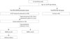
The study protocol adhered to the Declaration of Helsinki and was approved by the Institutional Review Board of Yonsei University Health System. All patients provided written informed consent. This study was a single-center retrospective comparison study, and patients were matched 1:1 by age, sex, and AF type. The study included patients with AF (76.5% males; 57.4±11.1 years old) who underwent RFCA between March 2011 and December 2012. The exclusion criteria were as follows: 1) permanent AF refractory to electrical cardioversion; 2) AF with valvular disease (>grade II); 3) associated structural heart disease other than left ventricular hypertrophy; and 4) prior AF ablation. IRAPB provocation tests were conducted in 100 consecutive patients, who were compared with 100 age-, sex-, and AF-type-matched patients who completed ablation without any induction or mapping test (No-Test group) (Fig. 1). Among them, 67.0% of patients had PAF, and 33.0% had PeAF. 3D spiral computerized tomography (CT) scans (64 Channel, Light Speed Volume CT, Philips, Brilliance 63, the Netherlands) were performed to visually define atrial and PV anatomy in all patients. The presence of a left atrial (LA) thrombus was excluded by trans-esophageal echocardiography. All antiarrhythmic drugs were discontinued for a period corresponding to at least five half-lives. In total, 58 patients (29.0%) were taking amiodarone, which was discontinued for at least 4 weeks prior to the procedure. Anticoagulation therapy was maintained before catheter ablation.

Electrophysiologic mapping and radiofrequency catheter ablation

Intracardiac electrograms were recorded using the Prucka Cardio Lab™ electrophysiology system (General Electric Health Care System Inc., Milwaukee, WI, USA), and catheter ablation was performed in all patients using 3D electroanatomical mapping (St. Jude Medical Inc., Minnetonka, MN, USA) merged with 3D spiral CT. We used an open irrigated-tip catheter (Celsius, Johnson & Johnson Inc., Diamond Bar, CA, USA; Coolflex, St. Jude Medical Inc., Minnetonka, MN, USA; 30-35 W; 47℃) to deliver RF energy for ablation (Stockert generator, Biosense Webster Inc.; Diamond Bar, CA, USA). All patients initially underwent circumferential PV isolation and bi-directional block of the cavo-tricuspid isthmus. For patients with PeAF, we added the roof line, posterior inferior line, and anterior line10 as a standard lesion set. All procedures were conducted by two operators with over 10 years of experience.

Post-procedural protocol for IRAPB provocation

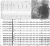
Fig. 1 summarizes the study protocol. After a standard procedure, AF or AT was induced by 10-second high-current burst pacing (10 mA, pulse width 5 ms, Bloom Associates, Denver, CO, USA) from the high right atrial (RA) electrodes. This commenced at a pacing cycle length of 250 ms and was gradually reduced to 120 ms as previously described (Fig. 2A).11 We initiated isoproterenol (5 µg/min) infusion for at least 3 minutes before induction and maintained this for 3 minutes after the induction of AF or AT. Then, internal cardioversion was performed by utilizing biphasic shock (2-15 J) with R wave synchronization (Lifepak12, Physiocontrol Ltd., Redmond, WA, USA) using an anodal decapolar catheter (WOVEN diagnostic catheter, Bard Electrophysiology, Lowell, MA, USA) in the RA and a cathodal duo-decapolar catheter (St. Jude Medical Inc., Minnetonka, MN, USA) in the coronary sinus (CS) (Fig. 2B). After successful electrical cardioversion, we stopped isoproterenol infusion and waited for 10 minutes to map IRAPBs or immediate recurrence of AF originating from the non-PV foci (Fig. 2C). All mappable IRAPBs (≥6/min) or APBs triggering AF were mapped and ablated during the waiting period in the IRAPB provocation group. If AF recurred immediately (8 of 100) or during the mapping of IRAPBs (5 of 100) after cardioversion, we repeated internal cardioversion and attempted to ablate all mappable frequent IRAPBs.

Post-ablation follow-up

Patients were discharged without any anti-arrhythmic drugs and asked to visit the outpatient clinic at 1, 3, 6, and 12 months after RFCA and every 6 months thereafter for follow-up. Electrocardiography (ECG) was performed at each visit or anytime the patient reported palpitations. A Holter ECG (24-hour or 48-hour) and/or event recorder was evaluated at 3, 6, and 12 months after RFCA and every 6 months thereafter for at least 2 years, according to the 2012 HRS/EHRA/ECAS Expert Consensus Statement guidelines.12 We defined recurrence of AF as any episode of AF or AT with a duration of at least 30 sec.13 If any ECG documented an AF episode within the 3-month blanking period during follow-up, the patient was diagnosed with an early recurrence. Any AF recurrence thereafter was diagnosed as clinical recurrence,13 and antiarrhythmic medications were prescribed.

Data analyses

We compared clinical and electroanatomical markers in patients who experienced recurrence of AF with those in patients who remained in sinus rhythm. Data are expressed as mean±standard deviation. Statistical significance of the comparisons was assessed using Student's t-test, Fisher's exact test, and Kaplan-Meier analysis. A p-value of <0.05 was considered statistically significant.

RESULTS

Mapping and ablation of post-procedural IRAPBs

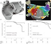
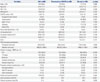
Fig. 1 summarizes the study protocol. Among the 100 patients in the IRAPB provocation group (70 PAF, 30 PeAF), we mapped and ablated IRAPBs in 33 patients, and 67 did not show post-procedural IRAPB after stopping the isoproterenol infusion (Table 1). The patients who tested positive for IRAPB had a higher CHADS2 score (p=0.003), higher prevalence of previous stroke/transient ischemic attack (p=0.002), higher frequency of sinus node dysfunction (p=0.025), and a larger left atrial volume index (LAVI; p<0.001) than those who did not. Fig. 3A shows the 44 sites for IRAPBs and additional ablation in 33 patients with positive IRAPB, with locations as follows: superior vena cava (n=12), interatrial septum (n=11), CS ostium (n=10), ligament of Marshall (n=6), crista terminalis (n=3), and LA posterior wall (n=2, one PAF and one PeAF). The RA volume index of the patients with RA-origin IRAPB (21.1±5.6 mL/m2) tended to be greater than those with LA origin IRAPB (19.5±4.6 mL/m2) without statistical significance (p=0.462). In patients with frequent and uni-focal IRAPBs, 3D-electroanatomical mapping was utilized for accurate mapping of non-PV foci or recovered conduction at linear ablation sites (Fig. 3B). The patients who underwent IRAPB ablation required a significantly longer procedure time (p=0.001) and ablation time (p=0.038) than those without IRAPB (Table 1). However, additional ablation for IRAPBs did not increase the procedure-related complication rate (p=0.732).

Presence of post-procedural IRAPBs is associated with higher clinical recurrence rate

During 18.0±6.6 months of follow-up, the early recurrence rate and clinical recurrence rate were 19% and 15%, respectively, for the IRAPB-provocation group. Among 51 patients with positive post-procedural inducibility before isoproterenol infusion, there was no significant difference in recurrence rates with respect to the type of induced atrial tachyarrhythmias (AF vs. AT; p=0.694) (Table 2). However, the patients with positive IRAPB showed a significantly higher early recurrence rate (36.4% vs. 10.4%; p=0.002) and clinical recurrence rate (27.3% vs. 9.0%; p=0.016) than those with negative IRAPBs, despite additional ablation and a longer procedure time (p=0.001) (Table 1). On multivariate logistic regression analysis, IRAPB (β=1.398; odds ratio=4.046; 95% confidence interval 1.14-14.39; p=0.031) was independently associated with clinical recurrence after RFCA of AF (Table 3). Table 3 also shows that the type of induced atrial tachyarrhythmias, AT (p=0.308) or AF (p=0.422), was not associated with clinical recurrence. Kaplan-Meier analysis showed consistently worse clinical outcome in patients with positive IRAPBs after the procedure (log rank, p=0.007) (Fig. 3C).

IRAPB provocation and ablation results in better clinical outcome of AF ablation

We compared patient characteristics, procedure time, procedure-related complication rate, and clinical outcome between the patients with IRAPB provocation and age-, sex-, and AF-type-matched patients who completed the procedure without an induction test (No-Test group) (Table 4). The baseline characteristics, LA diameter (p=0.187), LAVI (p=0.105), procedure times (p=0.540), ablation times (p=0.848), and procedure-related complication rates (p=0.702), were not significantly different between the two groups. However, the clinical recurrence rate (15.0% vs. 28.0%; p=0.025) was significantly lower in the IRAPB provocation and ablation group than in the No-Test group. Ka-plan-Meier AF-free survival analysis indicated a consistently better clinical outcome in the IRAPB provocation group than in the No-Test group (log rank, p=0.038) (Fig. 3D).

DISCUSSION

In the current study, we reported the potential usefulness of post-procedural IRAPB provocation and additional ablation during AF catheter ablation. The patients who showed post-procedural IRAPB under isoproterenol had a clearly higher clinical recurrence rate of AF than those without IRAPB. However, clinical outcome was better in patients with routine post-procedural IRAPB provocation and additional ablation than in those who completed the procedure without an induction test. This study revealed the importance of remaining non-PV foci as a mechanism of recurrence after AF catheter ablation. Therefore, a further prospective study with a larger number of patients is warranted to prove whether routine IRAPB provocation facilitates an improved clinical outcome of AF catheter ablation.

Role of post-procedural detection of AF triggers or substrate

AF is generally a progressive disease, and the mechanism for generation of AF is not yet fully understood.114 Coumel1516 suggested that trigger factors or trigger foci initiate AF, and arrhythmic substrate leads to its persistence. In this study, we explored the clinical significance of post-procedural tests for AF triggers or IRAPB. It is known that 70-90% of AF triggers exist around PVs in patients with PAF; however, non-PV foci are more common in those with PeAF.456 Generally, PV isolation at the level of the PV antrum is a cornerstone of catheter ablation for AF.171819 In this study, by isolating PVs during the procedure, all post-procedural IRAPBs arose from non-PV foci. Although the presence of IRAPBs from these non-PV foci clearly worsened the clinical outcome of AF ablation, additional ablation targeting IRAPB might result in a lower recurrence rate than that of the No-Test group. Therefore, remaining non-PV-origin IRAPB may play an important role in the recurrence mechanism of AF in the No-Test group.

Clinical significance of non-PV foci

APBs that trigger AF frequently arise from PVs,20 and cardiac autonomic nerves mainly connect to the heart along the PV antral area.42122 Therefore, bi-antral PV isolation is effective for the elimination of PV triggers,2324 cardiac autonomic denervation,25 and substrate modification. However, PV isolation may not be sufficient to control AF due to the presence of non-PV foci or atrial substrate remodeling, particularly in patients with PeAF.2627 Clinically, complex fractionated atrial electrograms (CFAEs) are known to play a role in maintaining AF2829 and act as a target for AF catheter ablation.2930 Verma, et al.31 considered CFAE ablation to be a non-PV substrate and a trigger of ablation, and Lemery, et al.32 co-localized the area of CFAE and cardiac autonomic ganglionated plexi by nerve stimulation. However, CFAE includes both the active driver of AF and passive wave breakers, and CFAE-guided AF ablation carries the risk of unnecessary cardiac tissue damage. In this study, we ablated non-PV foci targeting post-cardioversion IRAPBs and demonstrated a better clinical outcome.

Mapping and ablation technique for non-PV foci

We previously reported that an excessively long ablation dura-tion for substrate modification results in a poor clinical outcome.3334 Therefore, limited and sophisticated ablation for non-PV foci based on post-procedural IRAPB might be beneficial for a better clinical outcome. However, detection and ablation of non-PV foci are not simple. We targeted non-PV foci immediately after cardioversion under isoproterenol (5 µg/min) infusion. As we adhered to the linear ablation strategy, including the anterior line10 in patients with PeAF, re-initiation of AF was uncommon (8%) after cardioversion and cessation of isoproterenol infusion, and most non-PV foci remained as frequent APBs. We were able to map those non-PV IRAPBs based on the areas defined by linear ablation lines.11 Quick and limited 3D activation mapping utilizing a multi-electrode catheter was useful for accurate mapping and limited ablation of frequent non-PV-origin APBs in certain patients. We mapped and ablated IRAPBs during the isoproterenol cooling-down period until no APBs appeared.

Limitations

The patients included in this study were a highly selective group referred for RFCA, and the number of patients was limited. As this was a retrospective single-center study, there might have been a selection bias, despite comparing two groups matched for age, sex, and AF type. Considering that most IRAPBs were mapped and ablated within 10 minutes after stopping isoproterenol infusion, infrequent IRAPBs, multi-focal APBs, and catecholamine-insensitive APBs may not have been eliminated in this study protocol. In addition, we did not challenge isoproterenol again after IRAPB ablation.

Conclusion

The patients who showed post-procedural IRAPB under isoproterenol had a clearly higher clinical recurrence rate of AF. However, careful mapping and ablation for post-procedural IRAPB safely resulted in a better clinical outcome without requiring a longer procedure time compared to those who completed the procedure without an induction test. Therefore, a future prospective study with a larger number of patients is warranted to determine whether routine IRAPB provocation facilitates an improved clinical outcome of AF catheter ablation.

Figures and Tables

Fig. 1

Diagram of study protocol. We compared the clinical efficacy of post-procedural provocation for immediate recurrence of atrial premature beats (IRAPBs) under isoproterenol and additional ablation targeting these APBs (n=100) with 100 age-, sex-, and AF-type-matched patients who completed the procedure without an induction test (No-Test group). CL, cycle length; RFCA, radiofrequency catheter ablation; AF, atrial fibrillation; AT, atrial tachycardia.

ymj-57-72-g001
Fig. 2

(A) Inducibility test by high-current HRA pacing for 10 sec. AF or AT maintenance longer than 3 min was considered positive inducibility. (B) Catheter position for mapping and cardioversion. For the IRAPB test, we delivered internal cardioversion shock from HRA to CS. (C) An example of IRAPB after cardioversion under isoproterenol infusion. CS, coronary sinus; HRA, high right atrium; LAO, left anterior oblique view; RV, right ventricle; AF, atrial fibrillation; AT, atrial tachycardia; IRAPB, immediate recurrence of atrial premature beat.

ymj-57-72-g002
Fig. 3

(A) Locations of post-procedural IRAPBs and additional ablation sites (black circles). White circles indicate the standard ablation sites for PAF. (B)Mapping and ablation of post-procedural IRAPB utilizing 3D activation map. Quick activation mapping by multipolar catheter revealed non-PV foci located on LA posterior wall (white dots) and conduction gap at posterior inferior linear ablation site. We eliminated IRAPBs and ablation gap on posterior inferior line concomitantly. (C) Kaplan-Meier curve for AF-free survival comparing patients with post-procedural IRAPBs to those without IRAPBs. (D) Kaplan-Meier graph of AF-free survival comparing the IRAPB provocation group to the No-Test group. IRAPBs, immediate recurrence of atrial premature beats; 3D, three-dimensional; PV, pulmonary vein; LA, left atrial; AF, atrial fibrillation; PAF, paroxysmal AF.

ymj-57-72-g003
Table 1

Characteristics of IRAPB Provocation Group Depending on the Presence of IRAPBs and Additional Ablation

ymj-57-72-i001
Variables IRAPB (+) (n=33) IRAPB (-) (n=67) p value
Male, n (%) 22 (66.7) 54 (80.6) 0.128
Patient age (yrs) 57.6±9.5 56.6±11.7 0.692
PAF, n (%) 20 (60.6) 50 (74.6) 0.153
BMI (kg/m2) 25.0±3.0 24.1±2.6 0.183
CHADS2 score, n (%) 1.1±1.1 0.6±0.8 0.003
 Congestive heart failure 0 (0.0) 2 (3.0) 0.321
 Hypertension 17 (51.5) 26 (38.8) 0.232
 Age ≥75 yrs 1 (3.0) 1 (1.5) 0.610
 Diabetes 6 (18.2) 5 (7.5) 0.109
 Stroke/TIA 7 (21.2) 2 (3.0) 0.002
Sinus node dysfunction, n (%) 5 (15.2) 2 (3.0) 0.025
LA diameter (mm) 41.1±4.1 39.1±4.8 0.052
LA volume index (mL/m2) 36.2±11.2 28.2±8.5 <0.001
LV EF (%) 64.5±6.0 64.1±8.1 0.806
E/Em 9.7±3.6 8.9±3.0 0.278
Procedure time (min) 201.0±34.8 174.3±30.7 0.001
Ablation time (sec) 5280.0±1470.9 4617.0±1304.4 0.038
Procedure-related complications, n (%) 1 (3.0) 3 (4.5) 0.732
Follow-up duration (months) 17.4±5.7 18.1±7.4 0.643
Early recurrence rates (%) 36.4 10.4 0.002
Clinical Recurrence rates (%) 27.3 9.0 0.016

BMI, body mass index; E/Em, ratio of early mitral inflow peak velocity and Doppler-derived early diastolic mitral annular velocity; IRAPB, immediate recurrence of atrial premature beats after cardioversion; LA, left atrium; LV EF, left ventricular ejection fraction; PAF, paroxysmal atrial fibrillation; TIA, transient ischemic attack.

Table 2

Clinical Outcomes Depending on the Type of Post-Procedural Atrial Tachyarrhythmias (AT/AF) Induced by High-Current Rapid Atrial Pacing

ymj-57-72-i002
Variables AT induction (n=35) AF induction (n=16) p value
Follow-up duration (month) 18.2±6.1 17.3±4.9 0.625
Early recurrence rates (%) 28.6 31.3 0.849
Clinical recurrence rates (%) 20.0 25.0 0.694

AF, atrial fibrillation; AT, atrial tachycardia.

Table 3

Factors Associated with Clinical Recurrence after Radiofrequency Catheter Ablation (Logistic Regression Analyses)

ymj-57-72-i003
Variables Univariate analysis Multivariate analysis
OR 95% CI p value OR 95% CI p value
PAF 0.590 0.19-1.84 0.363 0.986 0.26-3.74 0.983
Male 1.312 0.34-5.10 0.695 1.373 0.29-6.56 0.691
Age (yrs) 1.014 0.96-1.07 0.596 1.001 0.94-1.07 0.967
Diabetes 4.052 1.02-16.13 0.047 3.205 0.71-14.41 0.129
Pre-RFCA LA volume index (mL/m2) 1.046 0.99-1.11 0.132
Pre-RFCA LV EF (%) 1.044 0.96-1.13 0.304 1.018 0.93-1.12 0.711
Pre-RFCA E/Em 1.009 0.85-1.21 0.919 0.983 0.79-1.22 0.878
AT or AF induction (+) 3.094 0.91-10.49 0.070
 AT induction (+) 1.781 0.59-5.41 0.308
 AF induction (+) 1.625 0.50-5.31 0.422
IRAPB (+) 3.812 1.22-11.87 0.021 4.046 1.14-14.39 0.031

AF, atrial fibrillation; AT, atrial tachycardia; CI, confidence interval; E/Em, ratio of early mitral inflow peak velocity and Doppler-derived early diastolic mitral annular velocity; IRAPB, immediate recurrence of atrial premature beats; LA, left atrial; LV EF, left ventricular ejection fraction; OR, odds ratio; PAF, paroxysmal atrial fibrillation; RFCA, radiofrequency catheter ablation.

Table 4

Characteristics Comparing IRAPB Provocation/Ablation Group vs. No-Test Group

ymj-57-72-i004
Variables All (n=200) Provocation of IRAPB (n=100) No test (n=100) p value
Male, n (%) 153 (76.5) 76 (76.0) 77 (77.0) 0.868
Age (yrs) 57.4±11.1 57.0±11.0 57.9±11.2 0.551
PAF, n (%) 134 (67.0) 70 (70.0) 64 (64.0) 0.369
BMI (kg/m2) 24.9±3.9 24.5±2.8 25.3±4.7 0.233
CHADS2 score, n (%) 0.8±1.0 0.8±0.9 0.9±1.0 0.309
 Congestive heart failure 6 (3.0) 2 (2.0) 4 (4.0) 0.410
 Hypertension 92 (46.0) 43 (43.0) 49 (49.0) 0.397
 Age ≥75 yrs 4 (2.0) 2 (2.0) 2 (2.0) 1.000
 Diabetes 28 (14.0) 11 (11.0) 17 (17.0) 0.223
 Stroke/TIA 18 (9.0) 9 (9.0) 9 (9.0) 1.000
Sinus node dysfunction, n (%) 10 (5.0) 7 (7.0) 3 (3.0) 0.196
LA diameter (mm) 40.2±4.8 39.8±4.6 40.7±5.0 0.187
LA volume index (mL/m2) 32.0±10.2 30.8±10.1 33.2±10.2 0.105
LV EF (%) 63.6±7.7 64.2±7.5 63.0±7.9 0.275
E/Em 9.7±4.3 9.2±3.2 10.2±5.1 0.107
RFCA procedural parameters
 Procedure time (min) 180.9±34.5 182.5±34.2 179.4±34.8 0.540
 Ablation time (sec) 4803.5±1286.4 4822.8±1384.4 4786.4±1199.7 0.848
Ablation strategy, n (BDB rate, %)
 PVI 200 (100.0) 100 (100.0) 100 (100.0) >0.999
 CTI block 192 (100.0) 94 (100.0) 98 (100.0) 0.155
 Roof line 66 (97.0) 30 (100.0) 36 (94.4) 0.369
 Posterior-inferior line 51 (71.9) 29 (66.7) 22 (76.5) 0.258
 Anterior linear line 53 (67.9) 26 (57.1) 27 (78.6) 0.873
Procedure-related complications, n (%) 7 (3.5) 4 (4.0) 3 (3.0) 0.702
 Hemopericardium 4 (2.0) 1 (1.0) 3 (3.0) 0.315
 Mild PV stenosis 1 (0.5) 1 (1.0) 0 (0.0) 0.319
 Groin hematoma 2 (1.0) 2 (2.0) 0 (0.0) 0.157
Follow-up duration (months) 18.0±6.6 17.9±6.9 18.2±6.4 0.694
Early recurrence rates (%) 30.5 19.0 42.0 <0.001
Clinical recurrence rates (%) 21.5 15.0 28.0 0.025

BMI, body mass index; E/Em, ratio of early mitral inflow peak velocity and Doppler-derived early diastolic mitral annular velocity; IRAPB, immediate recurrence of atrial premature beats after cardioversion; LA, left atrium; LV EF, left ventricular ejection fraction; PAF, paroxysmal atrial fibrillation; PV, pulmonary vein; PVI, pulmonary vein isolation; TIA, transient ischemic attack; BDB, bidirectional block; RFCA, radiofrequency catheter ablation; CTI, cavotricuspid isthmus.

ACKNOWLEDGEMENTS

This work was supported by a grant (A085136) from the Korea Health 21 R&D Project; Ministry of Health and Welfare; and from the National Research Foundation of Korea (NRF) funded by the Ministry of Science, ICT&Future Planning (MSIP; 7-2013-0362).

Notes

The authors have no financial conflicts of interest.

References

1. Inoue K, Kurotobi T, Kimura R, Toyoshima Y, Itoh N, Masuda M, et al. Trigger-based mechanism of the persistence of atrial fibrillation and its impact on the efficacy of catheter ablation. Circ Arrhythm Electrophysiol. 2012; 5:295–301.
crossref
2. Camm AJ, Lip GY, De Caterina R, Savelieva I, Atar D, Hohnloser SH, et al. 2012 focused update of the ESC Guidelines for the management of atrial fibrillation: an update of the 2010 ESC Guidelines for the management of atrial fibrillation. Developed with the special contribution of the European Heart Rhythm Association. Eur Heart J. 2012; 33:2719–2747.
3. European Heart Rhythm Association. European Association for Cardio-Thoracic Surgery. Camm AJ, Kirchhof P, Lip GY, Schotten U, et al. Guidelines for the management of atrial fibrillation: the Task Force for the Management of Atrial Fibrillation of the European Society of Cardiology (ESC). Eur Heart J. 2010; 31:2369–2429.
4. Pak HN, Hwang C, Lim HE, Kim JW, Lee HS, Kim YH. Electroanatomic characteristics of atrial premature beats triggering atrial fibrillation in patients with persistent versus paroxysmal atrial fibrillation. J Cardiovasc Electrophysiol. 2006; 17:818–824.
crossref
5. Lee SH, Tai CT, Hsieh MH, Tsao HM, Lin YJ, Chang SL, et al. Predictors of non-pulmonary vein ectopic beats initiating paroxysmal atrial fibrillation: implication for catheter ablation. J Am Coll Cardiol. 2005; 46:1054–1059.
crossref
6. Oral H, Crawford T, Frederick M, Gadeela N, Wimmer A, Dey S, et al. Inducibility of paroxysmal atrial fibrillation by isoproterenol and its relation to the mode of onset of atrial fibrillation. J Cardiovasc Electrophysiol. 2008; 19:466–470.
crossref
7. Oral H, Chugh A, Good E, Sankaran S, Reich SS, Igic P, et al. A tailored approach to catheter ablation of paroxysmal atrial fibrillation. Circulation. 2006; 113:1824–1831.
crossref
8. Oral H, Chugh A, Lemola K, Cheung P, Hall B, Good E, et al. Noninducibility of atrial fibrillation as an end point of left atrial circumferential ablation for paroxysmal atrial fibrillation: a randomized study. Circulation. 2004; 110:2797–2801.
crossref
9. Gaita F, Caponi D, Pianelli M, Scaglione M, Toso E, Cesarani F, et al. Radiofrequency catheter ablation of atrial fibrillation: a cause of silent thromboembolism? Magnetic resonance imaging assessment of cerebral thromboembolism in patients undergoing ablation of atrial fibrillation. Circulation. 2010; 122:1667–1673.
crossref
10. Pak HN, Oh YS, Lim HE, Kim YH, Hwang C. Comparison of voltage map-guided left atrial anterior wall ablation versus left lateral mitral isthmus ablation in patients with persistent atrial fibrillation. Heart Rhythm. 2011; 8:199–206.
crossref
11. Hwang ES, Nam GB, Joung B, Park J, Lee JS, Shim J, et al. Significant reduction of atrial defibrillation threshold and inducibility by catheter ablation of atrial fibrillation. Pacing Clin Electrophysiol. 2012; 35:1428–1435.
crossref
12. Cappato R, Calkins H, Chen SA, Davies W, Iesaka Y, Kalman J, et al. Updated worldwide survey on the methods, efficacy, and safety of catheter ablation for human atrial fibrillation. Circ Arrhythm Electrophysiol. 2010; 3:32–38.
crossref
13. European Heart Rhythm Association (EHRA). European Cardiac Arrhythmia Scoiety (ECAS). American College of Cardiology (ACC). American Heart Association (AHA). Society of Thoracic Surgeons (STS). Calkins H, et al. HRS/EHRA/ECAS expert Consensus Statement on catheter and surgical ablation of atrial fibrillation: recommendations for personnel, policy, procedures and follow-up. A report of the Heart Rhythm Society (HRS) Task Force on catheter and surgical ablation of atrial fibrillation. Heart Rhythm. 2007. 4:p. 816–861.
crossref
14. Wazni O, Wilkoff B, Saliba W. Catheter ablation for atrial fibrillation. N Engl J Med. 2011; 365:2296–2304.
crossref
15. Coumel P. Paroxysmal atrial fibrillation: a disorder of autonomic tone? Eur Heart J. 1994; 15:Suppl A. 9–16.
crossref
16. Coumel P. Autonomic influences in atrial tachyarrhythmias. J Cardiovasc Electrophysiol. 1996; 7:999–1007.
crossref
17. Haïssaguerre M, Jaïs P, Shah DC, Takahashi A, Hocini M, Quiniou G, et al. Spontaneous initiation of atrial fibrillation by ectopic beats originating in the pulmonary veins. N Engl J Med. 1998; 339:659–666.
crossref
18. Pappone C, Rosanio S, Oreto G, Tocchi M, Gugliotta F, Vicedomini G, et al. Circumferential radiofrequency ablation of pulmonary vein ostia: a new anatomic approach for curing atrial fibrillation. Circulation. 2000; 102:2619–2628.
crossref
19. Kumar S, Kalman JM, Sutherland F, Spence SJ, Finch S, Sparks PB. Atrial fibrillation inducibility in the absence of structural heart disease or clinical atrial fibrillation: critical dependence on induction protocol, inducibility definition, and number of inductions. Circ Arrhythm Electrophysiol. 2012; 5:531–536.
crossref
20. Chen SA, Hsieh MH, Tai CT, Tsai CF, Prakash VS, Yu WC, et al. Initiation of atrial fibrillation by ectopic beats originating from the pulmonary veins: electrophysiological characteristics, pharmacological responses, and effects of radiofrequency ablation. Circulation. 1999; 100:1879–1886.
crossref
21. Pappone C, Santinelli V, Manguso F, Vicedomini G, Gugliotta F, Augello G, et al. Pulmonary vein denervation enhances long-term benefit after circumferential ablation for paroxysmal atrial fibrillation. Circulation. 2004; 109:327–334.
crossref
22. Krul SP, Driessen AH, van Boven WJ, Linnenbank AC, Geuzebroek GS, Jackman WM, et al. Thoracoscopic video-assisted pulmonary vein antrum isolation, ganglionated plexus ablation, and periprocedural confirmation of ablation lesions: first results of a hybrid surgical-electrophysiological approach for atrial fibrillation. Circ Arrhythm Electrophysiol. 2011; 4:262–270.
crossref
23. Edgerton JR, Brinkman WT, Weaver T, Prince SL, Culica D, Herbert MA, et al. Pulmonary vein isolation and autonomic denervation for the management of paroxysmal atrial fibrillation by a minimally invasive surgical approach. J Thorac Cardiovasc Surg. 2010; 140:823–828.
crossref
24. Oh S, Zhang Y, Bibevski S, Marrouche NF, Natale A, Mazgalev TN. Vagal denervation and atrial fibrillation inducibility: epicardial fat pad ablation does not have long-term effects. Heart Rhythm. 2006; 3:701–708.
crossref
25. Schauerte P, Scherlag BJ, Pitha J, Scherlag MA, Reynolds D, Lazzara R, et al. Catheter ablation of cardiac autonomic nerves for prevention of vagal atrial fibrillation. Circulation. 2000; 102:2774–2780.
crossref
26. Haissaguerre M, Hocini M, Shah AJ, Derval N, Sacher F, Jais P, et al. Noninvasive panoramic mapping of human atrial fibrillation mechanisms: a feasibility report. J Cardiovasc Electrophysiol. 2013; 24:711–717.
crossref
27. Haïssaguerre M, Hocini M, Sanders P, Sacher F, Rotter M, Takahashi Y, et al. Catheter ablation of long-lasting persistent atrial fibrillation: clinical outcome and mechanisms of subsequent arrhythmias. J Cardiovasc Electrophysiol. 2005; 16:1138–1147.
crossref
28. Konings KT, Smeets JL, Penn OC, Wellens HJ, Allessie MA. Configuration of unipolar atrial electrograms during electrically induced atrial fibrillation in humans. Circulation. 1997; 95:1231–1241.
crossref
29. Lo LW, Lin YJ, Tsao HM, Chang SL, Hu YF, Tsai WC, et al. Characteristics of complex fractionated electrograms in nonpulmonary vein ectopy initiating atrial fibrillation/atrial tachycardia. J Cardiovasc Electrophysiol. 2009; 20:1305–1312.
crossref
30. Nademanee K, McKenzie J, Kosar E, Schwab M, Sunsaneewitayakul B, Vasavakul T, et al. A new approach for catheter ablation of atrial fibrillation: mapping of the electrophysiologic substrate. J Am Coll Cardiol. 2004; 43:2044–2053.
crossref
31. Verma A, Mantovan R, Macle L, De Martino G, Chen J, Morillo CA, et al. Substrate and Trigger Ablation for Reduction of Atrial Fibrillation (STAR AF): a randomized, multicentre, international trial. Eur Heart J. 2010; 31:1344–1356.
crossref
32. Lemery R, Birnie D, Tang AS, Green M, Gollob M. Feasibility study of endocardial mapping of ganglionated plexuses during catheter ablation of atrial fibrillation. Heart Rhythm. 2006; 3:387–396.
crossref
33. Shim J, Joung B, Park JH, Uhm JS, Lee MH, Pak HN. Long duration of radiofrequency energy delivery is an independent predictor of clinical recurrence after catheter ablation of atrial fibrillation: over 500 cases experience. Int J Cardiol. 2013; 167:2667–2672.
crossref
34. Kim JB, Yang DH, Kang JW, Jung SH, Choo SJ, Chung CH, et al. Left atrial function following surgical ablation of atrial fibrillation: prospective evaluation using dual-source cardiac computed tomography. Yonsei Med J. 2015; 56:608–616.
crossref
TOOLS
Similar articles