

 PDF
PDF ePub
ePub Citation
 Print
Print


INTRODUCTION
Non-steroidal anti-inflammatory drugs (NSAIDs) provide several benefits in intraocular and refractive surgery. Because NSAIDs are potent inhibitors of cyclooxygenase (COX) enzymes, they inhibit production of pro-inflammatory prostaglandins, which cause vasodilation and increased vascular permeability.1 Therefore, NSAIDs are used to maintain pupillary dilation during surgery, to reduce postoperative pain, to control inflammation during and after surgery, and to inhibit development of cystoid macular edema (CME) after cataract surgery.234
Topical NSAID eye drops gained attention as an alternative treatment to topical steroids for postoperative pain and inflammation because they are associated with fewer complications, such as increased intraocular pressure, infection, and delayed corneal epithelial wound healing.5678 Concurrent administration of corticosteroids and NSAIDs has shown potential synergistic activity in some studies.5910 Food and Drug Administration (FDA)-approved NSAIDs currently used for cataract surgery include diclofenac, ketorolac, nepafenac, and bromfenac.5
Bromfenac sodium 0.1% ophthalmic solution (Bronuck®, Senju Pharmaceutical Co., Ltd., Osaka, Japan) has clinical indications for treating postoperative inflammation, blepharitis, conjunctivitis, and scleritis.5 Preservative-free ketorolac 0.45% solution (Acuvail®, Allergan Inc., CA, USA) is also targeted for postoperative use after cataract surgery.11
Several reports identified corneal complications of using early-generation topical NSAIDs, including corneal melting caused by the generic form of diclofenac, as well as ketorolac and nepafenac; these reports prompted subsequent decline in the use of topical NSAIDs after ocular surgery.1213 However, Flach13 demonstrated that not only simple drug toxicity but also coexistent factors, such as concurrent ocular and systemic disease, inconsistent and variable doses, and other medications used simultaneously, were attributable to corneal melting. Since then, many clinical trials have established the efficacy and safety of other topical NSAIDs1415 and they are being used again in ophthalmic surgery.
In this study, we report the results of a prospective, randomized clinical trial that compared two kinds of NSAID eye drops, with different application schedules, for use in combination therapy with topical steroid for cataract surgery.
MATERIALS AND METHODS
This single-center, randomized study was approved by the Severance Hospital Institutional Review Board, Seoul, South Korea. Participants consisted of 91 patients who were scheduled for cataract surgery between November 2013 and June 2014. All patients gave written informed consent before enrollment, and clinical research was performed in accordance with the tenets of the Helsinki Declaration.
Inclusion criteria for enrollment were as follows: males or non-pregnant females aged between 20- to 80-years-old. Exclusion criteria were as follows: poor general condition, including high blood pressure, poor blood glucose control, or renal failure; history of ocular trauma or disease; history of intraocular surgery; systemic or topical NSAIDs or corticosteroids use within 4 weeks of enrollment; known hypersensitivity to salicylates or other NSAIDs; and use of alpha-1 adrenergic antagonist or other analogous systemic medications that may increase the tendency for miosis during the operation (intraoperative floppy iris syndrome).
Patients were enrolled in the study during a preoperative visit after initial screening. Preoperatively, all patients underwent a complete ophthalmic examination, including visual acuity, slit-lamp examination, indirect ophthalmoscopy, and optical coherence tomography (OCT). Patients were randomized into three groups according to the administered study drugs, and the study medications were masked to both the surgeon and examiners. The administration of the topical NSAIDs was determined based on the suggested regimen of each pharmaceutical company. Group 1 patients were instructed to apply 1 drop of bromfenac sodium hydrate ophthalmic solution 0.1% twice daily beginning 3 days before surgery and two drops at 20 minute intervals at 2 hours before surgery. They continued using bromfenac 0.1% twice-daily with other postoperative eye drops for 4 weeks. Group 2 patients were instructed to instill 1 drop of ketorolac 0.45% ophthalmic solution twice-daily beginning 1 day prior to cataract surgery and 2 drops at 20 minute intervals at 2 hours before surgery, with continued application twice-daily throughout the first 2 postoperative weeks. Control eyes in Group 3 did not receive any additional medications. Postoperatively, patients in all three groups were prescribed topical gatifloxacin 0.3% and prednisolone acetate 1% eye drops that were to be applied 4 times daily for 4 weeks.
Surgical technique
All cataract surgeries were phacoemulsification with intraocular lens (IOL) implantation after topical anesthesia with proparacaine hydrochloride 0.5% eye drops by a single surgeon (T.I. Kim). The surgical procedure consisted of a clear cornea incision on the steep axis, capsulorrhexis, phacoemulsification, and IOL placement in the capsular bag.
Outcome measures
Outcome measures comprised changes in pupil size during surgery, postoperative anterior chamber inflammation, macular thickness and volume, and change in the ocular surface status. Patients were evaluated on postoperative days 1, 7, and 28. Horizontal pupillary diameters were measured in 0.5 mm units using a sterile caliper over the cornea and measurements were taken under a microscope upon the initiation of the surgery and before the insertion of the ophthalmic viscosurgical device (OVD) for IOL implantation. Ocular inflammation was assessed, and the summed ocular inflammation score (SOIS) was calculated by adding the subject's anterior chamber cells and flare grades (range, 0-4) at 1 week and 1 month postoperatively.16
All OCT images were obtained using the Spectralis SD-OCT (Heidelberg Engineering, Carlsbad, CA, USA), and the macular retinal thickness and volume map analysis protocol was used to evaluate the macular state.17 The central foveal subfield (CSF) thickness was defined as the average of all points in the inner circle within 1 mm radius from the fovea, and macular thickness was determined as the average thickness of five subfields, including the CSF and nasal, temporal, superior, and inferior inner segments in the central 3 mm area. We used the term macular edema (ME) to describe an increase in macular thickness or volume of >30% on OCT, 1 month after cataract surgery.18 CME was defined as presence of cystoid changes associated with substantial (≥40 µm) retinal thickening.19 Evaluation of ocular surface status, including Schirmer test without anesthesia, tear breakup time (TBUT), and fluorescein corneal staining (range, 0-5),20 was performed preoperatively and 1 month postoperatively. Adverse events were monitored and recorded.
Statistical analysis
The results are presented as mean±SD, or as patient number and percentage. Comparisons of the outcome measures among patient groups were performed using one-way analysis of variance with Scheffé's test. Comparisons between the preoperative and postoperative data were analyzed by Wilcoxon ranksum tests. Statistical analyses were performed using SPSS statistical software (version 20.0; SPSS Inc., Chicago, IL, USA), and p-values less than 0.05 were considered statistically significant.
RESULTS
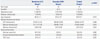
A total of 91 eyes from 91 subjects (41 men and 50 women) were included in the study, and all patients underwent uncomplicated phacoemulsification with IOL implantation. Mean age of the subjects was 66.9±8.5 years (range, 33-78 years). Table 1 shows the demographics and preoperative characteristics of subjects in all three groups. Overall, baseline demographics and clinical characteristics, including preoperative ocular surface status and macular thickness and volume, were similar among the groups. There were 24 subjects (26.4%) with diabetes mellitus (DM) without retinopathy.
Maintenance of pupil dilation during the cataract surgery
In group 1, pupil diameters at the beginning of surgery were larger than those of Groups 2 and 3 (p=0.043 and 0.046, respectively). Pupil diameters before inserting the OVD for IOL implantation were significantly different between Groups 1 and 3 (p=0.003). Both NSAID groups (Groups 1, 2) showed small decreases in pupil diameter during the surgical procedure, compared with Group 3 (p=0.025 and 0.013, respectively) (Table 2). However, there was no difference in pupil diameter change between Group 1 and Group 2. No patients required intraoperative additive medication for pupil dilation, such as intracameral epinephrine.
Postoperative anterior chamber inflammation after cataract surgery
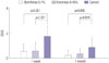
One week after surgery, the total scores of cells and flare, SOISs, were 0.53±0.61, 0.58±0.69, and 1.74±0.81 in Group 1, 2, and 3, respectively. This score was similar (p=0.974) and significantly lower in both NSAID groups compared with controls (p<0.001 in both comparisons). The inflammatory score at 1 month was similar in Group 1 and Group 2 (0.21±0.42 and 0.32±0.48, respectively; p=0.853). In contrast, the SIOS of Group 3 was 0.84±0.76 at 1 month, which was significantly higher than that for both NSAID groups (p=0.005, 0.024, respectively) (Fig. 1).
Postoperative macular edema after cataract surgery
Mean CSF, macular thickness, and macular volume significantly increased after cataract operation in all groups (p<0.05 for all groups). The CSF thickness increased from before the operation to postoperative 1 month by 4.30±4.25, 4.87±6.03, and 12.47±12.24 µm in Groups 1, 2, and 3, respectively. Both NSAID groups showed similar increases in CSF thickness, which were significantly less than the control group (p<0.05 for both comparisons). Increased macular thickness of both NSAID groups was significantly less than that in the controls (p<0.05 for both comparisons). The total macular volume increased by 0.13±0.17, 0.20±0.14, and 0.26±0.19 mm3 in Group 1, 2, and 3, respectively, one month after the operation. A significant difference in macular volume existed between the bromfenac 0.1% group and the control (Table 3). No eyes in any group exhibited ME or CME at 1 month after operation.
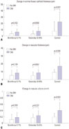
Fig. 2 shows that the macular changes after cataract operation were different between subgroups according to the presence of DM. In NSAID-treated Groups 1 and 2, the changes in CSF thickness, macular thickness, and macular volume before the operation and at postoperative 1 month were not significantly different between patients without DM and those with DM (p>0.05). However, in Group 3, the changes in CSF thickness were 7.60±6.43 µm in patients without DM and 22.20±15.42 µm in patients with DM (p=0.001). The changes in macular thickness were 9.40±3.75 µm in patients without DM and 17.30±10.94 µm in patients with DM (p=0.006) and the changes in macular volume were 0.19±0.12 mm3 in patients without DM and 0.40±0.25 mm3 in patients with DM (p=0.003).
Postoperative ocular surface status after cataract surgery
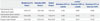
At 1 month postoperatively, TBUT was significantly worse than that before cataract surgery in all groups (p<0.05). Although all three parameters, especially corneal staining, were worse in the bromfenac 0.1% group than the ketorolac 0.45% group, these differences were not significant (Table 4).
DISCUSSION
Several topical NSAIDs are currently approved for controlling postoperative inflammation after cataract surgery. Ophthalmic NSAID eye drops maintain pupil dilation, reduce postoperative inflammation, pain and discomfort, and prevent and treat ME after cataract operation.5 Each type of NSAID ophthalmic solutions has its own characteristics, thus treatment effects may differ among various drugs. The purpose of this prospective study was to compare the effects of two different NSAIDs with different application schedules, when added to concurrent steroid treatment.
Two NSAID ophthalmic solutions, bromfenac sodium hydrate 0.1% with 0.005% benzalkonium chloride (BAK) and preservative-free ketorolac tromethamine 0.45% have recently become indicated for the control of postoperative inflammation in people undergoing cataract surgery in Korea. The anti-inflammatory effect of twice-daily bromfenac sodium 0.1% has been reported,2421 and twice-daily administered preservative-free ketorolac 0.45% was well-tolerated and effectively treated inflammation and pain after cataract surgery.11
Additive use of both NSAID eye drops effectively maintained pupillary dilation during the operation and reduced postoperative ocular inflammation in this study, compared to the steroid-only control group. Previous studies reported that NSAIDs potentiate concomitant topical steroid effects of postoperative ocular inflammation59 and inhibit miosis during the operation,2223 as in our results. One difference between previous reports2223 and ours was the results of preoperative pupil size. Since bromfenac sodium 0.1% was used prior to 3 days before surgery in Group 1, preoperative pupil size might be larger than in other groups. However, because we did not measure pupil size before the use of the medication, we cannot conclude this definitely.
In this study, although macular thickness and volume increased in all groups after cataract operation, no patient presented with clinically important ME or CME during the 1 month postoperative follow-up period. A previous study24 has demonstrated that topical NSAID treatment was effective in prevention or treatment of ME or CME after cataract surgery. In our study, topical NSAIDs added to corticosteroid also reduced these changes more effectively than corticosteroid alone. Spectralis OCT has a very high repeatability and reproducibility, with a very small measurable change (1.5 µm) and coefficient of variation (0.53%);25 therefore, small changes and differences between groups in our study can be considered as meaningful findings.
A randomized study by Wittpenn, et al.19 reported that a combination of ketorolac 0.4% and steroid, compared to steroid alone, reduced CME incidence rate (0.0% versus 2.4%) and postoperative mean retinal thickening (3.9 µm versus 9.6 µm) in cataract surgery patients. Our results demonstrated the effects of NSAIDs on ME after cataract surgery in patients without DM and in early phase of DM patients without retinopathy (24/91, 26.4% of total patients). In the control group, CSF, macular thickness, and macular volume significantly increased after cataract procedure in the subgroup of patients with DM. In contrast, in both NSAIDs groups, macular changes were minimized in both subgroups with DM or without DM. Diabetic patients who have no diabetic retinopathy or definite ME show partial damage to the blood-retinal barrier.26 Prostaglandins released by surgical trauma cause vasodilation, increased vascular permeability, and breakdown of the blood-retinal barrier, and consequently, postoperative ME or CME can potentially develop.27 Thus, prophylactic treatment of topical NSAIDs would be recommended in DM patients with a weak barrier, even if they do not exhibit diabetic retinopathy.
There was no superiority between two NSAID ophthalmic solutions in inhibiting miosis and reducing inflammation and macular change. Topical NSAIDs inhibit ocular COX-1 and COX-2 enzyme activities, thereby reducing prostaglandin production and providing both analgesic and anti-inflammatory effects. One report demonstrated that administration of ketorolac 0.4% four times a day may provide better control of prostaglandin-mediated inflammation than administration of bromfenac 0.09% twice a day, by showing the inhibition of prostaglandin E2 in aqueous level.28 Meanwhile, other studies reported that ketorolac inhibited COX-1 more strongly, while bromfenac had greater COX-2 inhibitory activity than ketorolac.52930 Because COX-2 is the primary mediator of ocular inflammation, COX-2 inhibitory activity is thought to be more critical. Our study compared for the first time, the clinical effects of two NSAID eye drops administered with the same frequency and at different durations of application based on manufacturer suggested guidelines, and found no significant differences in effectiveness between these two NSAID regimens. The use of ketorolac 0.45% for 2 weeks showed comparable effects in controlling inflammation to the use of bromfenac 0.1% for 4 weeks.
Acuvail® (ketorolac 0.45%) contains no BAK and contains carboxy-methylcellulose, which preserves ocular surface stability and promotes corneal reepithelialization.1231 The BAK in Bronuck® (bromfenac 0.1%) is 0.005%, which is as low as or lower than that in other commercially available preserved ophthalmic eye drops. The eyes in the bromfenac 0.1% group showed worsening of the ocular surface, especially corneal staining, than those of the ketorolac group, although this difference did not achieve statistical significance. BAK has been shown to cause delays in epithelial wound healing due to increase in inflammation and trans-epithelial permeability, as well as decreases in epithelial vitality.15323334 Therefore, although we expected to observe a difference in change in ocular surface status between the two NSAIDs groups, we did not. The possible reasons for this may be related to the relatively short-term and twice per day application or use of a low BAK concentration of bromfenac 0.1%. There were no adverse events except for a mild burning sensation in one patient in the ketorolac group; the symptom was tolerable and did not lead to discontinuation of the medication.
This study had several limitations, including its relatively small sample size, which limited the statistical power. Because the follow-up time was only 1 month, late onset CME patients were not detected.
In conclusion, we believe that combination therapy of a topical steroid and bromfenac 0.1% or ketorolac 0.45% NSAID ophthalmic solution for cataract surgery is superior to steroid alone in suppressing intraoperative miosis, ocular inflammation, and macular changes after cataract operation.


 XML Download