

 PDF
PDF ePub
ePub Citation
 Print
Print


INTRODUCTION
Chemotherapy-related toxicity can lead to neutropenia, undermining a patient's immune system, and, in turn, leaving the patient highly susceptible to a plethora of infections. In such patients, the most common causes of right lower quadrant abdominal pain include appendicitis and typhlitis, and the physician's ability to differentiate between these etiologies is critical. Typhlitis, also known as neutropenic colitis, is an inflammation of the ileocecal region and ascending colon, and may be observed after chemotherapy. The primary therapy thereof is conservative and surgery may be considered when the condition is aggravated or when complications begin to occur.1,2 In contrast, appendicitis is a localized inflammation of the appendix, which is usually treated with surgery in immune competent patients. Such surgical treatment is used with caution in leukemia patients, who are at an increased rerisk of complications and death. As a result, there is currently little consensus as to the optimum treatment method for appendicitis in these immune compromised patients.2-6 This report is a single institution study that spanned 12 years of experience with leukemia patients who developed appendicitis; we report the disease incidence, clinical characteristics and treatment methods thereof.
MATERIALS AND METHODS
The study population consisted of 7 patients who were diagnosed with appendicitis out of 1209 patients who were treated for leukemia between 1996 and June, 2008 at Saint Mary's Hospital, The Catholic University of Korea. The patients' records were retrospectively reviewed for the patient's age, gender, underlying disease, clinical hematologic status at the onset of symptoms, signs and symptoms, laboratory and radiologic data, treatment modalities and outcomes. Neutropenia was defined as an absolute neutrophil count below 1×10⁹/L. The ultrasound (US) or computed tomography (CT) criteria for the diagnosis of appendicitis included a blind-ending incompressible tubular structure in the right lower quadrant of the abdomen, an overall diameter of the appendix >6 mm and increased echogenicity of the surrounding mesenteric tissues with or without free fluid or abscess in the right lower quadrant of the abdomen. The criterion for typhlitis was a certain circumferential segmental cecal or terminal ileal wall thickness greater than 3 mm.3
RESULTS
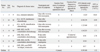
In the 12 year period between 1996 and June, 2008 a total of 1209 pediatric patients were treated for acute leukemia, and seven of these patients developed appendicitis for an incidence of 0.57%. Of the 1209 patients, 845 had acute lymphocytic leukemia (ALL) and 364 had acute myelocytic leukemia (AML) with appendicitis developing in 3 (0.35%) and 4 (1.09%) patients, respectively, in each disease group.
The median age at the time of the diagnosis of appendicitis was 12 years old (range: 3-15 years); 3 patients were male and 4 were female. Of the 3 ALL patients, 1 patient had just completed remission induction chemotherapy, 1 patient was receiving maintenance chemotherapy and the other patient had completed delayed intensification chemotherapy. All 4 AML patients developed acute appendicitis during the period of neutropenia that occurred after intensive chemotherapy: 3 and 1 patients developed acute appendicitis during the period of neutropenia that occurred after consolidation chemotherapy and reinduction chemotherapy following post-transplantation relapse, respectively (Table 1). None of them presented an extramedullary lesion.
The median absolute neutrophil count (ANC) at the time of diagnosis and operation were 0.99×10⁹/L (range: 0-3×10⁹/L) and 1.32×10⁹/L (range: 0-18.64×10⁹/L), respectively. The median platelet count at the time of diagnosis was 160×109/L (range: 17-225×109/L) (Table 1). C-reactive proteins were above normal levels in 4 of the 7 patients, but the others were not checked. Lactate dehydrogenase increased in 3 patients.
Among the leukemia patients, the median length of the duration of total neutropenia at the time of symptom onset was 20 days (range: 11-71 days). One patient was on oral maintenance chemotherapy and the rest were on either intravenous chemotherapy or they had completed chemotherapy within the past 30 days. Four patients were in a neutropenic state at the start of symptoms (range: 2-47 days). The remaining 3 patients also were in a neutropenic state at less than 1 week before the start of symptoms (1-6 days). For the leukemia patients, the mean time from the onset of symptoms to the diagnosis of acute appendicitis was 4 days (range: 1-9 days) (Table 1).
All the patients displayed abdominal pain except for one (86%), and other symptoms included fever in 5 patients (71%), vomiting in 3 patients (43%), diarrhea in 1 patients (14%) and abdominal distension in 1 patient (14%). On physical examination, 6 patients (86%) showed signs of peritoneal irritation such as direct tenderness or rebound tenderness.
Perforated appendicitis was diagnosed in 4 patients and appendicitis without perforation was diagnosed in 3 patients. In 2 patients subphrenic free air was evident on plain film, which obviated the need for further diagnostic procedures before surgery, and of the remaining 5 patients, 1 underwent abdominal sonography, 2 underwent CT imaging and 2 underwent both procedures (Table 1). On the preoperative radiologic study, 1 patient was tentatively diagnosed with acute appendicitis without appendicolith, 2 patients were tentatively diagnosed with acute appendicitis with appendicolith and 2 patients were tentatively diagnosed with periappendiceal abscess due to appendicitis. All 5 patients were differentiated from having typhlitis.
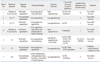
All 7 leukemia patients received surgery, 2 with laparotomy and 5 with laparoscopic appendectomy. Six patients received surgery either on the day of diagnosis or one day after. The ANCs assessed on the day of surgery in 6 patients were almost the same as the ANCs assessed at the time of symptom presentation. Only one patient among these 6 patients preoperatively received transfusions with 1 unit of packed red blood cells and 1 unit of single donor platelets. One patient received laparoscopic interval appendectomy 6 weeks after the diagnosis. This patient presented pancytopenia that did not respond well to transfusions for preoperative management, and the patient was started on conservative care that included broad spectrum antibiotics. We decided to perform the interval appendectomy 6 weeks later. The symptoms had subsided and the pancytopenia recovered 9 days after the diagnosis; however, despite the usage of antibiotics for 6 weeks, follow-up CT showed a remaining 3.5×2.4×4.2 cm sized pelvic abscess that necessitated laparoscopic appendectomy and drainage. On the operation day, this patient's ANC was 2.78×10⁹/L. All the patients received pre and post-operative antibiotics as indicated under the Infectious Diseases Society of America guidelines.7 G-CSF (granulocyte colony stimulating factor) was injected into 5 patients, of whom 2 were further treated with antithrombin III and intravenous immune globulin because of sepsis with abnormal levels of antithrombin III, D-Dimer, fibrinogen degradation products and fibrinogen. All 7 patients recovered well after surgery. An oral diet was commenced at a mean of 5.1 days after surgery (range: 2-10 days). Umbilical wound infection was observed in 1 patient, and this responded well to supportive care (Table 2). All the cases were confirmed to have appendicitis with neutrophil infiltration by surgical pathology. None of the appendices showed evidence of leukemic infiltration on H & E stain and immunohistochemical study (Table 2).
DISCUSSION
Appendicitis is the most common ailment requiring surgery in the pediatric population, with an incidence of 1% in children below the age of 15. However, the only studies of appendicitis in patients with underlying hematologic illnesses such as leukemia are limited to case reports, and only a few single institution studies with a large study population have been conducted over a substantial period of time.2,3,6
The incidence of appendicitis among the patients with hematologic malignancy has been reported to be 0.5-4.4%. In 1992, Angel, et al.6 reported on 6099 patients with leukemia and other malignancies. The patients were accrued over a span of 27 years with 16 patients experiencing appendicitis (0.5%). In 2005, Hobson, et al.2 reported on 7 patients with appendicitis (1.5%) out of 464 patients with hematologic malignancies in a study that compared appendicitis and typhlitis. In 2007, Alioglu, et al.8 reported on 2 patients who were diagnosed with appendicitis out of 118 leukemia patients (1.7%), and in 2008 Wiegering, et al.3 reported on 5 patients who were diagnosed with appendicitis out of a group of 113 leukemia patients (4.4%). In our study, 7 patients were diagnosed with appendicitis out of a group of 1209 pediatric acute leukemia patients, representing an incidence of 0.57%.
The etiology of appendiceal disease is luminal obstruction and the most common cause of obstruction is a fecalith. Not all cases of appendicitis are associated with a fecalith, but in most, some form of obstruction occurs. Lymphoid tissure, which is found in the wall of the appendix, may become hyperplastic in response to viral infections of the gut or respiratory tract, resulting in obstruction of the lumen of the appendix. Extramedullary involvement in leukemia is a rare complication, and only a few reports have demonstrated leukemic cell infiltration of appendix. Acute appendicitis can be presented as the initial manifestation or the relapse of acute myelogenous leukemia.9-11 However, such cases have only been recorded in adults and there is no case report of leukemic cell infiltration of the appendix in pediatric hematologic malignancies in the literature. In our study, none showed leukemic cell infiltration of the appendix.
In immune competent children, appendicitis usually begins with periumbilical pain that migrates to the right lower quadrant (RLQ). Appendiceal distension leads to nausea and vomiting in 85% of these patients and continued inflammation eventually leads to fever.12 These signs may not be so evident in neutropenic or immune compromised patients and for whom fever may be the first sign13 and also for whom RLQ pain and the signs of peritoneal irritation may not be so obvious.6 Hobson, et al.2 indicated that diarrhea and a high fever above 38.5℃ are more characteristic of typhlitis than appendicitis. In our group, abdominal pain and peritoneal irritation of the RLQ were the most commonly reported symptoms of the patients (86%), and fever was a presenting symptom in 2 patients. Symptoms of vomiting and diarrhea (43% and 14% respectively) were not as frequent in our patients and 1 patient complained only of diarrhea and abdominal distension without abdominal pain. Free air in this patient was evident on simple X-ray leading up to the operation.
So often, the gastrointestinal complications seen in neutropenic patients are difficult to diagnose on the basis of clinical findings alone. The average time from the onset of symptoms to diagnosis was 4 days for acute leukemia patients. Difficulty with differentiating abdominal pain due to relyappendicitis from the pain resulting from chemotherapy toxicity, and the use of corticosteroids, which mask the precise presentation of symptoms, delayed the accurate diagnosis of these patients.2,6,13 Also, in our study, over 50% of the cases were perforated upon diagnosis.
Imaging may aid in forming a diagnosis in these cases. Although Hobson, et al.2 noted that the diagnostic accuracy of CT for appendicitis is only about 33%, Stroman, et al.14 reported that the sensitivity and overall accuracy of CT for appendicitis in immune competent patients was about 92% and 90% respectively. Other authors have also noted that either CT or ultrasound is appropriate for diagnosing appendicitis in neutropenic patients.2,3,6,13 However, it remains unclear what criteria were used to make the differentiation of acute appendicitis from typhlitis. Typhlitis is a poorly understood gastrointestinal complication of neutropenia. Pathologically, typhlitis is described as a compromisation of bowel wall integrity with subsequent bacterial or fungal invasion. Although pathologic abnormalities can involve any segment of the small or large bowel, the cecum is the most common location of abnormality. CT findings of typhlitis include right-sided colon wall thickening, pericolonic stranding, ascites, and cecal pneumatosis. Kirkpatrick and Greenberg15 compared mean bowel wall thickening for the differentiation of specific gastrointestinal complications including C difficile colitis, neutropenic colitis and graft versus-host disease in neutropenic patients, and found that bowel wall thickening was significantly more prominent in C difficile colitis (mean wall thickness, 12 mm; range, 8-20 mm) than in neutropenic enterocolitis (mean thickness, 7 mm; range, 4-15 mm; p<0.01) and graft-versus-host disease (mean thickness, 5 mm; range, 3-7 mm; p<0.01).
In our study, the criterion for typhlitis was a cecal or terminal ileal wall thickness greater than 3 mm.3 However, depending on the severity or portion of the inflammation of the appendix, the cecal wall around the base of the appendix can be thickened by inflammation. So, although wall thickness is important, the circumferential thickened length of the cecum and ascending colon wall could be a more important finding. However, it remains unclear as to how long of a segment would have to be involved for the diagnosis of typhlitis because large periappendiceal abscess formation can cause the circumferential thickening of certain segmental cecal walls, too. In 3 of 4 of our cases wall thickness greater than 3 mm was present only around the appendix base of the cecum. In one patient, who underwent the interval appendectomy with a large periappendiceal abscess, a circumferential thickened cecal wall of 39 mm in length was present.
The optimum method of treatment remains a controversy. The reasons why some authors have a negative opinion about early appendectomy revolve around postoperative complications and mortality. Sbragia, et al.5 recommended an initial nonoperative approach in which surgery is only considered after a rise in neutrophil count or with an aggravation of clinical symptoms when these patients are managed by conservative care alone. Wiegering, et al.3 noted that even perforated appendicitis may improve with conservative care alone. They reported that 1 case was complicated by perforation during the conservative care and the median time to normalization of the US findings was 14 days. Moreover, although imaging evidence for recurrence of appendicitis was absent, 3 cases experienced recurrent right lower abdominal pain during a subsequent episode of febrile neutropenia after the following cycle of chemotherapy. Notwithstanding, conservative care alone requires a lengthy period for full recovery and incurs risks of progression, perforation and recurrence. In immune competent children, an inflamed appendiceal mass may be successfully treated with percutaneous drainage and antibiotics in 90-97% of the cases with a recurrence of symptoms only happening in 5-14%, so interval appendectomy may not be required in all patients.16-20 However, immune compromised children may tend towards a different clinical course. In our study, 1 AML patient who presented with a perforated appendicitis along with a 5 cm sized pelvic abscess with bilateral obstructive ureteral dilatations clinically improved after conservative management alone and was started on a diet on the 15th day after the appearance of the symptoms, even as the patient's ANC rose to 1×10⁹/L on the 7th day after the start of symptoms. Despite the usage of antibiotics for 6 weeks, follow-up CT showed a remaining right pelvic abscess 3.5×2.4×4.2 cm in size without ureteral dilatation, and this required laparoscopic appendectomy and drainage. Furthermore, delayed recovery from infection leads to delay in the continuation of chemotherapy, which could possibly lead to aggravation of the patient's underlying illness and escalation of medical costs. Therefore, conservative care alone is not the most effective method of treatment.
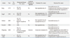
Since the initial reports on surgical treatment for pediatric leukemia patients who present with an acute abdomen were put forward by Björnsson, et al.21 and Exelby, et al.22 numerous studies have reported on improved outcomes resulting from surgery (Table 3).2,3,6,13,23 Exelby, et al.22 reported that the conditions that led to a 100% mortality when relying on non-surgical methods improved to 50% overall survival when these patients were given surgical treatment, while Angel, et al.6 reported on a series of 16 leukemia patients with appendicitis, of whom 14 were treated with surgery with only 1 death (7.1%). Hobson, et al.2 reported 100% overall survival after appendectomy for leukemia patients. In our study, the mortality was 0% and the only complication was a minor wound infection. All the patients who received surgery recovered rapidly, despite an ANC of 0×109/L and they were started on an oral diet at a mean of 5.1 days, and no later than 10 days. The relationship between ANC on the operation day and postoperative recovery time was obscure, but a long duration from onset of symptoms to diagnosis of appendicitis tended to show longer durations from treatment to start of a soft diet.
Laparoscopic appendectomy in children is a procedure that we have been performing in our institution since 2000. This procedure has the advantages of a small wound, rapid recovery, decreased pain and fewer wound infections, and these qualities may gain importance in immune compromised pediatric leukemia.
In conclusion, when right lower quadrant abdominal pain occurred in pediatric leukemia patients, early diagnosis of acute appendicitis by US and/or CT and early surgical resection before perforation may be the more effective treatment option for leukemia patients with appendicitis. Even when perforation has occurred and the patient has an ANC of 0×109/L, surgical treatment may improve overall survival without incurring significant complications.
 XML Download