Abstract
We encountered a patient with hemolytic uremic syndrome (HUS) with persistent isolation of shiga toxin-producing Escherichia coli (STEC) for 3 weeks despite of having no clinical symptoms. STEC has been recognized as an important food-borne pathogen that causes severe diseases such as HUS. We characterized this STEC strain via a polymerase chain reaction, reverse-passive latex agglutination and the slide agglutination method. In this STEC strain, stx2 (shiga toxin), eaeA, tir, iha (adherence genes), espADB (type III secretion genes), and hlyA, ehxA, clyA (hemolysin genes) were present. The O antigen of the strain was non-typable.
Shiga toxin-producing Escherichia coli (STEC) O157:H7 has been recognized as an important food-borne pathogen that causes severe diseases such as a hemolytic uremic syndrome (HUS).1 The majority of cases of this disease are caused by strains of the serotype O157:H7, but infections by enterohemorrhagic Escherichia coli (EHEC) strains belonging to serogroups other than O157, such as O26, O103, O111and O145, have been reported with increasing frequency.1,2
Shiga toxins (stxs) are the virulence factor of EHEC. The two groups consist of stx1 and stx2.3 Apart from stxs, there are various virulence factors of EHEC, eae (Intimin), tir (translocated intimin receptor), hlyA (EHEC hemolysin), and espADB (type III secretion proteins).1,4 The locus for enterocyte effacement (LEE) is associated with intimate adherence to epithelial cells.5 Several protein were proposed to be adhesion factors in LEE-negative strains, including iha (an adherence-conferring protein),6 efa1 (an EHEC factor for adherence),7 Saa (an autoagglutinating adhesin)8 and toxB (a protein from 93-kbplasmid pO157), which are required for O157:H7 strain Sakai expression of adherence.9 E. coli hemolysin (hlyA), enterohemorrhagic E. coli toxin (ehx) and cytolysin A (clyA) are well known as repeats in the toxin (RTX) family.10-12
In Korea, we encountered a patient who had been hospitalized for a long-time, due to long-term isolation of STEC. We conducted molecular and serotype analyses on this patient.
A three-year-old child was admitted to Uijeongbu St. Mary's Hospital with abdominal pain and watery/bloody diarrhea 3-4 days after eating boiled fish paste. Physical examination upon admission was unremarkable. The abdomen computerized tomography image showed prominent submucosal edema of the total colon. Hematological tests revealed-: hemoglobin 12.6 g/dL, leukocyte count 43.5×10⁹/L with neutrophilia (90%), platelet count 33×109/L. The results of a blood chemistry study of the serum samples were blood urea nitrogen, 39.1 mg/dL, albumin 1.7 g/dL and CRP 5.03 mg/dL. The serum creatinine level was increased from 0.44 mg/dL to 2.33 mg/dL. Urinalysis showed four positive tests for hematuria and proteinuria. A diagnosis of HUS was made and the patient was transferred to another hospital, where she underwent hemodialysis.
She was admitted to our hospital a second time due to persistent STEC isolation, although her clinical symptoms were improved. We isolated the STEC strain from the patient's stool. However, no other pathogenic bacteria such as Salmonella spp., Shigella spp., and Vibrio spp. were detected in the stool. The isolate was biochemically characterized using the API20E system (Biomerieux, Marcy l'Etoile, France). The isolate was directly inoculated into 3 mL of Luria-Bertani (LB) broth, and this was incubated overnight at 37℃. After incubation, the enriched broth culture was centrifuged at 13,000 rpm (Sorvall® Biofuge Pico, Germany) for 1 min and the pellet was heated at 100℃ for 10 min. Following centrifugation, 5 µL of the supernatant was used in the PCR assays. The PCR assays were performed using the primers shown in Table 1. The PCR assays were carried out in a 50 µL volume with 2 U DNA Taq polymerase (Takara Ex Taq™, Otsu, Japan) in a thermal cycler (PTC-100; MJ Research, Watertown, MA, USA). Positive DNA and distilled water were used as positive and negative controls.
Production of stx1 and stx2 was determined using a reversed passive latex agglutination kit (VTEC-RPLA; Denka Seiken Co., Ltd., Tokyo, Japan). One mL of the overnight culture was centrifuged and the titer of the supernatant was determined using the VTEC-RPLA test, which was performed at dilutions up to 1 : 256. The PCR and RPLA test showed that the strain produced only stx2. The adherence genes, eaeA and tir genes and non-LEE adhesion genes such as the iha, saa, efa1 and toxB genes, were analyzed in the STEC strain. The eaeA, tir, and iha genes were present in the strain (Table 1). All the genes tested for type III secretion proteins, i.e., espADB were found in the strain in the PCR analysis. PCR analysis showed that the hlyA, ehxA, and clyA genes for hemolysin production were detected in the strain (Table 1).
The presence of O antigens was determined by slide agglutination with the available O (O1-O181) antisera (Universidad de Santiago de Compostela, Lugo, Spain).13 We performed serotyping of the stain with the antisera. However, the serotype of the strain was non-typable with the tested antisera.
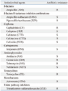
Antimicrobial susceptibility of the isolate to the following 16 antibiotics was determined by agar disk diffusion (Kirby-Bauer method) using Mueller-Hinton agar (Difco) : ampicillin/sulbactam (SAM), ampicillin (AM), tetracycline (TE), aztreonam (ATM), cefotetan (CTT), cefepime (FEP), cefoxitin (FOX), cefotaxime (CTX), tobramycin (NN), trimethoprim/sulfamethoxazole (SXT/TM), cephalothin (CF), imipenem (IPM), gentamicin (GM), amikacin (AN), piperacillin/tazobactam (TZP), netilamicin (NET). E. coli ATCC 25922 and E. coli ATCC 35218 were used as quality controls. As shown in Table 2, the isolate showed resistance to Ampicillin, Tetracycline and Trimethoprim-sulfamethoxazole, while it was sensible to two antibiotics of β-lactam/β-lactamase inhibitor combinations and Imipenem. Among the Cephems, the isolate showed resistance only to Cephalothin. On hospital day (HD) 17, cefuroxime was started due to persistent STEC isolation. On HD38, cefepime was started for four days. On HD 49, she was discharged after having negative results for stool STEC isolation for a week.
In the present study, we characterized the virulence genes of the STEC strain. Our results showed that the strain in this study possessed many virulent factors for infection, for example, toxins, adhesins and hemolysins.
The Shiga-toxin genotype of the infecting strain may influence the risk of developing microangiopathic sequelae and the outcome of infection. Patients infected with STEC 0157 possessing stx2 but not stx1 had significantly developed systemic sequelae, including HUS, than patients infected with STEC 0157 harboring stx1 alone or both stx1 and stx2.14 The strain in this study possessed only stx2 gene among stx genes (Table 1).
The stx2 toxin has been described as being 1,000 times more cytotoxic than the stx1 toxin, and was related with high virulence in STEC strains from HUS patients.15 The adherence of bacteria is important for the infection. Enteropathogenic E. coli (EPEC) and EHEC produce an adherence factor, intimin encoded by the eae gene.16 This gene is detected in the locus of enterocyte effacement (LEE), which is required for attachment-effacement lesions. Another study showed that the most widely distributed STEC adhesin gene was the iha gene.17 The LEE gene encodes the type III secretion system.4 The strain possessed all the tested genes related with the hemolysin. HlyA lyses cells via the creation of pores in the cell membrane and this affects erythrocytes, leukocytes, and renal tubularcells.12 The HlyA is frequently produced by E. coli strains that cause urinary tract infections, while ehxA is found in the EHEC strains of serogroupO157.10 ClyA is the prototype of pore-forming cytotoxins.18
Although STEC O157:H7 is known to be the predominant serotype associated with most STEC-related outbreaks worldwide, previous epidemiological studies have implied that non-O157 STEC infection is becoming problematic in public health.19 Among those non-O157 STEC associated with human disease are the serotypes O8:H-, O26:H11, O91:H21, O103:H2, O111:H-, O113:H21, and O128:H2.1 The O antigen of the strain was non-typable. From this result, we suggest that a non-typable serotype might play an important role in causing a long-term carrier state.
There have been several reports of Stx producing E. coli from asymptomatic human carriers.20,21 The patient may have produced antibodies against the STEC strain during the long-term infection. However, further study of the mechanisms of long-term isolation in the patient is needed.
In conclusion, we suggest that this result provides epidemiological information for a prototype of a molecular mechanism showing how the STEC strain can remain in a patient for a long time without causing symptoms.
Figures and Tables
ACKNOWLEDGEMENTS
This work was supported by a grant of the National Institute of Health, Ministry of Health, and Welfare, Republic of Korea (NIH 4800-4845-300). We also acknowledge the financially supporting of Gyeonggi-do (Local Government), Republic of Korea and Catholic University, Republic of Korea.
References
1. Nataro JP, Kaper JB. Diarrheagenic Escherichia coli. Clin Microbiol Rev. 1998. 11:142–201.
2. Tozzi AE, Caprioli A, Minelli F, Gianviti A, De Petris L, Edefonti A, et al. Shiga toxin-producing Escherichia coli infections associated with hemolytic uremic syndrome, Italy, 1988-2000. Emerg Infect Dis. 2003. 9:106–108.

3. Melton-Celsa AR, O'Brien AD. Animal models for STEC-mediated disease. Methods Mol Med. 2003. 73:291–305.

4. Makino S, Tobe T, Asakura H, Watarai M, Ikeda T, Takeshi K, et al. Distribution of the secondary type III secretion system locus found in enterohemorrhagic Escherichia coli O157:H7 isolates among Shiga toxin-producing E. coli strains. J Clin Microbiol. 2003. 41:2341–2347.

5. Clarke SC, Haigh RD, Freestone PP, Williams PH. Virulence of enteropathogenic Escherichia coli, a global pathogen. Clin Microbiol Rev. 2003. 16:365–378.

6. Tarr PI, Bilge SS, Vary JC Jr, Jelacic S, Habeeb RL, Ward TR, et al. Iha: a novel Escherichia coli O157:H7 adherence-conferring molecule encoded on a recently acquired chromosomal island of conserved structure. Infect Immun. 2000. 68:1400–1407.
7. Nicholls L, Grant TH, Robins-Browne RM. Identification of a novel genetic locus that is required for in vitro adhesion of a clinical isolate of enterohaemorrhagic Escherichia coli to epithelial cells. Mol Microbiol. 2000. 35:275–288.

8. Paton AW, Srimanote P, Woodrow MC, Paton JC. Characterization of Saa, a novel autoagglutinating adhesin produced by locus of enterocyte effacement-negative Shiga-toxigenic Escherichia coli strains that are virulent for humans. Infect Immun. 2001. 69:6999–7009.

9. Tatsuno I, Horie M, Abe H, Miki T, Makino K, Shinagawa H, et al. toxB gene on pO157 of enterohemorrhagic Escherichia coli O157:H7 is required for full epithelial cell adherence phenotype. Infect Immun. 2001. 69:6660–6669.

10. Welch RA, Forestier C, Lobo A, Pellett S, Thomas W Jr, Rowe G. The synthesis and function of the Escherichia coli hemolysin and related RTX exotoxins. FEMS Microbiol Immunol. 1992. 5:29–36.
11. Bauer ME, Welch RA. Characterization of an RTX toxin from enterohemorrhagic Escherichia coli O157:H7. Infect Immun. 1996. 64:167–175.

12. König B, Ludwig A, Goebel W, König W. Pore formation by the Escherichia coli alpha-hemolysin: role for mediator release from human inflammatory cells. Infect Immun. 1994. 62:4611–4617.

13. Guinée PA, Agterberg CM, Jansen WH. Escherichia coli O antigen typing by means of a mechanized microtechnique. Appl Microbiol. 1972. 24:127–131.

14. Ostroff SM, Tarr PI, Neill MA, Lewis JH, Hargrett-Bean N, Kobayashi JM. Toxin genotypes and plasmid profiles as determinants of systemic sequelae in Escherichia coli O157:H7 infections. J Infect Dis. 1989. 160:994–998.
15. Tarr PI, Gordon CA, Chandler WL. Shiga-toxin-producing Escherichia coli and haemolytic uraemic syndrome. Lancet. 2005. 365:1073–1086.
16. Jerse AE, Gicquelais KG, Kaper JB. Plasmid and chromosomal elements involved in the pathogenesis of attaching and effacing Escherichia coli. Infect Immun. 1991. 59:3869–3875.
17. Toma C, Martínez Espinosa E, Song T, Miliwebsky E, Chinen I, Iyoda S, et al. Distribution of putative adhesins in different seropathotypes of Shiga toxin-producing Escherichia coli. J Clin Microbiol. 2004. 42:4937–4946.
18. Oscarsson J, Westermark M, Löfdahl S, Olsen B, Palmgren H, Mizunoe Y, et al. Characterization of a pore-forming cytotoxin expressed by Salmonella enterica serovars typhi and paratyphi A. Infect Immun. 2002. 70:5759–5769.

19. Coombes BK, Wickham ME, Mascarenhas M, Gruenheid S, Finlay BB, Karmali MA. Molecular analysis as an aid to assess the public health risk of non-O157 Shiga toxin-producing Escherichia coli strains. Appl Environ Microbiol. 2008. 74:2153–2160.
20. Mukherjee J, Chios K, Fishwild D, Hudson D, O'Donnell S, Rich SM, et al. Human Stx2-specific monoclonal antibodies prevent systemic complications of Escherichia coli O157:H7 infection. Infect Immun. 2002. 70:612–619.
21. Stephan R, Untermann F. Virulence factors and phenotypical traits of verotoxin-producing Escherichia coli strains isolated from asymptomatic human carriers. J Clin Microbiol. 1999. 37:1570–1572.


 PDF
PDF ePub
ePub Citation
 Print
Print


 XML Download