Journal List > Yonsei Med J > v.52(4) > 1030829

Jang: Somatotopic Arrangement and Location of the Corticospinal Tract in the Brainstem of the Human Brain

Abstract

The corticospinal tract (CST) is the most important motor pathway in the human brain. Detailed knowledge of CST somatotopy is important in terms of rehabilitative management and invasive procedures for patients with brain injuries. In this study, I conducted a review of nine previous studies of the somatotopical location and arrangement at the brainstem in the human brain. The results of this review indicated that the hand and leg somatotopies of the CST are arranged medio-laterally in the mid to lateral portion of the cerebral peduncle, ventromedial-dorsolaterally in the pontine basis, and medio-laterally in the medullary pyramid. However, few diffusion tensor imaging (DTI) studies have been conducted on this topic, and only nine have been reported: midbrain (2 studies), pons (4 studies), and medulla (1 study). Therefore, further DTI studies should be conducted in order to expand the literature on this topic. In particular, research on midbrain and medulla should be encouraged.

INTRODUCTION

The corticospinal tract (CST) is the major neuronal pathway for motor function in the human brain.1-4 The CST is known to be critical in recovery of motor weakness following brain injury, particularly with regard to fine motor activity of the hands.2,4-7 Many previous studies have reported that the CST has somatotopy along the pathway of the human brain.8-21 Detailed knowledge of CST somatotopy would be helpful in establishing scientific rehabilitative strategies, estimating rehabilitative period, establishing guidelines for invasive procedures, and predicting final outcomes for patients with brain injury.
The CST descends through an area at the brainstem that is narrower than the supratentorial area in the human brain.1,22 Therefore, even a small lesion at the brainstem can cause severe motor weakness. The anatomical location of CST at the brainstem has been well-documented in neuroanatomy textbooks and studies.1,9,10,12,15,22-33 However, little is known about the somatotopic location and arrangement of the CST at the brainstem.8,11,16-19,21,34,35 Recent developments in diffusion tensor tractography (DTT), which is derived from diffusion tensor imaging (DTI), have contributed to research on somatotopy at the supratentorial level.9,10,12,15,32,33 However, only a few DTT studies have reported on somatotopy in the human brainstem.11,16,18
In the current study, I reviewed the literature on somatotopic location and arrangement at the brainstem of the human brain. Relevant studies were identified using the following electronic databases (Pubmed and MEDLINE) from 1966 to 2011. The following key words were used: CST, somatotopy, brainstem, cerebral peduncle (CP), midbrain, pons, and medulla. I limited this review to human studies of the CST and excluded studies of the corticonuclear or the corticobulbar tract. Ultimately, nine studies were selected for this review.8,11,16-19,21,34,35

MIDBRAIN

Many studies using the Wallerian degeneration phenomenon on brain CT or MRI,23-25,28-31,36 direct brain stimulation study during surgery,27 pedunculotomy for control of involuntary movements,37,38 or DTT18,39,40 have demonstrated the location of the CST at the CP of the human brain. Studies reporting that the CST was located in the middle portion of the CP have been a general trend;23,24,29,30,36 on the other hand, some textbooks or studies have suggested more specific data: the middle three-fifths,1 middle half,31 or middle two-thirds.22 However, three recent DTT studies provided visual data suggesting that the CST was located in the mid to lateral portion of the CP.18,39,40
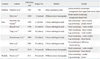
As for the somatotopic location and arrangement of the CST, many neuroanatomy textbooks have shown mediolateral arrangement of somatotopies for the arm and leg.1,22 However, there has been a shortage of studies that elucidated somatotopic anatomy in detail.18,34 In 1967, Mantinez, et al.34 investigated somatopic anatomy using a direct brain stimulation study during stereotactic surgery in more than 700 patients. They found face predominance from 7 to 12 mm behind the anterior commissure, and maximal response for the upper limb at 14 mm and at 19 mm for the lower limb. They reported an antero-posterior somatotopic arrangement from the face to the upper and lower limbs, rather than a medio-lateral arrangement at the upper CP. Since the introduction of DTT, one DTT study has reported on the somatotopic arrangement of the CST at the CP.18 In 2008, Park, et al.18 attempted to demonstrate the somatotopic arrangement at the CP of nine normal human brains. They found that the CST showed transverse orientation in the mid to lateral portion of the CP, and that hand fibers were located medial to foot fibers. Therefore, they concluded that somatotopic arrangement for the hand and leg showed medio-lateral orientation at the CP (Table 1).

PONS

The CST is located in the center of the pontine basis, which is surrounded by trasnspontine fibers. Several clinico-radiological correlation studies have reported on the somatotopic arrangement and location of the CST at the pons.8,17,19,21,35,41 In 1994, Schneidder and Gautier35 discovered that fibers for arm motor function were located antero-medially, while those for leg motor function were located more postero-laterally at the pons in 17 stroke patients with brainstem lesions. During the same year, Bassetti, et al.8 reported that 10 patients among 12 patients with ventro-medial pontine infarcts showed moderate to severe weakness of the distal hand; in contrast, nine patients with ventrolateral pontine infarct showed no weakness or mild weakness. However, Tohgi, et al.21 reported different results, showing hand dominant weakness in patients with infarct in the dorso-medial region of the pontine basis, and leg dominantweakness in patients with infarct in the ventro-medial region of the pontine basis in 36 patients with pontine basis infarct. In 2004, Schmahmann, et al.19 demonstrated that hand representation was located ventro-medially at the upper and mid-pons, and that leg representation was located dorso-laterally at the lower pons in 25 patients with focal infarcts on the pontine basis. Soon after, using a three-dimensional brain mapping technique from brain MRI and motor-evoked potential, Marx, et al.17 revealed that lesion location was more dorsal in patients with hemiparesis affecting the proximal muscles and more ventral in patients with distal limb weakness, among 41 patients with pontine infarct. However, they concluded that the arms and legs did not show significant somatotopical differences. Recently, using DTT, Hong, et al.11 investigated the anatomical location of CST somatotopies for the hand and leg at the pons in 25 normal human brains. In the group analysis, they found that hand somatotopy descended through the ventro-medial portion of the pontine basis and that leg somatotopy was located dorso-laterally to the hand somatotopy of the CST. Individual DTI data showed that the relative average location of the CST for the hand was 47.70% with the standard from the midline to the most lateral point of the upper pons, and 35.87% in the lower pons. For the leg, the average location of the CST was 56.82% in the upper pons and 40.63% in the lower pons. For the anteroposterior direction from the most anterior point of the pons to the most anterior point of the fourth ventricle, the location of the CST for the hand was 42.30% in the upper pons and 36.18% in the lower pons. For the leg, the location of the CST was 45.68% in the upper pons and 39.01% in the lower pons.

MEDULLA

The CST descends through the medullary pyramid (MP) in the medulla. The MP is the narrowest area through which the CST descends in the human brain.1,22 The location of the whole CST in the MP can be easily identified on a conventional MRI or a color map of DTI. However, little is known about somatotopy in the MP. To the best of our knowledge, only one study has been reported on this topic. Using normalized DTT, Kwon, et al.16 investigated the somatotopic arrangement of the CST at the MP in 30 normal human brains. They found that the hand somatotopy of the CST was located in the medial portion of the MP; in contrast, the leg somatotopy occupied the lateral portion of the MP.

CONCLUSIONS

In the current review, I reviewed previous studies of somatotopy of the CST in the brainstem of the human brain. Detailed knowledge of CST somatotopy is important in terms of rehabilitative management and invasive procedures for patients with brain injury; however, few DTI studies have been conducted on this topic. To the best of my knowledge, only nine studies on this topic have been reported: midbrain (2 studies), pons (6 studies), and medulla (1 study). Therefore, further DTI studies should be conducted in order to expand the literature on this topic. In particular, research on midbrain and medulla should be encouraged, because only three studies have been reported. Recent developments in DTI allow depiction of various cranial nerves in the human brain; therefore, studies of the corticonuclear tract should also be encouraged.42,43

Figures and Tables

Table 1
Previous Studies on Somatotopical Location and Arrangement in the Brainstem of the Human Brain
ymj-52-553-i001

ACKNOWLEDGEMENTS

This work was supported by a National Research Foundation of Korea Grant funded by the Korean Government (KRF-2008-314-E00173).

Notes

The author has no financial conflicts of interest.

References

1. Afifi AK, Bergman RA. Functional neuroanatomy: text and atlas. 2005. 2nd ed. New York: Lange Medical Books/McGraw-Hill.
2. Cho SH, Kim SH, Choi BY, Cho SH, Kang JH, Lee CH, et al. Motor outcome according to diffusion tensor tractography findings in the early stage of intracerebral hemorrhage. Neurosci Lett. 2007. 421:142–146.
crossref
3. Davidoff RA. The pyramidal tract. Neurology. 1990. 40:332–339.
crossref
4. Jang SH. The role of the corticospinal tract in motor recovery in patients with a stroke: a review. NeuroRehabilitation. 2009. 24:285–290.
crossref
5. Ahn YH, Ahn SH, Kim H, Hong JH, Jang SH. Can stroke patients walk after complete lateral corticospinal tract injury of the affected hemisphere? Neuroreport. 2006. 17:987–990.
crossref
6. Binkofski F, Seitz RJ, Arnold S, Classen J, Benecke R, Freund HJ. Thalamic metbolism and corticospinal tract integrity determine motor recovery in stroke. Ann Neurol. 1996. 39:460–470.
crossref
7. Hallett M, Wassermann EM, Cohen LG, Chmielowska J, Gerloff C. Cortical mechanisms of recovery of function after stroke. Neurorehabilitation. 1998. 10:131–142.
crossref
8. Bassetti C, Bogousslavsky J, Barth A, Regli F. Isolated infarcts of the pons. Neurology. 1996. 46:165–175.
crossref
9. Han BS, Hong JH, Hong C, Yeo SS, Lee D, Cho HK, et al. Location of the corticospinal tract at the corona radiata in human brain. Brain Res. 2010. 1326:75–80.
crossref
10. Holodny AI, Gor DM, Watts R, Gutin PH, Ulug AM. Diffusion-tensor MR tractography of somatotopic organization of corticospinal tracts in the internal capsule: initial anatomic results in contradistinction to prior reports. Radiology. 2005. 234:649–653.
crossref
11. Hong JH, Son SM, Jang SH. Somatotopic location of corticospinal tract at pons in human brain: a diffusion tensor tractography study. Neuroimage. 2010. 51:952–955.
crossref
12. Ino T, Nakai R, Azuma T, Yamamoto T, Tsutsumi S, Fukuyama H. Somatotopy of corticospinal tract in the internal capsule shown by functional MRI and diffusion tensor images. Neuroreport. 2007. 18:665–668.
crossref
13. Jang SH. A review of corticospinal tract location at corona radiata and posterior limb of the internal capsule in human brain. NeuroRehabilitation. 2009. 24:279–283.
crossref
14. Kim JS, Pope A. Somatotopically located motor fibers in corona radiata: evidence from subcortical small infarcts. Neurology. 2005. 64:1438–1440.
crossref
15. Kim YH, Kim DS, Hong JH, Park CH, Hua N, Bickart KC, et al. Corticospinal tract location in internal capsule of human brain: diffusion tensor tractography and functional MRI study. Neuroreport. 2008. 19:817–820.
crossref
16. Kwon HG, Hong JH, Lee MY, Kwon YH, Jang SH. Somatotopic arrangement of the corticospinal tract at the medullary pyramid in the human brain. Eur Neurol. 2011. 65:46–49.
crossref
17. Marx JJ, Iannetti GD, Thömke F, Fitzek S, Urban PP, Stoeter P, et al. Somatotopic organization of the corticospinal tract in the human brainstem: a MRI-based mapping analysis. Ann Neurol. 2005. 57:824–831.
crossref
18. Park JK, Kim BS, Choi G, Kim SH, Choi JC, Khang H. Evaluation of the somatotopic organization of corticospinal tracts in the internal capsule and cerebral peduncle: results of diffusion-tensor MR tractography. Korean J Radiol. 2008. 9:191–195.
crossref
19. Schmahmann JD, Ko R, MacMore J. The human basis pontis: motor syndromes and topographic organization. Brain. 2004. 127:1269–1291.
crossref
20. Song YM. Somatotopic organization of motor fibers in the corona radiata in monoparetic patients with small subcortical infarct. Stroke. 2007. 38:2353–2355.
crossref
21. Tohgi H, Takahashi S, Takahashi H, Tamura K, Yonezawa H. The side and somatotopical location of single small infarcts in the corona radiata and pontine base in relation to contralateral limb paresis and dysarthria. Eur Neurol. 1996. 36:338–342.
crossref
22. Carpenter MB. Core text of neuroanatomy. 1985. 3rd ed. Baltimore: Williams & Wilkins.
23. Bouchareb M, Moulin T, Cattin F, Dietemann JL, Racle A, Verdot H, et al. Wallerian degeneration of the descending tracts. CT and MRI features of the brain stem. J Neuroradiol. 1988. 15:238–252.
24. Kobayashi S, Hasegawa S, Maki T, Murayama S. Retrograde degeneration of the corticospinal tract associated with pontine infarction. J Neurol Sci. 2005. 236:91–93.
crossref
25. Mark VW, Taub E, Perkins C, Gauthier LV, Uswatte G, Ogorek J. Poststroke cerebral peduncular atrophy correlates with a measure of corticospinal tract injury in the cerebral hemisphere. AJNR Am J Neuroradiol. 2008. 29:354–358.
crossref
26. Mori S, Crain BJ, Chacko VP, van Zijl PC. Three-dimensional tracking of axonal projections in the brain by magnetic resonance imaging. Ann Neurol. 1999. 45:265–269.
crossref
27. Quiñones-Hinojosa A, Lyon R, Du R, Lawton MT. Intraoperative motor mapping of the cerebral peduncle during resection of a midbrain cavernous malformation: technical case report. Neurosurgery. 2005. 56:E439.
crossref
28. Stovring J, Fernando LT. Wallerian degeneration of the corticospinal tract region of the brain stem: demonstration by computed tomography. Radiology. 1983. 149:717–720.
crossref
29. Uchino A, Imada H, Ohno M. MR imaging of wallerian degeneration in the human brain stem after ictus. Neuroradiology. 1990. 32:191–195.
crossref
30. Uchino A, Onomura K, Ohno M. Wallerian degeneration of the corticospinal tract in the brain stem: MR imaging. Radiat Med. 1989. 7:74–78.
31. Warabi T, Miyasaka K, Inoue K, Nakamura N. Computed tomographic studies of the basis pedunculi in chronic hemiplegic patients: topographic correlation between cerebral lesion and midbrain shrinkage. Neuroradiology. 1987. 29:409–415.
crossref
32. Westerhausen R, Huster RJ, Kreuder F, Wittling W, Schweiger E. Corticospinal tract asymmetries at the level of the internal capsule: is there an association with handedness? Neuroimage. 2007. 37:379–386.
crossref
33. Yamada K, Kizu O, Kubota T, Ito H, Matsushima S, Oouchi H, et al. The pyramidal tract has a predictable course through the centrum semiovale: a diffusion-tensor based tractography study. J Magn Reson Imaging. 2007. 26:519–524.
crossref
34. Martinez SN, Bertrand C, Botana-Lopez C. Motor fiber distribution within the cerebral peduncle. Results of unipolar stimulation. Confin Neurol. 1967. 29:117–122.
35. Schneider R, Gautier JC. Leg weakness due to stroke. Site of lesions, weakness patterns and causes. Brain. 1994. 117:347–354.
crossref
36. Waragai M, Watanabe H, Iwabuchi S. The somatotopic localisation of the descending cortical tract in the cerebral peduncle: a study using MRI of changes following Wallerian degeneration in the cerebral peduncle after a supratentorial vascular lesion. Neuroradiology. 1994. 36:402–404.
crossref
37. Bucy PC, Keplinger JE, Siqueira EB. Destruction of the "Pyramidal Tract" in man. J Neurosurg. 1964. 21:285–298.
crossref
38. Jane JA, Yashon D, Becker DP, Beatty R, Sugar O. The effect of destruction of the corticospinal tract in the human cerebral peduncle upon motor function and involuntary movements. Report of 11 cases. J Neurosurg. 1968. 29:581–585.
crossref
39. Mori S, van Zijl PC. Fiber tracking: principles and strategies - a technical review. NMR Biomed. 2002. 15:468–480.
crossref
40. Newton JM, Ward NS, Parker GJ, Deichmann R, Alexander DC, Friston KJ, et al. Non-invasive mapping of corticofugal fibres from multiple motor areas--relevance to stroke recovery. Brain. 2006. 129:1844–1858.
crossref
41. Kumral E, Bayülkem G, Evyapan D. Clinical spectrum of pontine infarction. Clinical-MRI correlations. J Neurol. 2002. 249:1659–1670.
42. Chen DQ, Quan J, Guha A, Tymianski M, Mikulis D, Hodaie M. Three dimensional in vivo modelling of vestibular schwannomas and surrounding cranial nerves using diffusion imaging tractography. Neurosurgery. 2011.
43. Hodaie M, Quan J, Chen DQ. In vivo visualization of cranial nerve pathways in humans using diffusion-based tractography. Neurosurgery. 2010. 66:788–795.
crossref
TOOLS
Similar articles