

 PDF
PDF ePub
ePub Citation
 Print
Print


INTRODUCTION
In 1975, Reid et al.1 discovered catecholaminergic polymorphic ventricular tachycardia (CPVT). CPVT is known to cause syncope or sudden cardiac death, and the three distinguishing features of CPVT has subsequently been described by Coumel and associates.2 These features of CPVT are as follows: 1) exercise- or emotion-induced severe ventricular tachyarrhythmias; 2) a typical pattern of bidirectional ventricular tachycardia with a normal resting ECG; and 3) a structurally normal heart. Recent reports suggest that CPTV is a genetic disease related to the mutation of 2 genes: mutations in cardiac ryanodine receptor gene (RYR2) or calsequestrin 2 gene (CASQ2) lead to an increase in the intracellular Ca++ concentration, resulting in arrhythmia due to a cascade of delayed afterdepolarization and triggered activity.3-5
CASE REPORT
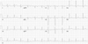
An 11-year-old female patient with a prior history of cardiac arrest arrived at the emergency room (ER) 35 minutes after losing consciousness. Her mother had immediately administered basic life support. The patient was born in the 40th gestational week by normal spontaneous vaginal delivery and weighed 4.0 kg. There was no specific family history of syncope or sudden cardiac death. Six years ago, she experienced a brief syncopal episode. Electroencephalography (EEG) at that time showed no specific abnormality. Six months after this first event, she was admitted to the ER for sudden loss of consciousness. Ventricular fibrillation (VF) was noted, and the normal sinus rhythm was restored by defibrillation. Neither delta wave nor QT interval prolongation was observed on a resting electrocardiogram (ECG) (Fig. 1). Echocardiography did not reveal any structural abnormalities, and Holter monitoring did not reveal specific arrhythmias. Brain magnetic resonance imaging (MRI) revealed no abnorzmalities. EEG was remarkable for dysrrhythmic waves and low amplitude voltage compatible with hypoxic encephalopathy. Although the definite cause of VF was not determined, it was recommended that the patient takes a beta-blocker (atenolol, 12.5 mg bid). She then underwent rehabilitation for cognitive function defects which resulted from hypoxic brain damage. She did not suffer any more attacks and stopped taking atenolol after several years.
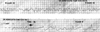
Upon this ER arrival, the patient was unconscious, pulseless, and without spontaneous respirations. Blood pressure was undetectable, and her body temperature was 36.5℃. The initial ECG showed VF. Normal sinus rhythm was restored after 2 monophasic direct current defibrillations at 200 J (Fig. 2). On chest X-ray, no cardiomegaly or pulmonary congestion was observed. The laboratory test results were as follows: white blood cell count, 12,400/mm3; hemoglobin, 14.5 g/dL; platelet count, 188,000/mm3; sodium, 138 mmol/L; potassium, 5.7 mmol/L; chloride, 111 mmol/L; ionized calcium, 1.42 mmol/L; ionized magnesium, 1.06 mmol/L; creatine kinase, 124 U/L; and creatine kinase-MB, 0.9 ng/mL. An arterial blood gas test revealed the following: pH 7.13; PaCO2, 55.5 mmHg; PaO2, 59.8 mmHg; HCO3, 17.7 mmol/L; and SaO2, 80.1%.
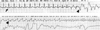
After defibrillation and intubation, the patient was moved to the coronary care unit (CCU). During nasogastric tube insertion, VF redeveloped. Sinus rhythm was successfully restored by 2 defibrillations at 200 J. A review of telemetry monitoring in the CCU revealed that sinus tachycardia precipitated bidirectional ventricular tachycardia that in turn degenerated into VF (Fig. 3).
The patient was diagnosed with CPVT accompanied by typical bidirectional ventricular tachycardia which was triggered by sinus tachycardia. After intravenously administered beta-blocker (labetalol, 10 mg), tachyarrhythmia was no longer observed. After stabilization, echocardiography revealed an ejection fraction of 68% without any structural abnormalities. She was prescribed an oral beta-blocker (metoprolol, 100 mg bid) and discharged from hospital. She is currently being followed up.
DISCUSSION
Since it was first discovered in 1975, CPVT has been reported as a cause of syncope, ventricular arrhythmias and sudden cardiac death. CPVT typically manifests as syncope between 7 and 9 years of age,6 but sudden death may be the first presentation. In 30% of CPVT patients, there is family history of sudden death before the age of 40.7 Patients with RYR2 mutation become symptomatic earlier, and men are at higher risk of cardiac events.8 CPVT is associated with two genetic mutations, RYR2 and CASQ2. RYR2 is inherited in an autosomal dominant pattern and mediates the release of calcium from the sarcoplasmic reticulum that is required for myocardial contraction.9 The RYR2 mutation increases calcium release and can trigger life-threatening ventricular arrhythmias. A second genetic form of CPVT, with an autosomal recessive inheritance, involves CASQ2. The CASQ2 protein, which serves as the major calcium reservoir within the sarcoplasmic reticulum, has an ability to bind extremely large amounts of calcium. The mutated protein may alter the calcium content within the sarcoplasmic reti-culum, alter the function of the ryanodine receptor, or impair the calcium release process.10
CPVT is difficult to diagnose, because ECG is normal in the absence of symptoms and echocardiography shows no specific findings. A typical finding on ECG is ventricular tachycardia with 180-degree alternation of the QRS axis (bidirectional tachycardia). CPVT is not inducible by programmed electrical stimulation.6 In patients suspected to have this disease, the arrhythmia must be recorded by Holter monitoring or induced by exercise treadmill testing. In the present patient, sinus tachycardia appeared to have been induced by the insertion of the nasogastric tube, and this in turn might have given rise to the bidirectional ventricular tachycardia that finally degenerated into VF. This is the typical course of CPVT, and activity triggering by a burst in the sympathetic tone is the main mechanism for this process. Hence, the focus of treatment is to suppress the adrenergic activity, therefore, beta-blockers are the most important drugs in the treatment of CPVT. Beta-blockers are effective for acute phase and maintenance treatment.11 However, if the symptoms recur despite the administration of a sufficient dose of beta-blockers, an implantable cardioverter/defibrillator must be employed. Our patient received a beta-blocker. To optimize the dosage of beta-blocker, the isoproterenol challenge test was planned for the patient's neurologic sequela, but the test was not performed because the patient's parents did not approve. A genetic study for the patient and her family was also refused.
Syncope or sudden cardiac death in childhood might occur due to other arrhythmogenic entities. These include arrhythmogenic right ventricular cardiomyopathy (ARVC), Brugada syndrome, long QT syndrome (LQT), particularly LQT type 1, pre-excitation syndrome, commotio cordis, and Andersen-Tawil syndrome (ATS). ARVC has structural abnormalities of heart and does not usually exhibit ventricular arrhythmias when provoked by an exercise stress test. Brugada syndrome is characterized by electrocardiographic findings of right bundle branch block and ST-segment elevation in leads V1 to V3 at resting ECG. In LQT, some patients do not develop QT prolongation, and it may be explained by repolarization reserve (variable redundancy of repolarizing currents), and resemble to CPVT.12,13 However, differential diagnosis is possible because LQT type 1 does not usually exhibit progressive polymorphic ventricular arrhythmias during graded exercise. Furthermore, there exist the delta wave in pre-excitation syndrome and a history of blunt chest trauma in commotio cordis. ATS is an inherited arrhythmogenic disorder caused by mutations in the KCNJ2 gene. Patients may develop bidirectional ventricular tachycardia, similar to that in CPVT. Although ATS may be similar to CPVT, it is considered as a distinct disorder, because of extracardiac manifestations (e.g., periodic paralysis and distinctive facial features), the low risk of sudden cardiac death, and the lack of relationship between arrhythmia and elevated sympathetic tones.
CPVT is a potentially fatal disorder that is usually observed in childhood. As in our case, patients presenting with sudden cardiac arrest can be mistaken as idiopathic VF and suffer from the sequelae of resuscitation. Therefore, clinicians should carefully analyze the triggering of factors VF in healthy individuals.
 XML Download