

 PDF
PDF ePub
ePub Citation
 Print
Print


INTRODUCTION
Pathological gambling has been classified as an impulse control disorder or an obsessive-compulsive spectrum disorder.1,2 It is a debilitating disorder with a chronic and progressive course, and co-morbid psychiatric conditions are common in pathological gamblers. However, despite estimates of significant prevalence and associations with adverse consequences, relatively little is known about pathological gambling, particularly its neurobiological underpinnings.3
Gamblers Anonymous is the most popular intervention for problematic gambling. The literature suggests that cognitive-behavioral therapy, pharmacotherapy, and family therapy may be useful in the treating the disorder.4 Among pharmacotherapies, several studies have suggested the efficacy and tolerability of the selective serotonin reuptake inhibitor (SSRI), especially fluvoxamine, in the treatment of pathological gambling.5
We hypothesized that functional MRI (fMRI), which has been shown to provide some clues to the underlying neurobiology of pathological gambling,6 might be useful in monitoring the results of pharmacotherapy. Herein, we report a pathological gambling patient whose improvement with fluvoxamine treatment was monitored by using fMRI.
CASE REPORT
A 36-year-old married man was admitted for his maladaptive gambling behavior, suicide attempt, and depressive mood. He had taken over his father's business and had worked successfully as the president of a ready-mixed concrete cooperation until he fell into gambling. This happened when he lost more than 13 billion won (about 1.4 million US$) because of a downward trend in the stock market. He struggled and made every effort to recover his loss. When he went to a casino while vacationing with a friend, he gambled forty million won that he had, and he succeeded twice. Since that time, he began to visit the casino after finishing business early in the morning. The frequency of his visits increased rapidly, and he eventually visited this casino almost daily by the end of the year 2000. He played mostly 'baccarat' and rarely played slot machines or any other type of game. His family members and employees did not know about his repeated and maladaptive gambling behavior because he concealed it from them. As his gambling continued, he lost money more frequently. However, he could not control his desire to recover all of his losses at once. As time passed, his financial losses continued to increase, and he lost not only part of his father's property, but also fell into debt to his friends. He attempted suicide by hanging himself in his house in the autumn of 2001, at which point his family became aware of his maladaptive gambling behavior. His gambling behavior continued for more two years, and his suicidal thoughts, attempts, and depressive mood caused his mother and brother to take him to the Depressive Disorder Clinic, Department of Psychiatry, Chonbuk National University Hospital, Jeonju, Korea. He was voluntarily admitted to the psychiatric ward at Chonbuk National University Hospital in February 2002.
There were no psychiatric disorders including pathological gambling, substance abuse, or depression among his family and relatives. In the first interview after admission, he appeared physically healthy, but somewhat tense and confused by his abrupt admission. He felt guilty about his gambling behavior, especially toward his parents, In an unreasonable belief, however, he insisted that he could regain his gambling losses by gambling again. He had not been addicted to alcohol or other substances and demonstrated no other impulsive behaviors (e.g. sexual, aggressive). However, he was a heavy smoker at two packs a day.
Immediately after admission, his laboratory results were found to be in the normal range; complete blood count (CBC), liver function test (LFT), urine analysis, thyroid function test (TFT), venereal disease research laboratory (VDRL) test, electrocariogram (EKG), chest X-ray, and other routine laboratory tests. His blood pressure was 130/80 mmHg; and respiration and other physical findings were in the normal range.
Without any medication, he was examined for mood, thought, behavior, and craving for gambling one week after admission. He had a score of 10 in the Korean South Oaks Gambling Screening (KSOGS),7 and a score of 18 in the Gambling Symptom Assessment Scales (GSAS). According to the DSM-IV-TR, he met the criteria for pathological gambling. He did not meet the DSM-IV-TR criteria for major depressive disorder, but he met the criteria for dysthymic disorder.
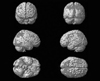
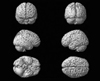
Baseline imaging was obtained by fMRI on the seventh day after admission (Fig. 1). On the eighth day, fluvoxamine (50 mg/day) was prescribed for him with no concomitant mediations. In the three weeks after admission, the dose of fluvoxamine was raised to 200 mg/day and maintained during a follow-up period; there were no significant adverse effects of the medication. Beck's Depression Inventory (BDI), State-Trait Anxiety Inventory (STAI), and GSAS had been administered before medication, and they were reexamined on the seventh day after medication. The second fMRI scan was also obtained on the seventh day after medication (Fig. 2).
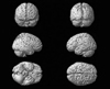
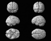
Three weeks after discharge (six weeks after beginning of the fluvoxamine prescription), BDI, STAI, and GSAS were again administered, and the same assessments were also carried out at six and nine months after beginning of the medication (Table 1). Additionally, fMRI scans took place at six weeks and six months after medication (Figs. 3 and 4).
The patient did not undergo individual or group cognitive behavioral therapy, nor did he participate in Gamblers Anonymous during or after his admission to the hospital. He had not gone to gamble during the follow-up period. He also had reported that his desire to gamble had decreased, and he was disabused of his unreasonable belief that he could regain his losses by gambling. He presently maintained his job and social relationships stably after treatment.
fMRI was performed on a 1.5T magnetic resonance scanner (Magnetom VISION, Siemens, Erlangen, Germany). For functional imaging, echo-planar imaging (EPI) was used (matrix = 64×64). Five rest conditions were alternated with five activation conditions, and each period consisted of 120 images at 22-second intervals (repetition time). These 1,200 images were collected and analyzed according to the time. During the activation period, the stimuli consisted of card pictures showing a full house, four-of-a-kind, and a royal straight flush. During the resting period, the patient was shown landscape pictures. Analysis of the images from the activation period was performed using SPM 99 statistical parametric mapping software to establish reference vectors corresponding to activation and resting periods. Among regions in which changes in signal intensity were cross-correlated with the reference vector, only regions meeting a threshold of p = .0001 and an extent threshold of 30 voxels (size 2×2×2 mm) were considered as significant signals. The anatomical locations of the activated foci were assigned using the Talairach atlas and were then presented as the appropriate Brodmann's areas.
DISCUSSION
Sociological and individual factors have been implicated in the development and maintenance of pathological gambling behavior. Recently, the etiology of pathological gambling has been suggested to involve abnormalities in certain neurotransmitters. Although an understanding of the neurobiology of pathological gambling is still in its infancy, several recent studies support these theories. Dysfunction of the serotonergic system has been linked to impulsive and compulsive traits in pathological gamblers.8 In addition, serotonin is associated with behavioral initiation and disinhibition, which may disrupt one's ability to stop gambling, thus playing an important role in the gambling cycle.9,10 Ventral prefrontal regions may be an important interface between the cognitive and emotional components of risk-taking behaviors.11 Also, functional imaging studies using infusions of nicotine or cocaine have shown associations between the rewarding effects of these drugs and the neurons in the nucleus accumbens, brainstem, amygdala, and prefrontal cortices.12,13 These regions may be linked to the addictive symptoms of pathological gambling. In our case, the significant decrease in the generally activated regions after administration of fluvoxamine coincided with the decreased desire to gamble, despite subjective measures of a slightly depressive mood according to BDI. It should be demonstrated in further studies whether the significant decrease of activation in different brain regions is correlated with the therapeutic effects of fluvoxamine.
Examination of the neural correlates of treatment of pathological gambling with selective serotonin reuptake inhibitors seems particularly intriguing because of data suggesting the efficacy and tolerability of these drugs in the short-term treatment of pathological gambling, and because of the effects of these drugs on cortico-basal-ganglionic-thalamic activity in OCD.14,15
In this case, the responses to pharmacotherapy with fluvoxamine in a patient suffering from pathological gambling were observed not only by symptomatic self-report, but also by the objective measure of brain functional MRI. However, a question of whether the improvement of pathological gambling symptoms in this case was secondary to improvement of a co-morbid condition remains unanswered. Furthermore, fMRI findings could not be evaluated on the basis of precise correlation between regions showing differences and symptoms of pathological gambling. In spite of these limitations, the present study combined brain fMRI and pharmacotherapeutical findings of pathological gambling, providing an indication of the usefulness of fMRI as a diagnostic tool and predictor of treatment response in a pathological gambler. More research testing this hypothesis may deepen our knowledge about the prevention, diagnosis, and treatment of pathological gambling.

 XML Download