

 PDF
PDF ePub
ePub Citation
 Print
Print


INTRODUCTION
The ponticulus posticus means "little posterior bridge" in Latin.1,2 It was defined as an abnormal small bony bridge formed between the posterior portion of the superior articular process and the posterolateral portion of the superior margin of the posterior arch of the atlas.2,3 This condition had not been a matter of concern for spine surgeons until its surgical significance in the insertion of screws into the lateral mass of the atlas was recently reported.2 Some spine surgeons have recently reported that it could cause a severe complication such as a vertebral artery injury during C1 lateral mass screws insertion.2
The sulcus situated on the posterolateral margin of the atlas forms a groove for the vertebral artery which varies in size and depth, rangimg from merely an impression to a clear groove or sulcus for the passage of the artery. At times, the sulcus is bridged by an anomalous ossification and a posterior ponticulus; a lateral ponticulus is occasionally formed.4 It forms the arcuate foramen that contains the vertebral artery and the suboccipital nerve. It can be a possible cause of posterior circulation ischemia and cervicogenic headache.5 Its prevalence has been reported to be between 5.14% and 37.83% in the western population.1,2,5 Most prevalence studies had been performed by plain radiographs or dried atlas specimens.3,5-8 However, it is difficult to find ponticulus in inspection of plain radiographs because of poor visualization of the posterior arch of the atlas due to overlapping of surrounding bones and limitation of plain X-ray. 3-dimensional (3-D) CT reconstruction technique demonstrated excellent detail of its morphologic feature as well as its existence. Through the review of literatures, we have found that its prevalence in Korean has not yet been studied. We, therefore, investigated the prevalence and morphologic features of ponticulus posticus in Koreans using 3-D cervical CT scanning.
PATIENTS AND METHODS
This study was approved by the institutional review board of our institution. We retrospectively reviewed 200 cervical 3-D CT scanning digital images of 200 consecutive patients over twenty years of age who had visited our hospital due to cervical problem from January 2006 to December 2006. We also examined digital plain lateral cervical radiographs of the same patients and 200 CT scanning images (1-mm interval digital) were obtained. There were 100 men and 100 women, and the overall mean age was 45.0 ± 19.65 years (range; 23 - 68). The age of male patients ranged from 23 to 64 years (mean ages ± standard deviation; 40.2 years ± 15.26) and from 26 to 68 years (47.4 years ± 18.74) in females. Computed tomography cervical images were taken at 1 mm intervals from inion of skull to C7 by a scanner (GE 9800 CT; General Electric, Milwaukee, WI, USA). Collimation was set at 1 mm with a table speed of 0.5mm/sec. All the images were retrieved in digital format from a PACS (picture archiving and communication system) server and 150 to 250 sections were calculated at an inter-slice distance of 1 mm with a standard reconstruction algorithm. Three-D surface rendering reconstructions (V-works, Cybermed, Seoul, Korea) were performed for each case. A threshold was set at 150 HU in shaded-surface display. These 3-D images were displayed with axial and vertical rotational views at every 5°. They were carefully inspected by a radiologist for the presence and types of ponticuli and arcuate foramen. Complete type was defined as clear bony bridge between the superior articular process and the posterior arch of the atlas in 3-D CT images. We did not include bony protrusion due to noticeable impression for vertebral arteries as partial type. Partial type was considered as partial posterior ponticulus which was noted as a distinct bony spicule extending from the superior articular facet overhanging the dorsal arch.
We could not tell that the prevalence of ponticuli in the patients analyzed above would represent that of the normal Korean population, because this studied patient group had the cervical problem. However, through the investigation of cervical CT scanning, we took a step closer to real prevalence and morphologic features of ponticuli in Koreans. In each scanning, the presence of a ponticulus posticus and also whether it was complete or partial were carefully inspected. Statistical analysis was conducted with chi-square test, using SPSS statistical package (ver. 12.0; SPSS, Chicago, IL, USA). The level of significance was set at p < 0.05.
RESULTS
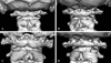
Analysis of 200 3-D CT scan images revealed ponticulus posticus on 1 or both side(s) in 31 patients; consist of 15.5% (Table 1). There was no significant difference in the prevalence between men (14/100, 14%) and women (17/100, 17%; p = 0.42). Total 31 patients identified consisted of bilateral in 17 patients, on the right side alone in 6 patients, and on the left side alone in 8 patients. The difference in the frequency of laterality was not statistically significant (p = 0.65). Among 48 ponticuli posticus observed on one or both side(s), s23 had complete bony bridge formation and 25 were partial (Figs. 1A-F). Total 31 patients identified consisted of bilateral complete in 7, bilateral partial in 4, bilateral mixed in 6, unilateral complete in 3, and unilateral partial in 11 (Table 2).
In plain cervical radiograph, 45 of 200 images (22.5%) were excluded because poor visualization of the posterior arch of the atlas due to overlapping of the mastoid process or the occiput. Analysis of the digital plain radiographs of 155 patients revealed 3 complete and 5 partial ponticuli posticus. Thus, the overall prevalence of ponticulus posticus in this patient population was 6.95%, comprising 2.61% complete and 4.34% partial. This was significantly smaller than the prevalence in the cervical spine patients in whom CT scans were analyzed (p = 0.009).
DISCUSSION
Ponticulus posticus has become an important anomaly of the atlas, as the use of lateral mass screws for the fixation of the atlas has become common for the treatment of atlantoaxial instability. However, it can sometimes be a difficult procedure, as the region contains venous plexuses as well as the greater occipital nerve.2 To avoid these difficulties, some surgeons have recommended that, in the presence of a broad posterior arch of the atlas, the insertion of the screw be started in the dorsal aspect of the posterior arch instead of at the base of the lateral mass or at the junction of the posterior arch and the lateral mass.2 A broad dorsal arch of the atlas is the best indication for this modified screw trajectory. However, in patients with ponticulus posticus, and resulting arcuate foramen carrying vertebral artery, it can be mistaken for a broad dorsal arch and the surgeon may insert the screw into the ponticulus posticus.2 This can result in an injury to the vertebral artery, and lead to stroke or even death by thrombosis, embolism, or arterial dissection.2
In the western population, the prevalence of ponticulus posticus has been reported to be between 5.1% and 37.8%.1,4,5 Varying incidences of posterior ponticuli (bridges) and their study methods are shown in Table 3.8 The study on its prevalence in Korean has not yet been done and 3-D CT scan has not been used.
In our study, it was of interest to note that as much as 15.5% of the patients had ponticulus posticus on one or both sides/complete or partial. We have not anticipated that its prevalence was relatively high in Korean: This may be due to high diagnostic values of CT scans about bony anomaly and selection of patients group with cervical problems. In any case, we found that the ponticulus posticus is not a rare anomaly in Koreans. Therefore, the presence of this anomaly should carefully be checked before screw placement in the lateral mass of the atlas in order to avoid vertebral artery injury. As mentioned above, plain cervical radiograph is not suitable for screening the ponticuli. In the present study, we noted that many plain radiographs did not even identify the existence of ponticuli and bilaterality and completeness of ponticuli were not identified either. Most previous studies have been based on lateral radiographs, therefore, it is not usually possible to determine if the anomaly is unilateral or bilateral. In our study, ponticulus posticus was almost equally detected in both left and right sides. This was not clearly identified in the previous plain radiographic studies, because the plain film studies fail to differentiate the right and left. In the previous cadaveric studies, there was no difference between the 2 sides.1,8-10
Most of these patients took a CT scan because of cervical spine problems. Therefore, these patients did not necessarily represent the whole population. Nevertheless, we could infer the prevalence of ponticulus posticus in Koreans from this retrospective study. Examination of the 3-dimensional CT images revealed the laterality, various spectra of shape and size of ponticulus posticus. When ponticulus posticus is observed or suspected on a lateral radiograph of a patient who requires lateral mass screw placement in the atlas, 3-D CT scan and check of the size and shape of the ponticulus on each side would be helpful for preoperative planning. Additionally, since these patients require screw fixation of other levels of the cervical spine or the occiput, 3-D CT scans will be helpful for planning these screws as well.
Further studies, including lateral cephalometric head radiographs, which had been taken at the department of dentistry for evaluation of dental conditions, regardless of any cervical symptoms, are needed to investigate of the exact prevalence of ponticulus posticus in Koreans.
Ponticulus posticus is not an uncommon anomaly in Korean people, although its prevalence in other Asian races remains to be investigated. Therefore, the presence of this anomaly should be carefully be inspected on lateral cervical radiographs before screw insertion in the lateral mass of the atlas in order to avoid vertebral artery injury. Because it has a wide and various spectra of shape and size which cannot exactly be determined using other studies, we recommend that 3-D CT scan should be taken when a ponticulus posticus is suspected or observed on radiographs of a patient who is about to undergo lateral mass screw placement in the posterior arch of the atlas.


 XML Download