

 PDF
PDF ePub
ePub Citation
 Print
Print


INTRODUCTION
Enthesitis and syndesmophytosis are pathological hallmarks of AS. Progressive stiffening of the spine often results from new bone formation induced by chronic inflammation in patients with AS. In an apparent contradiction, vertebral osteoporosis is also commonly seen in cases of AS.1,2 Bone loss predominates at the spine and femurs, and vertebral fractures constitute a non- negligible cause of morbidity. However, it is not certain which subsets of patients with AS are susceptible to the progression of new bone formation or osteoporosis, and few laboratory markers have proven to reliably indicate either disease activity or loss of BMD in patients with AS.3
Although syndesmophytosis represents increased new bone formation in patients with AS, previous reports show that the majority of patients with advanced AS have decreased BMD.4-6 The reason for this discrepancy is not clear, nevertheless, it has been suggested that the bone quality of patients with AS may be influenced by various confounders, such as the degree of inflammation, and that bone growth and bone loss occur in parallel. Besides the coexistence of both new bone formation and bone resorption in patients with AS, the pathology of this disease differs from that of other articular disorders in that it is characterized by a greater tendency for involvement of cartilaginous joints, including intervertebral discs and manubrosternal joints.7 In this regard, biochemical markers that reflect bone and cartilage turnover may be useful tools in monitoring disease activity, osteoporosis, and radiographic damage in patients with AS as well as in identifying the subset of patients at particular risk for serious morbidities associated with the disease.
Assays for the proteins produced by osteoblasts, such as BALP and osteocalcin, have commonly been used to assess the degree of bone formation.8 In addition, measurement of CTX-I is one of the most valuable assessments of osteoclastic activity,9 whereas urinary CTX-II specifically reflect cartilage degradation.10 Previous studies have shown that the levels of these bone and cartilage turnover markers can be used to monitor changes of BMD and predict the progression of joint damage in several articular diseases.11-14 The value of tracking bone formation and resorption markers in patients with AS is well recognized, and several previous studies have established that increased bone degradation markers are associated with increased disease activity and loss of BMD in patients with AS.15-18 The association between bone degradation markers and radiographic damage of the spine has not clearly been elucidated, however, the role of cartilage degradation markers has not yet been reported in patients with AS.
To evaluate the potential uses of bone and cartilage turnover markers in understanding disease pathogenesis and in monitoring the morbidities associated with AS, we determined the levels of these markers in patients with AS and investigated their association with disease activity, BMD, and radiographic damage of the spine.
PATIENTS AND METHODS
Study subjects
This cross-sectional study included 35 men with newly diagnosed AS (mean age, 28.8 - 8.6 years; range, 18 - 37 years) who visited the Division of Rheumatology at Severance Hospital, Yonsei University Medical Center, between January 2003 and January 2004. All patients fulfilled the modified New York criteria for AS,19 and the mean disease duration from symptom onset to study enrollment was 20.1 - 15.0 months (range, 6.0 - 61.0 months). Patients who had been treated with calcium, vitamin D, bisphosphonate, glucocorticoids, anticonvulsants, or anticoagulants prior to study enrollment, and those with a medical illness or spinal disease other than AS, such as a herniated spinal disc, were excluded. Patients with peripheral arthritis were also excluded because the presence of peripheral arthritis can influence the levels of bone and cartilage turnover markers. Seventy age-matched healthy men (mean age, 29.2 - 8.8 years; range, 19.0 - 37.0 years) were enrolled as control subjects. This study was approved by the ethics committee of our institute, and signed informed consent was obtained from all study participants.
Clinical assessments
BMD of men with AS was measured at the posteroanterior lumbar spine (L1-4) and femoral neck using dual energy X-ray absorptiometry (DXA). Results are expressed as BMD in g/cm2 and T scores using a Hologic QR4500 densitometer. T score was defined as the standard deviation by which a patient's BMD differed from the mean BMD of normal Korean young controls, provided by the Korean branch of the densitometer's manufacturer. Validated clinical criteria from the AS assessment working group were also measured, including BASDAI for disease activity20 and BASRI of spine for radiological damage.21
Biochemical assessments
Biochemical assays were performed on fasting morning blood samples and fasting second morning void urine samples obtained concurrently with assessments of clinical parameters. Serum BALP and osteocalcin levels were determined using enzyme immunoassay (EIA, Metra Biosystems, Mountain View, CA, USA), according to the manufacturer's protocol. Both intra- and interassay coefficients of variation for tests were lower than 7%.
Urinary CTX-I levels were measured by urine CrossLaps enzyme-linked immunosorbent assay (ELISA; Nordic Bioscience Diagnostics, Herlev, Denmark), which uses a polyclonal rabbit antiserum containing an antibody against a sequence in the C-terminal telopeptide α1 chain of human type I collagen.9 Both intra- and inter-assay coefficients of variation were both than 9%. Urinary CTX-II levels were determined by urine CartiLap ELISA (Nordic Bioscience Diagnostics) based on a monoclonal antibody raised against the EKGPDP sequence of the human C-terminal telopeptide fragment of type II collagen,8 with intra- and inter-assay coefficients of variation below 6% and 8%, respectively.
ESR was measured by a modified Westergren method (reference range < 15 mm/hr in men and < 20 mm/hr in women), and CRP level was determined by immunonephelometry (reference range < 0.8 mg/dL in all subjects; BN-II nephelometer, Dade-Behring, Marburg, Germany).
Statistical analysis
All data are expressed as mean ± SD, and data from biochemical assays represent duplicate measurements. Results were analyzed using Mann-Whitney U test, and correlations were evaluated using Pearson's correlation test. P values less than 0.05 were considered statistically significant.
RESULTS
Comparison between patients and controls
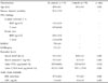
The demographic, clinical, and laboratory characteristics of patients with AS and of control subjects are summarized in Table 1. Mean age of patients with AS was not different from that of controls. Mean ESR and CRP levels of patients with AS were significantly higher than those of the controls as were urinary CTX-I (p = 0.014) and CTX-II (p < 0.001) levels. The differences between patients with AS and controls for BALP and osteocalcin were not statistically significant.
Correlations between biochemical and clinical parameters
Table 2 shows correlations between biochemical and clinical parameters in patients with AS. The age and disease duration were not significantly related to any of the clinical or biochemical parameters, although CRP levels correlated well with BASDAI (r = 0.612, p < 0.001) while ESR did not. Among the clinical parameters, significant inverse correlations were found between BASDAI and femoral neck BMD (r = - 0.493, p = 0.003) and between BASDAI and femoral neck T scores (r = - 0.568, p < 0.001) in patients with AS.
Serum BALP and osteocalcin levels did not correlate with any of the clinical or laboratory parameters. Urinary CTX-I levels showed significantly inverse correlations with femoral neck BMD (r = - 0.414, p = 0.030) and femoral neck T scores (r = - 0.399, p = 0.037), but not with spinal BMD or T score. There were significantly positive correlations between urinary CTX-I levels and BASDAI (r = 0.445, p = 0.016) as well as between urinary CTX-I and CRP levels (r = 0.491, p = 0.006). A significantly positive correlation was also found between urinary CTX-II levels and spinal BASRI (r = 0.472, p = 0.010).
DISCUSSION
In the present study, an elevated urinary CTX-I level, a marker for bone resorption, was found to reflects increased disease activity and decreased BMD in men with AS. Although this study enrolled young men (mean 28.8 years) so as to exclude confounding factors of osteoporosis, such as age, sex, and menopause, both BMD and T scores measured in men with AS in this study were relatively lower than in normal Korean young men. In addition, a strong correlation was found between disease activity and loss of femoral neck BMD. Previous studies have demonstrated that patients with AS have normal bone formation marker levels and increased bone resorption markers, and that bone resorption markers reflect disease activity and the development of osteoporosis in patients with AS.15-18 In agreement with these previous findings, our patients with AS had normal bone formation markers and an increased bone resorption marker, and the increased bone resorption marker showed significantly positive correlation with disease activity and significantly inverse correlation with BMD in men with AS. These data support the theory that patients with AS have unbalanced bone turnover, and that inflammatory activity can alter bone metabolism and lead to osteoporosis.15-18
In our study, urinary CTX-I levels were significantly correlated only with loss of femoral neck BMD but not with loss of spinal BMD. Previously, Bronson et al. showed that BMD was significantly lower at the femur neck but not the posteroanterior lumbar spine in patients with AS than in control subjects,22 and they suggested that ankylosis of the posterior elements of the spine might contribute to the false elevation of posteroanterior lumbar BMD measured by DXA. Baek et al. also showed that the progression of bone loss may be reflected more reliably at proximal femur sites than at the lumbar spine.23 Thus, the possibility exists that syndesmophytosis, a process of new bone formation, caused an over-estimation of BMD in our study since it was measured at the posteroanterior lumbar spine.
The role of CTX-II measurement has been emphasized in several articular diseases other than AS. Our data demonstrate that urinary CTX-II levels, which specifically indicate degradation of cartilage, are also increased and related to radiographic damage of the spine in patients with AS. Significant associations of urinary CTX-II level with long-term progression of joint destruction in rheumatoid arthritis and with joint damage in severe osteoarthritis have been reported.9-11 However, the role of this cartilage degradation marker has not been determined in patients with AS, and our study is the first to report a significant correlation between increased urinary CTX-II levels and radiographic damage of the spine in patients with AS.
It is interesting to note that only the cartilage degradation marker showed a significant association with the parameter reflecting spinal damage in our patients. Radiographic damage of the spine, as assessed in this study by BASRI-spine, reflects syndesmophytosis,21 and syndesmophytosis represents pathological new bone formation in the spine. But bone formation markers were not found to be associated with BASRI-spine, and their serum levels in patients with AS were not different from those in controls. The significant association between a cartilage degradation marker and radiographic damage of the spine observed in this study suggests a role for cartilage in the pathogenesis of syndesmophytosis in patients with AS.
The pathogenic mechanisms responsible for the spinal damage and ankylosis observed in patients with AS are unknown. Although enthesitis is a distinctive feature of AS, its relevance to the pathogenesis of synovitis and its significance to the pathogenesis of disease at various other articular and extra-articular sites typically affected by AS, such as the sacroiliac joint, hip joint, and anterior uvea, are questionable. The disparate clinical, pathological, radiological, and immunological data in AS are more consistent with an alternative hypothesis of primary autoimmunity to cartilage and, in particular, to fibrocartilage.7 A peculiar predilection at fibrocartilaginous sites, including the intervertebral discs, manubriosternal joints, and symphysis pubis, is widely appreciated in AS.24,25 Proteoglycans present in cartilage and fibrocartilage have been shown to be involved in the immune response in spondyloarthritides, 26,27 and a histopathological study of sacroiliitis showed that a proliferative process of cartilage metaplasia and endochondral ossification, fibrosis, and formation of woven bone led to ankylosis.28 These observations suggest that cartilage, particularly fibrocartilage, may be another target of inflammation in AS and may be involved in syndesmophytosis. In this regard, the significant association between a cartilage degradation marker and spinal damage observed in this study supports the role of cartilage and its degradation in the pathogenesis of AS. Furthermore, our finding suggests that measurement of CTX-II levels can be useful in monitoring spinal damage in patients with AS. Further investigations enrolling a larger number of patients are needed to confirm the relationship found in this study and to gain better understanding of the role of cartilage in the pathogenesis of AS, particularly in the process of syndesmophytosis.
In summary, urinary CTX-I and CTX-II were elevated in men with AS and correlated well with disease activity, osteoporosis of the femur neck, and radiographic damage of the spine. Our findings suggest that these markers can be used to monitor disease activity, loss of BMD, or radiographic damage of the spine as well as to identify patients with AS who are at particular risk for serious complications and would thus benefit from more aggressive treatment early in the disease.

 XML Download