

 PDF
PDF ePub
ePub Citation
 Print
Print


INTRODUCTION
Pathologic lesions in the axilla develop in a wide range of disease. Among them, lymph node abnormality is the most common pathologic finding. The causes of the lymph node abnormality also vary, ranging from benign inflammatory disorders to malignant reasons.1 Most cases of lymphadenopathies detected through imaging examinations are not palpable, and pretreatment pathologic diagnoses are demanded. Fine needle aspiration cytology under the ultrasound guidance is commonly employed and has been reported to be the easiest and accurate method.2-4 Jaffer et al. reported the sensitivity and the specificity of the ultrasound-guided fine-needle aspiration cytology (US-guided FNAC) as high as 100%,5 and another report presented that the overall sensitivity of the US-guided FNAC was 86.4%, the specificity was 100%, and the diagnostic accuracy was 79%.3 However, the false negative rate of FNAC was 11.6%, and 8 of 38 patients (21.0%) who did not receive preoperative chemotherapy because of false negative rate.3 Biopsies with 18-gauge needle of the small series have been reported, and several studies have recently reported about the ultrasound-guided larger 14-gauge needle core biopsy of the axilla lesions.6,7 An imaging-guided core biopsy has higher sensitivity than FNAC for assessing breast lesions, and the trend would be similar to the US-guided core biopsy of the axilla. The purpose of this study was to investigate the usefulness and safety of the US-guided core biopsy of axilla lesions and to evaluate the benefit of preoperative diagnosis of the nodal staging in breast cancers.
MATERIALS AND METHODS
From March 2004 to September 2005, 31 patients with suspicious lymph nodes in the axilla, visible in ultrasound, underwent the US-guided core biopsy. A waiver of informed consent from institutional review board before our study was obtained. All patients underwent clinical examinations and mammogram before their ultrasound examinations. The mean age of patients was 45 ± 15.7 (from 31 to 64 years). Among 31 patients, 6 patients complained of a palpable mass in the axilla, and suspicious lymph nodes were incidentally found in the remaining 25 patients during the breast ultrasound. Abnormal Lymph nodes were categorized according to their shape and the morphology of the cortex and hilum as follows: 1) there was the length to width ratio (l/w) with less than 1.5, since l/w higher than 2.0 indicates that the node is benign;6 2) The cortex of the nodes was concentrically or eccentrically thickened to more than 2 mm;6 3) Compression of the hilus and especially the absence of the hilus are thought to be highly suggestive of malignancy. If the axillary lymph nodes exhibited any of abnormal findings mentioned above, they were defined as suspicious.
Sixteen patients were associated with breast cancer, and 15 other patients had no lesion in the breast. Among 16 breast cancer patients, 13 cases had a previous history of breast cancer. Their follow-up breast ultrasound revealed suspicious lymph nodes in the axilla without breast lesions. Seven out of 13 cases were contralateral lymph node, and 6 cases were ipsilateral lymph node in the axilla. Remaining 3 out of 16 patients were diagnosed of BI-RADS (Breast Imaging Reporting and Data System) category 5 lesions in the breast and suspicious lymph nodes in the axilla at the same tim: Those were seen at ipsilateral in one case and contralateral in two cases. The TNM stages of the breast cancer patients were stage I (n = 0), IIA (n = 2), IIB (n = 6), IIIA (n = 3), IIIB (n = 2), and IV (n = 0) in previously diagnosed patients, and stage IIB (n = 2) and IIIA (n = 1) in currently diagnosed patients.
Ultrasound scanning of the axilla and breast was carried out with a 10 - 12 MHz transducer with HDI 5000 machine (Advanced Technology Laboratories, Bothell, WA, USA).
The core biopsies of nonpalpable axilla lymph nodes were performed on suspicious axilla lymph nodes which were found during breast ultrasound. The contralateral or ipsilateral suspicious axilla lymph nodes in patients with ACR BI-RADS category 4 or 5 lesion in their breast and those patients whose breast cancer had previously been treated were also indicated. The lesion size was 0.6 cm to 3.3 cm (mean = 1.59 ± 0.76 cm). All biopsies were ultrasound-guided with a free hand approach. Fourteen-gauge (2.1 mm) needles were used with an automatic biopsy gun, and 15 mm shooting was selected (Bard-Magnum Biopsy Instrument, Covington, GA, USA). The number of needle pass was 3 to 5. For the evaluation of any post-procedural complications at the site of the biopsy, all patients underwent clinical examinations a few days after the biopsy.
For the patients who underwent axillary dissection (n = 4), the number and size of the dissected and metastatic nodes were reported. The close follow-up was conducted with ultrasound in patients treated with chemotheraphy or radiotheraphy. The cases of benign pathology were examined by ultrasound over the next 2 years.
RESULTS
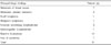
Among 31 cases of axilla lymph nodes biopsy, 11 cases showed malignant and 20 cases benign pathology. Seven of the 11 malignant nodes were metastatic lymph nodes from breast cancer; 2 were from unknown primary, and remaining 2 from lymphoma. Twenty benign histopathologic results were as follows: subacute necrotizing lymphadenitis in 2, dermatopathic lymphadenitis in 1, and reactive hyperplasia in 10 and free of carcinomas in 7 (Table 1). Among them, 8 patients had a previous history of breast cancer, and one patient was diagnosed with breast cancer at the current examination. There was no complication during and after the core biopsy procedure, and plenty of specimens were obtained even in a small lesion less than 1 cm in size.
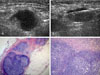
Out of 7 patients with breast cancer, 5 patients had a previous history of breast cancer. Only one patient showed an ipsilateral lymph node metastasis and 4 patients demonstrated a contralateral lymph nodes metastasis. All 5 patients received chemotheraphy, and 2 patients had a systemic metastasis, such as pericardium and supraclavicular lymph nodes metastases or a lung metastasis. One patient underwent contralateral axillary dissection, and metastasis in 5 out of 19 dissected lymph nodes was confirmed (Fig. 1).
Two of 7 patients were diagnosed with both breast cancer and metastatic lymph node by core biopsy. One of the 2 cases was crossed axillary metastasis, i.e., breast cancer in the right breast and a metastatic lymph node in the left axilla. Hence, she was taken for a modified radical mastectomy of the right breast and the contralateral axillay dissection and radiotherapy. The pathologic specimen showed positive in 14 of 19 lymph nodes. The other one patient received the modified radical mastectomy and the ipsilateral axillay dissection, which revealed 3 metastatic of 18 lymph nodes.
Two out of the 11 malignant pathologic results revealed metastatic lymph nodes (Fig. 2), however, primary site could not be identified through the mammogram, whole breast ultrasound, MRI and whole body PET. Thus, they underwent the axillary dissection and ipsilateral breast radiotherapy, and a close follow-up was performed for over 2 years.
Remaining 2 patients were diagnosed with Hodgkin's lymphoma and B-cell lymphoma, respectively. They were treated with chemotheraphy, and the outcomes were highly satisfactory.
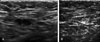
In the cases of chemotheraphy or radiotheraphy (n = 7), their follow-up ultrasound revealed that the pathologic lymph nodes were reduced in size and showed thinning of the cortex (Fig. 3). The mean of maximum diameters of lymph nodes before chemotherapy was 1.37 ± 1.1 cm and it was reduced to 0.72 ± 1.4 cm after the chemotherapy.
DISCUSSION
The percutaneous image-guided core biopsy increasingly becomes an alternative to a surgical biopsy for the histologic assessment of breast lesions.8 Guidance for the percutaneous biopsy is provided by stereotaxis or ultrasound. Especially, the ultrasound-guided 14-gauge automated core biopsy is safe, fast, accurate, and cost effective. Advantages of ultrasound as a guidance modality for the percutaneous biopsy include lack of ionizing radiation, use of nondedicated equipment, accessibility of all areas of the breast and the axilla, real- time visualization of the needle, multidirectional sampling, and low cost.9 The only disadvantage of the ultrasound guidance is that the lesion must be sonographically evident for implementing the ultrasound-guided biopsy. Therefore, we consider that the ultrasound guided axilla lymph nodes core biopsy can also be an alternative to the surgical biopsy for the histologic evaluation of axilla lesions.
The axillary nodal status is a very important factor in the preoperative staging in breast cancer patients, and it is also crucial in the pre-op decision to perform a sentinel node biopsy or axillary lymph node dissection. In a majority of cases, US-guided FNAC has been an initial procedure to detect the lymph node metastasis in the axilla. However, the US-guided FNAC exhibits relatively high false negative rate (11.6% to 21.0%),3 and this is most probably due to inadequate volume sampling, fibrosis, inflammatory reactions, and previous radiation therapies.10 Hence, several authors6,7 tried to replace the US-guided FNAC of axilla lymph nodes with the large needle core biopsy using a gun in the preoperative staging of breast cancer patients. To our best knowledge, previous studies performed the core biopsy by using 16-gauge or 18-gauge needles, and there are a few reports about the 14-gauge needle core biopsy of the axilla lymph node.6,11,12 In the case of the breast core biopsy, we routinely implement the 14-gauge needle for sampling more specimens. However, it is undoubtful whether the more specimens we obtain, the more precise diagnosis is obtainable. In our series, we acquired 11 cases of malignant pathology in the core biopsy. Except 4 patients who needed the axilla dissection for clearing the breast cancer treatment, the axillary dissection was unnecessary for remaining 7 patients. In cases of benign pathology in the axilla lymph node core biopsy, there was no case to suggest false negative results such as the progression of the lymph node size or the change of the morphology in the ultrasound follow-up. Therefore, we postulate that the ultrasound follow-up of pathologically confirmed benign lymph nodes in the core biopsy is a satisfactory without any additional imaging study or a further surgical intervention.
Various complications of the percutaneous core biopsy such as hemorrhage or infection have been reported. However, there were no cases of non-diagnostic specimens and no complications in our 31 patients who had received the large needle gun biopsy (14G) of axilla lymph nodes. Nevertheless, a special care should be taken for reducing unwanted complications by fully acknowledging detailed anatomy including vessels and nerves in the axilla. The nerves are not usually visible on ultrasound, while the axillary vein and artery can easily be recognized. Since the major nerves lie between the vein and artery, their path can be presumed through imaging the vessels.7 Lymph nodes, on the other hand, lie more superficially than the neurovascular structures. Thus, proper positioning of patients and perfect guiding by ultrasound are highly important to avoid traumatic injuries during the large needle core biopsy of the axilla.
In conclusion, the US-guided large needle (14G) core biopsy of the axilla lesion is safe and effective for the pathological evaluation. The core biopsy could easily be employed if suspicious lymph nodes or mass lesions were found in the axilla. Especially in breast cancer patients, the core biopsy of the axilla lymph node could be an effective alternative to surgical biopsy for the histological diagnosis.

 XML Download