

 PDF
PDF ePub
ePub Citation
 Print
Print


INTRODUCTION
Recently, several studies reported the usefulness of manganese-enhanced T1-weighted MR cholangiography in evaluating the biliary system, reflecting the dynamics of bile flow.1-6 We illustrate here a case report to show how manganese-enhanced T1- and T2-weighted MR cholangiography could differentiate cystic parenchymal lesions from cystic abnormalities which communicate with the bile ducts.
CASE REPORT
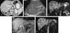
A 53-year-old-man was referred to our hospital because of abnormal findings on CT scan for health screening showing multiple, nonenhancing, small, hypodense nodules scattered throughout his entire liver. He did not have any malignant disease. Laboratory findings including liver function and blood chemistry were normal. To differentiate from multiple cysts, ultrasonography was performed. Hepatic US shows an inhomogeneous echo texture with some hypoechoic and hyperechoic nodules. We could not differentiate cystic lesions from solid nodules on CT and sonography. Bile duct was not dilated. MR cholangiography was performed with a 1.5-T superconducting unit (Magnetom Vision; Siemens Medical Systems, Erlangen, Germany) and a phased-array torso coil. The precontrast T2-weighted MR cholangiography using a half-Fourier rapid acquisition with relaxation enhancement (RARE) sequence with breath holds (TR/effective TE, infinite/95 msec; matrix, 240 × 256; field of view, 300 - 350 mm) showed multiple, small, and well-defined high signal intensity foci scattered throughout the liver, suggesting as multiple cystic lesions. And bile ducts appeared as high signal intensity structures. The relationship between the bile ducts and the multiple small cystic lesions could not be clarified in this study. Post-manganese 3D T1-weighted fat-saturated volumetric interpolated breath-hold images (TR/TE, 4.2/1.6; flip angle, 120; matrix 205 × 256; field of view, 300 - 350 mm; and 24 partitions interpolated to 48 slices with a thickness of 1.3 mm) showed only bile ducts as high signal intensity structures. Post-manganese enhanced T2-weighted MR cholangiography showed persistent high signals in the multiple small cystic lesions and the loss of bile duct signals. Manganese-enhanced T1- and T2-weighted MR cholangiography clearly showed that there was no communication between the bile ducts and cystic lesions (Fig. 1).
DISCUSSION
Recently, several studies reported the usefulness of manganese-enhanced T1-weighted MR cholangiography in evaluating the biliary system.1-6 Because manganese is a paramagnetic metal ion, it acts primarily on T1, resulting in T1 shortening, while also acting on and shortening T2.1-6 Enhanced liver and functioning bile ducts, therefore, have higher signal intensity on T1-weighted images and lower signal intensity on T2-weighted images.1-6 Therefore, mangafodipir trisodium is primarily used as a positive contrast agent of T1-weighted MR cholangiography. Additionally, it acts as a negative contrast agent on conventional heavily T2-weighted MR cholangiography. On T2-weighted MR cholangiography after manganese administration, signal intensity is lost in the functioning bile duct, but persists in the non-functioning bile duct or the non-communicating structure with the bile ducts. This characteristic property of combined T1- and T2-weighted MR cholangiography before and after administration of mangafodipir trisodium could be used as an excellent imaging tool in evaluating biliary dynamics.1-6
After mangafodipir trisodium injection, in our study, T1-weighted MR cholangiography only depicted the draining bile ducts as high signal intensity structures whereas T2-weighted MR cholangiography depicted the cystic lesions as well as the bile ducts as high signal intensity foci. They allowed us to separate the draining bile ducts from the cystic lesions, providing clear evidence of no communication between them. Therefore, manganese-enhanced T1- and T2-weighted MR cholangiography allow the differentiation of cystic parenchymal lesions from dilated biliary ducts or other bile duct anomalies communicating with the normal bile ducts. Mortele, et al.7 reported that MR cholangiography allowed differentiation of biliary hamartomas from dilated bilary ducts or other bile duct anomalies, such as Caroli's disease. They suggested that conventional T2-weighted MR cholangiography provided information on the communication between the hamartomas and the draining bile ducts.7 They also reported that the draining bile ducts in their subjects appeared normal.7 However, we disagree with their opinion, because conventional T2-weighted MR cholangiography cannot separate the draining bile ducts from cystic parenchymal lesions.
In conclusion, pre- and post-manganese-enhanced T1- and T2-weighted MR cholangiography makes it possible to differentiate cystic parenchymal lesions from cystic abnormalities which communicate with the bile ducts.
 XML Download